Open Journal of Ophthalmology, 2013, 3, 97-102
Published Online November 2013 (http://www.scirp.org/journal/ojoph)
http://dx.doi.org/10.4236/ojoph.2013.34023
Open Access OJOph
97
A Clinical Study of Pterygium and Results of Treatment by
Excision and Limbal Autograft or Augmented with
Post-Op Mitomycin C
Achyut N. Pandey1, Nishant Marken2, Ravinder Marken2, Bhuwan Chandra Pandey3
1Department of Ophthalmology, VCSG Government Medical College and Research Institute, Srinagar Garhwal, India; 2Ravi Eye
Hospital, Bhawanigarh, Punjab; 3Narayan Netralaya, Srinagar Garhwal, Uttarakhand.
Email: achyutpandey@gmail.com
Received July 16th, 2013; revised August 19th, 2013; accepted September 9th, 2013
Copyright © 2013 Achyut N. Pandey et al. This is an open access article distributed under the Creative Commons Attribution Li-
cense, which permits unrestricted use, distribution, and reproduction in any medium, provided the original work is properly cited.
ABSTRACT
Aim: To prospectively analyze the clinical profile of pterygium and to compare results of management by excision with
limbal conjunctival autograft or postoperative topical Mitomycin C drops. Methodology: Study was conducted over a
period of 23 months, at a tertiary eye care hospital including 80 eyes of 80 patients who underwent surgery, out of
which 40 underwent limbal conjunctival autograft and the remaining 40 underwent pterygium excision followed by
Mitomycin C after fulfilling the inclusion criteria. A detailed history was taken and recorded regarding the d isease with
reference to age, occupation, residence, exposure to dust and hot wind. The extent of corneal involvement by the ptery-
gium was noted. The patients were followed after one week and then monthly for a year. BCVA were noted on every
visit and slit lamp examination was done for recurren ce, sclera thinning and corneal vascularisation. Result: 80 eyes of
80 patients were enrolled with male preponderance, out of which 40 underwent limbal conjunctival autograft (gr. A)
and the remaining 40 underwent pterygium excision followed by Mitomycin C (gr. B). All patients were in the age
group of 23 to 70 years. The study showed a higher incidence of pterygium in the age group of 41 - 50 years with male
preponderance probably due to chronic dryness, and exposure to ultraviolet light, dust, and hot winds. The right eye was
more affected than the left eye, and nasal side was more involved than the temporal side. The recurrence among group
A was 2 out of 40 with a recurrence rate of 5% and among group B was 3 out of 40 with a recurrence rate of 7.5%.
Scleral thinning was seen in two cases (5%) in patients who underwent pterygium excision followed by Mitomycin C.
Conclusion: Conjunctival limbal autograft and postoperative MMC (0.02%) are both safe and effective adjuncts to
primary pterygium surg ery. The main prejudices against autografting are the expertise and time required for the proce-
dure. The recent use of biologic adhesives to fixate the autograft in place may simplify the procedure. Age of the pa-
tients was strongly associated with recurrence regardless of which procedure was used. More research needs to be done
to delve into this seemingly innocuous pathology of conjunctiva to effectively manage the disease condition.
Keywords: Pterygium; Autograft; Mitomycin C; Vascularisation
1. Introduction
Pterygium is a cause of diminution of vision when it en-
croaches on the pupillary area. The patient also has cos-
metic problems. Epidemiological surveys indicate that
the prevalence rates of pterygium vary depending on the
exact population [1-6]. Overall prevalence rates range
from 0.7 to 31% in various popu lations of the world [1-6].
As a general rule, prevalence rates for a pterygium in-
crease with age although a decline in prevalence rates has
been reported for patients over 60 to 70 years old [1,3]. It
typically develops in patients who have been living in hot
climates and may represent a response to chronic dryness
and exposure to u ltraviolet light, dust and ho t winds. It is
rarely seen in patients younger than the age of 20 years.
With the development of new methods of treatment, fre-
quency and severity of pterygium have declined. The
study was conducted to study the clinical profile of
pterygium and to compare results of recurrence by exci-
sion with limbal conjunctival autografts or postopera-
tive topical Mitomycin C drops.
A Clinical Study of Pterygium and Results of Treatment by Excision and Limbal
Autograft or Augmented with Post-Op Mitomycin C
98
2. Aim
To prospectively analyze a clinical study of pterygium
and results of treatment by excision and limbal autograft
surgery or augmented with post operative topical mito-
mycin C.
3. Materials and Methods
Present study was the prospective, comparative case
study involving 80 eyes of 40 patients who attended the
attended the tertiary eye care hospital in South India .The
duration of the study was 2 years. The study was re-
viewed by the institutional review board. Each eligible
patient provided informed consent prior to the enro lment
in the study.
3.1. Inclusion Criteria
All cases of pterygium attending the OPD including re-
current pterygium .
3.2. Exclusion Criteria
Patients coming from far away places who were not able
to come for regular follow up were excluded from this
study.
3.3. Methods
A detailed history was taken and recorded regarding the
disease with reference to age, occupation, residence, ex-
posure to dust and hot wind. The extent of corneal in-
volvement by the pterygium was noted.
Patient were assigned to two groups (A and B) ran-
domly. Group A underwent limbal conjuctival autograft.
Group B underwent pterygium excision augmented with
postoperative topical mitomycin C application (Figure 1
and 2). In patients belonging to Group A excision of
perygium was done followed by limbal conjuctival auto-
graft taken from upper temporal quadrant and sutured on
the bare sclera using 6.0 vicryl sutures (Figure 3 and 4).
Patients were given gatifloxacin or moxifloxacin with
dexamethasone eye drop 4 times a day for 2 weeks, then
Figure 1. Pterygium before excision.
Figure 2. Pterygium after excision.
Figure 3. Pterygium enroaching the pupillary area.
Figure 4. Flashy pterygium.
3 times a day for 2 weeks, and then 2 times a day for 2
weeks. Gatifloxacin or moxifloxacin with dexametha-
sone ointment was applied at night for 6 weeks. The pa-
tient were followed after one week and then monthly for
a year. Patients belonging to group B were treated with
mitomycin C in a strength of 0.01 mg/ml in carboxy
methyl cellulose. On first and second postoperative day
patient were told to use either gatifloxacin or moxiflox-
acin eye drops 6 times a day. From third to twenty first
postoperative day patients were given mitomycin C drops
4 times a day and gatifloxacin (3 mg) with dexametha-
sone (1 mg) eyedrops 4 times a day.
Patients were followed weekly for three weeks and
then monthly for a period of one year. Best corrected
visual acuity were noted on every visit. Patients were
asked for any pain, photophobia, redness, lacrimation.
Slit lamp examination was done and eye was looked
for:
1) Any reccurrence.
2) Scleral thinning.
Open Access OJOph
A Clinical Study of Pterygium and Results of Treatment by Excision and Limbal
Autograft or Augmented with Post-Op Mitomycin C 99
3) Corneal vascularisation.
4. Results
The present study was conducted at the attended the ter-
tiary eye care hospital in South India over a period of 23
months (October 2009 to September 2011).
80 eyes of 40 patients were enrolled in this study. Pa-
tients were assigned to two groups (A and B) randomly.
Group A underwent limbal conjuctival autograft, 40 pa-
tients. Group B underwent pterygium excision aug-
mented with postoperative topical mitomycin C applica-
tion included 40 patients.
4.1. Age Distribution
Out of the 80 patients maximum number of patients were
in the age group from 41 to 50 years in which the
youngest patient was 23 years of age and the oldest pa-
tient was 70 years of age.
4.2. Sex Distribution
Out of the 80 patients there was a male preponderance in
this study with 47 males (58.75%) as compared to 33
females (41.25%). The higher incidence in males could
be attributed to their greater exposure to hot, dry and
dusty climate.
4.3. Eye Involved
Out of 91 eyes in 80 patients right eye was involved in
54 patients (67.5%), left eye was involved in 37 patients
(46.25%).
4.4. Site of Pterygium
The site of pterygium is found to be nasal in 88 eyes
(96.70%) of eyes, while temporal in 3 (3.30%) of eyes.
4.5. Size of Pterygium
The cornea was encroached by pterygium between 2 - 3
mm in 88 eyes (96.70%) while the cornea was involved
greater than 3 mm by pterygium in 3 eyes (3.30%).
4.6. Recurrence Rates
One of the main aims of the study was to compare the
effectiveness of conjuctival limbal autografting and mi-
tomycin C in preventing the recurrence of pterygium. In
the present study it was found that recurrence of ptery-
gium occurred in 2 (5%) out of 40 eyes treated with lim-
bal conjuctival autograft and 3 (7.5%) out of 40 eyes in
whom pterygium excision was followed by postoperative
mitomycin C.
4.7. Recurrence Time
In cases where limbal conjuctival autografting was done
there was recurrence occurring in the first and third
month whereas in cases where pterygium excision fol-
lowed by mitomycin C was done recurrence occurred in
third, fourth and sixth month of follow up.
4.8. Age of Patients with Recurrence
In both group, patients showing recurrence are less than
50 years (5%).
4.9. Postoperative Complications
In Group B, 2 pts showed the sclera thinning while no
complication found in Group A.
5. Discussion
5.1. Age Distribution
In the study conducted by Alemwork Meseret et al. [7]
prevalence of pterygium was more in middle and old age
group. In the study conducted by Jang Sool Kwon [8]
also prevalence was more in middle and old age group.
The present study shows that maximum number of pa-
tients were in the age group from 41 to 50 years in wh ich
the youngest patient was 23 years of age and the oldest
was 70 years of age. So th e present stud y agrees with the
previous studie s.
5.2. Sex Distribution
Duke Elder [9] observed that pterygium is more likely to
occur in outdoor workers, and hence it is more common
in men than in women. In the study conducted by Rao et
al. [10] also prevalence was more in males.
The present study shows a male preponderance with
47 males (58.75%) as compared to 33 females (41.25%).
So the present study agrees with the previous studies.
5.3. Eye Involved
In the study conducted by McCoombes et al. [11] of the
258 cases in 135 right eye was involved as compared to
123 cases in which the left eye was involved.
The present study shows that right eye was involved in
54 patients (67.5%), left eye in 37 patients (46.25%). So
the present study agrees with the previous studies.
5.4. Site of Pterygium
Pterygia occurs more co mmonly on the nasal side proba-
bly due to greater exposu re of this area to actinic damage,
but it may occur temporally or at times bilaterally. In the
study conducted by Rachmiel et al. [12] all the pterygia
Open Access OJOph
A Clinical Study of Pterygium and Results of Treatment by Excision and Limbal
Autograft or Augmented with Post-Op Mitomycin C
100
were located nasally while in the study conducted by Rao
et al. [13] of the 53 eyes (51 patients) in 46 cases the
pterygia occurred nasally while in 4 cases there were
temporal ptery gi a .
The present study shows that the pterygium was nasal
in 88 eyes and temporal in 3 eyes (Table 1) which agrees
with the previous stud ies.
5.5. Size of Pterygium
In the study conducted by Shimazaki et al. [13] the size
of the pterygia ranged from 1.5 to 6 mm while in another
study conducted by Rachmiel [9] et al. the range was 2-
6.8 mm.
The present study shows that the cornea was en-
croached by pterygium between 2 - 3 mm in 88 eyes
(96.70%) while the cornea was involved greater than 3
mm by pterygium in 3 eyes (3.30%) (Table 2) which
agrees with the previous studies.
5.6. Recurrence Rates
Kenyon et al. [14] popularized the conjuctival autograft
technique in 1985, reporting retrospectively on 57 cases
of primary and recurrent pterygia, showing a recurrence
rate of only 5.3%. Allan et al. [15] reported a recurrence
of 6.5% in patients who underwent limbal conjuctival
autograft surgery. R. Rachmiel et al. [9] reported recur-
rence rate of 2.6% in patients with pterygium excision
followed by MMC. Cardillo et al. [16] reported recur-
rence between 4.0% and 6.6% in patients undergoing
pterygium excision followed by MMC.
In the present study it was found that recurrence of
pterygium occurred in 2 (5%) out of 40 eyes treated with
limbal conjuctival autograft and 3 (7.5%) out of 40 eyes
(Table 3) in whom pterygium excision was followed by
postoperative mitomycin C.
Differences in recurrence rates in various studies are
thought to be due to various factors including surgical
technique used, expertise of the surgeon, patient popula-
tion characterstics, latitude, race.
Table 1. Shows the site of pterygium involved.
Site No. of eyes Percentage
Nasal 88 96.70%
Temporal 3 3.30%
Total 91 100%
Table 2. Shows the size of pterygium.
Size No of eyes Percentage
2 - 3 mm 88 96.70
Greater than 3 mm 3 3.30
5.7. Recurrence Time after Surgery
Chen et al. [17] reported the mean time of recurrence
varied from 3.7 to 4.8 months with only 6% of recur-
rences occurring after the sixth postoperative month.
In the present study, cases where limbalconjuctival
autografting was done there was recurrence occurring in
the first and third month whereas in cases where ptery-
gium excision followed by mitomycin was done recur-
rence occurred in third, fourth and sixth month of follow
up (Table 4). So the present study agrees with previous
studies.
5.8. Age of Patients with Recurrence
In the study conducted by Figueiredo et al. [18] patients
with age less than 50 years had significantly more recur-
rences. It has been suggested that lipoid degeneration in
the cornea is an inhibiting factor to pterygium growth,
based on observations that pterygium does not cross an
arcus senilis to any great extent. The presence of in-
creasing amounts of lipoid degeneration with age might
explain in part, the strong association between age and
recurrence.
The present study (Table 5) shows that there were two
recurrences in eyes with pterygium excision followed by
conjunctival autograft with age less than 50 years of age
whereas in eyes with pterygium excision followed by
mitomycin C there was one recurrence in patients less
Table 3. Shows the recurrence rates.
Pterygium autograft
Pterygium excision
with mitomycin C
No of eyes treated 40 40
Recurrence no. % 2 (5%) 3 (7.5%)
Table 4. Shows the recurrence time after surgery.
Time of
recurrence
Group A
(conjuctival
autograft)
Group B (pterygium excision
followed by mitomycin C)
1 month 1 0
2 months 0 0
3 months 1 1
4 months 0 1
5 months 0 0
6 months 0 1
7 months 0 0
8 months 0 0
9 months 0 0
10 months 0 0
11 months 0 0
12 months 0 0
Open Access OJOph
A Clinical Study of Pterygium and Results of Treatment by Excision and Limbal
Autograft or Augmented with Post-Op Mitomycin C
Open Access OJOph
101
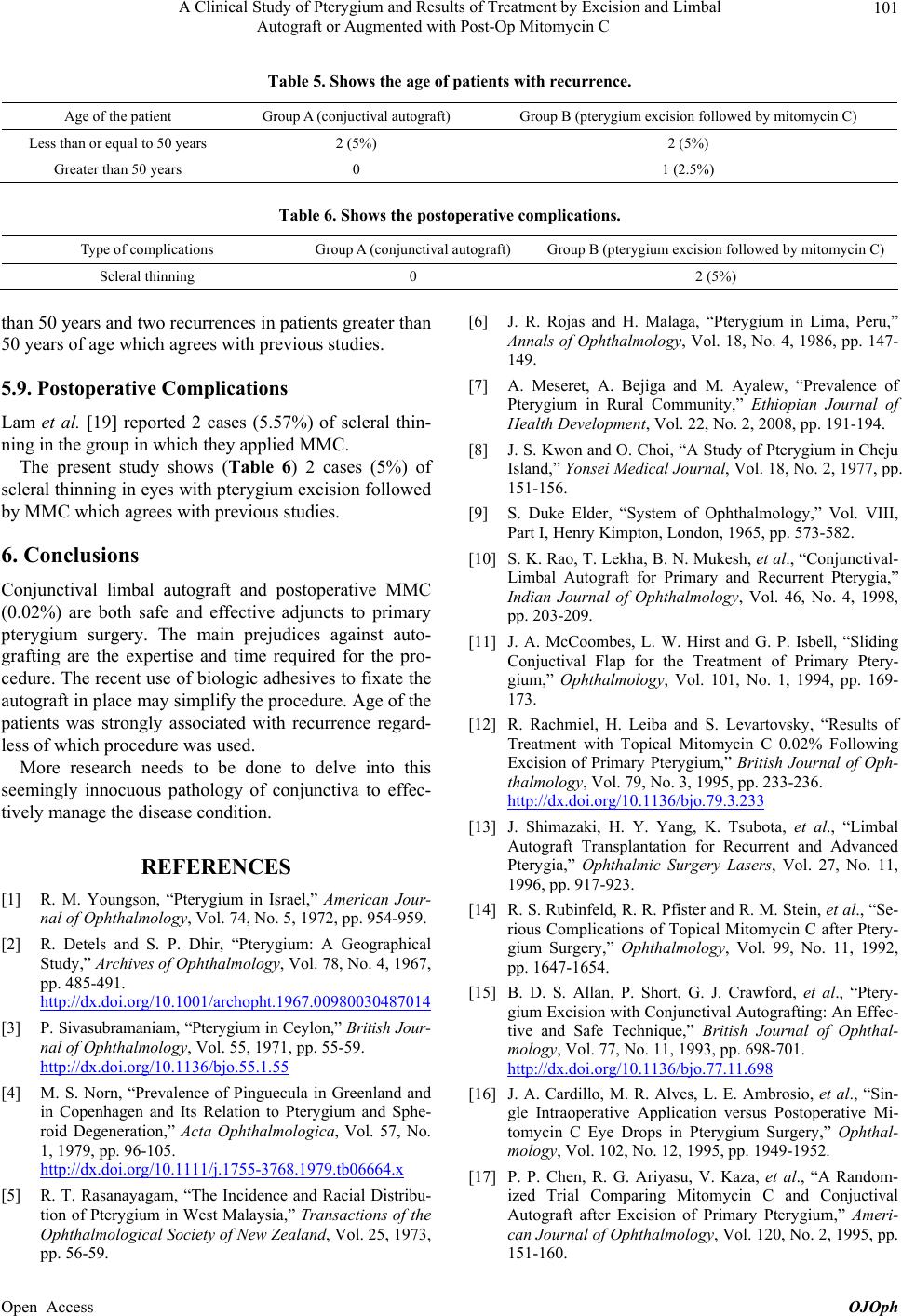
Table 5. Shows the age of patients with recurrence.
Age of the patient Group A (conjuctival autograft) Group B (pterygium excision f ollowed by mitomycin C)
Less than or equal to 50 years 2 (5%) 2 (5%)
Greater than 50 years 0 1 (2.5%)
Table 6. Shows the postoperative complications.
Type of complications Group A (conjunctival autograft) Group B (pterygium excision followed by mitomycin C )
Scleral thinning 0 2 (5%)
than 50 years and two recurrences in patients greater than
50 years of age which agrees with previous studies.
5.9. Postoperative Complications
Lam et al. [19] reported 2 cases (5.57%) of scleral thin-
ning in the group in which they applied MMC.
The present study shows (Table 6) 2 cases (5%) of
scleral thinning in eyes with pterygium excision followed
by MMC which agrees with previous studies.
6. Conclusions
Conjunctival limbal autograft and postoperative MMC
(0.02%) are both safe and effective adjuncts to primary
pterygium surgery. The main prejudices against auto-
grafting are the expertise and time required for the pro-
cedure. The recent use of biologic adhesives to fixate the
autograft in place may simplify the procedure. Age of the
patients was strongly associated with recurrence regard-
less of which procedure was used.
More research needs to be done to delve into this
seemingly innocuous pathology of conjunctiva to effec-
tively manage the disease condition.
REFERENCES
[1] R. M. Youngson, “Pterygium in Israel,” American Jour-
nal of Ophthalmology, Vol. 74, No. 5, 1972, pp. 954-959.
[2] R. Detels and S. P. Dhir, “Pterygium: A Geographical
Study,” Archives of Ophthalmology, Vol. 78, No. 4, 1967,
pp. 485-491.
http://dx.doi.org/10.1001/archopht.1967.00980030487014
[3] P. Sivasubramaniam, “Ptery gium in Ceylon,” British Jour-
nal of Ophthalmology, Vol. 55, 1971, pp. 55-59.
http://dx.doi.org/10.1136/bjo.55.1.55
[4] M. S. Norn, “Prevalence of Pinguecula in Greenland and
in Copenhagen and Its Relation to Pterygium and Sphe-
roid Degeneration,” Acta Ophthalmologica, Vol. 57, No.
1, 1979, pp. 96-105.
http://dx.doi.org/10.1111/j.1755-3768.1979.tb06664.x
[5] R. T. Rasanayagam, “The Incidence and Racial Distribu-
tion of Pterygium in West Malaysia,” Transactions of the
Ophthalmological Society of New Zealand, Vol. 25, 1973,
pp. 56-59.
[6] J. R. Rojas and H. Malaga, “Pterygium in Lima, Peru,”
Annals of Ophthalmology, Vol. 18, No. 4, 1986, pp. 147-
149.
[7] A. Meseret, A. Bejiga and M. Ayalew, “Prevalence of
Pterygium in Rural Community,” Ethiopian Journal of
Health Development, Vol. 22, No. 2, 2008, pp. 191-194.
[8] J. S. Kwon and O. Choi, “A Study of Pterygium in Cheju
Island,” Yonsei Medical Journal, Vol. 18, No. 2, 1977, pp.
151-156.
[9] S. Duke Elder, “System of Ophthalmology,” Vol. VIII,
Part I, Henry Kimpton, London, 1965, pp. 573-582.
[10] S. K. Rao, T. Lekha, B. N. Mukesh, et al., “Conjunctival-
Limbal Autograft for Primary and Recurrent Pterygia,”
Indian Journal of Ophthalmology, Vol. 46, No. 4, 1998,
pp. 203-209.
[11] J. A. McCoombes, L. W. Hirst and G. P. Isbell, “Sliding
Conjuctival Flap for the Treatment of Primary Ptery-
gium,” Ophthalmology, Vol. 101, No. 1, 1994, pp. 169-
173.
[12] R. Rachmiel, H. Leiba and S. Levartovsky, “Results of
Treatment with Topical Mitomycin C 0.02% Following
Excision of Primary Pterygium,” British Journal of Oph-
thalmology, Vol. 79, No. 3, 1995, pp. 233-236.
http://dx.doi.org/10.1136/bjo.79.3.233
[13] J. Shimazaki, H. Y. Yang, K. Tsubota, et al., “Limbal
Autograft Transplantation for Recurrent and Advanced
Pterygia,” Ophthalmic Surgery Lasers, Vol. 27, No. 11,
1996, pp. 917-923.
[14] R. S. Rubinfeld, R. R. Pfister and R. M. Stein, et al., “Se-
rious Complications of Topical Mitomycin C after Ptery-
gium Surgery,” Ophthalmology, Vol. 99, No. 11, 1992,
pp. 1647-1654.
[15] B. D. S. Allan, P. Short, G. J. Crawford, et al., “Ptery-
gium Excision with Conjunctival Autografting: An Effec-
tive and Safe Technique,” British Journal of Ophthal-
mology, Vol. 77, No. 11, 1993, pp. 698-701.
http://dx.doi.org/10.1136/bjo.77.11.698
[16] J. A. Cardillo, M. R. Alves, L. E. Ambrosio, et al., “Sin-
gle Intraoperative Application versus Postoperative Mi-
tomycin C Eye Drops in Pterygium Surgery,” Ophthal-
mology, Vol. 102, No. 12, 1995, pp. 1949-1952.
[17] P. P. Chen, R. G. Ariyasu, V. Kaza, et al., “A Random-
ized Trial Comparing Mitomycin C and Conjuctival
Autograft after Excision of Primary Pterygium,” Ameri-
can Journal of Ophthalmology, Vol. 120, No. 2, 1995, pp.
151-160.
A Clinical Study of Pterygium and Results of Treatment by Excision and Limbal
Autograft or Augmented with Post-Op Mitomycin C
102
[18] R. S. Figueiredo, E. J. Cohen, J. A. P. Gomes, et al.,
“Conjunctival Autograft for Pterygium Surgery: How
Well Does It Prevent Recurrence,” Ophthalmic Surgery
Lasers, Vol. 28, No. 10, 1997, pp. 99-104.
[19] D. S. C. Lam, A. K. K. Wong and D. S. P. Fan, “Intraop-
erative Mitomycin C to Prevent Recurrence of Pterygium
after Excision. A 30-Month Follow-Up Study,” Oph-
thalmology, Vol. 105, No. 5, 1998, pp. 901-905.
http://dx.doi.org/10.1016/S0161-6420(98)95034-5
Open Access OJOph