 American Journal of Plant Sciences, 2013, 4, 1954-1959 http://dx.doi.org/10.4236/ajps.2013.410242 Published Online October 2013 (http://www.scirp.org/journal/ajps) Microtropins J-P: 6’-O-(2”S,3”R)-2”-Ethyl-2”,3”- Dihydroxybutyrates of Phenolic Alcohol -D-Glucopyranosides from the Branches of Microtropis japonica Yuka Uemura1, Sachiko Sugimoto1, Katsuyoshi Matsunami1, Hideaki Otsuka1,2*, Yoshio Takeda2 1Department of Pharmacognosy, Graduate School of Biomedical and Health Sciences, Hiroshima University, Hiroshima, Japan; 2Department of Natural Products Chemistry, Faculty of Pharmacy, Yasuda Women’s University, Hiroshima, Japan. Email: *hotsuka@hiroshima-u.ac.jp, *otsuka-h@yasuda-u.ac.jp Received July 26th, 2013; revised August 26th, 2013; accepted September 15th, 2013 Copyright © 2013 Yuka Uemura et al. This is an open access article distributed under the Creative Commons Attribution License, which permits unrestricted use, distribution, and reproduction in any medium, provided the original work is properly cited. ABSTRACT From the branches of Microtropis japonica (Celastraceae), seven phenolic alcohol glucosides, named microtropins J-P (1-7), were isolated. The 6-position of glucose was esterified with 2-ethyl-2,3-dihydroxybutyric acid. Microtropin K (2) was hydrolyzed under a mild basic condition to give methyl (2S,3R)-2-ethyl-2,3-dihydroxybutyrate, whose absolute structure was determined by the comparison of NMR data and the optical rotation value with that reported. Keywords: Microtropis japonica; Celastraceae; Microtropin; (2S,3R)-2-Ethyl-2,3-Dihydroxybutyrate 1. Introduction Celastraceous plants appeared in the spotlight after a po- tent antileukemic ansa-type macrolide, maytansine, was isolated from an Ethiopian shrub, Maytenus serrata (for- merly M. ovatus) [1,2]. In continuous research on subtro- pical resource plants, a Celestraceous plant, Microtropis japonica, collected in Okinawa attracted our attention and its constituents were investigated. In previous papers, the isolation of ent-labdane diterpene glucosides, micro- troipiosides A-F [3,4], from the leaves of M. japonica, and the 2-ethyl-2,3-dihydroxybutyrate of nine aliphatic glucosides, microtropins A-I [4], from its branches has already been reported. Further extensive isolation work on a MeOH extract of the branches of M. japonica re- sulted in the isolation of seven new aromatic glucoside (2S,3R)-2-ethyl-2,3-dihydroxybutyrates (1-7), named mi- crotropins J-P, along with two known compounds, vanil- lic (8) [5] and salicylic (9) acids (Figure 1). Isolation of 2-ethyl-2,3-dihydroxybutyrate may be interesting in a chemotaxonomic point of view. 2. Results and Discussion Seven new 2-ethyl-2,3-dihydroxybutyrates of various phenolic glucosides, named microtropins J-P (1-7), were isolated from the 1-BuOH-soluble fraction of a MeOH extract of the branches of M. japonica by a combination of various separation procedures. Their structures were elucidated from spectroscopic evidence. Microtropin J (1), [ ]D 24 ‒59.8, was isolated as an amorphous powder and its elemental composition was determined to be C19H26O11 by high-resolution (HR)-electrospray ioniza- tion (ESI) mass spectrometry (MS). The IR spectrum exhibited absorption bands assignable to hydroxy (3380 cm‒1), ester carbonyl (1730 cm‒1), carboxylic acid (1705 cm‒1), aromatic ring (1606, and 1511 cm‒1), phenolic al- cohol (1239 cm‒1), and aliphatic alcohols (1073 cm‒1). The UV absorption band at 245 nm also supported the presence of an aromatic ring. In the 13C-NMR spectra, six signals assignable to 2-ethyl-2,3-dihydroxybutyrate were observed together with ones assignable to glucose, which had a substituent at the 6-position. The 1H-NMR spectrum of the aglycone moiety comprised seven signals, which included one for a para-substituted aromatic ring with a carboxyl functional group. Acid hydrolysis liberated D- glucose, which was identified by HPLC analysis with a chiral detector. In the heteronuclear multiple bond corre- lation spectrum (HMBC), the anomeric proton (δH 5.03) showed a correlation peak with C-4 (δC 162.7), and H-6’ *Corresponding author. Copyright © 2013 SciRes. AJPS  Microtropins J-P: 6’-O-(2”S,3”R)-2”-Ethyl-2”,3”-Dihydroxybutyrates of Phenolic Alcohol -D-Glucopyranosides from the Branches of Microtropis japonica 1955 COO R2 RO OCH3 RO OH OCH3 OH RO OCH3 RO OCH3 OH OH COO H HO OCH3 COO H OH 1 2 3 R1 R1R2 H OCH3 OCH3 H H CH3 456/ 89 7 1 2 2 1 12 1 2 7 O 7 OH HO HO O O OH HO 1' 1" 2" 3" 4" 6" R: Figure 1. Structures of compounds isolated. (δH 4.21 and 4.65) with the carbonyl signal at δC 176.2. Therefore, the structure of microtropin J (1) was elucida- ted to be p-hydroxybenzoic acid O- -D-glucopyranoside 6’-O-2”-ethyl-2”,3”-dihydroxybutyrate, as shown in Fig- ure 1. The absolute configuration of the acyl moiety was expected to be the same (2”S,3”R) as that of microtropin A [4]. Microtropin K (2), [ ]D 24 ‒50.6, was isolated as an amorphous powder and its elemental composition was determined to be C20H28O12 by HR-ESI-MS. The IR and UV spectra showed similar absorption bands to those of 1, and in the NMR spectra, a methoxy signal [δH 3.90 (3H, s) on δC 56.8] was observed. The AA’BB’ type cou- pled four protons observed in 1 were replaced by three aromatic protons coupled in an ABX system. In the HMBC spectrum, one of the aromatic protons [H-2, δH 7.625 (s)] showed correlation peaks with C-1 (δC 126.4), C-4 (δC 151.8), and C-7 (δC 169.5), and the anomeric proton (δH 5.03) with C-4. From the above evidence, the structure of aglycone was determined to be vanillic acid and the overall structure is shown as 2 in Figure 1. Since microtropin K (2) was isolated in a good quantity, it was hydrolyzed under a mild alkaline condition to give vanl- lic acid -D-glucopyranoside (2a) [5] and methyl 2- ethyl-(2S,3R)-dihydroxybutyrate (2b) [4]. Microtropin L (3), [ ]D 23 ‒31.6, was isolated an amor- phous powder and its elemental composition was deter- mined to be C21H30O12 by HR-ESI-MS. Spectroscopic data were almost superimposable on those of 2, except for the presence of an ester methoxy group (δH 3.89 on δC 52.6), and the molecular weight was 14 mass units larger than that of 2, which accounted for a labile hydrogen atom being replaced by a methyl group. Therefore, the structure of 3 was elucidated to be ester of 2, and it may be an artifact produced during the extraction and isola- tion procedures. Microtropin M (4), [ ]D 25 ‒96.7, was isolated as an amorphous powder and its elemental composition was determined to be C20H30O12 by HR-ESI-MS. The IR spectrum exhibited a strong absorption band for a car- bonyl functional group and two methoxy signals were observed in the NMR spectra (δH 3.73 on δC 61.1 and 3.80 on δC 56.5). Judging from the NMR data, the aro- matic ring had two meta-coupled protons [δH 6.27 (d, J = 2.7 Hz) and 6.32 (d, J = 2.7 Hz)] and thus was suggested to have unsymmetrically substituted benzene, that is, four electro-negative functional groups are substituted at the 1, 3, 4 and 5 positions. The relatively deshielded methoxy signal (δC 61.1) was implied that it was located between the substituents. The HMBC correlations from H-1' (δH 4.77) to δC 155.6 (C-1), those from H-2 and H-6 to δC 133.6, to which the methoxy signal at δH 3.73 on δC 61.1 also correlated, and those from H-2 and H-6 to C-6 and C-2 respectively were observed (Figure 2). Therefore, the structure of aglycone was expected to be 4,5-dime- thoxybenzene-1,3-diol 1-O-β-D-glucopyranoside, which was also supported by the NOESY spectrum in which the anomeric proton showed significant correlation cross peaks with H-2 and H-6. The structure was finally eluci- dated to be 4 shown in Figure 1. Figure 2.Microtropin N (5), [ ]D 25 ‒11.5, was isolated as an amorphous powder and its elemental composition was determined to be C19H28O11. In the NMR spectra, one methoxy carbon and three protons coupled in an ABX system were observed in the aglycone region. Other NMR spectral data for the aglycone were essentially the same as those of isotachioside isolated from Isotachis ja- ponica [6]. Therefore, the structure of 5 was elucidated to be isotachioside 6’-O-(2”S,3”R)-2”-ethyl-2”,3”-dihydroxy- butyrate, as shown in Figure 1. Microtropin O (6) and microtropin P (7), [ ]D 25 ‒32.1 and [ ]D 26 ‒49.0, respectively, were isolated as amor- phous powders and the elemental compositions of both compounds were C22H34O12. In the 1H-NMR spectrum, O O OH HO HO O O OH OCH3 OCH3 OH OH HC MHBC Figure 2. HMBC correlations of 4. Dual arrow curve denote HMBC correlations were observed in both directions. Copyright © 2013 SciRes. AJPS 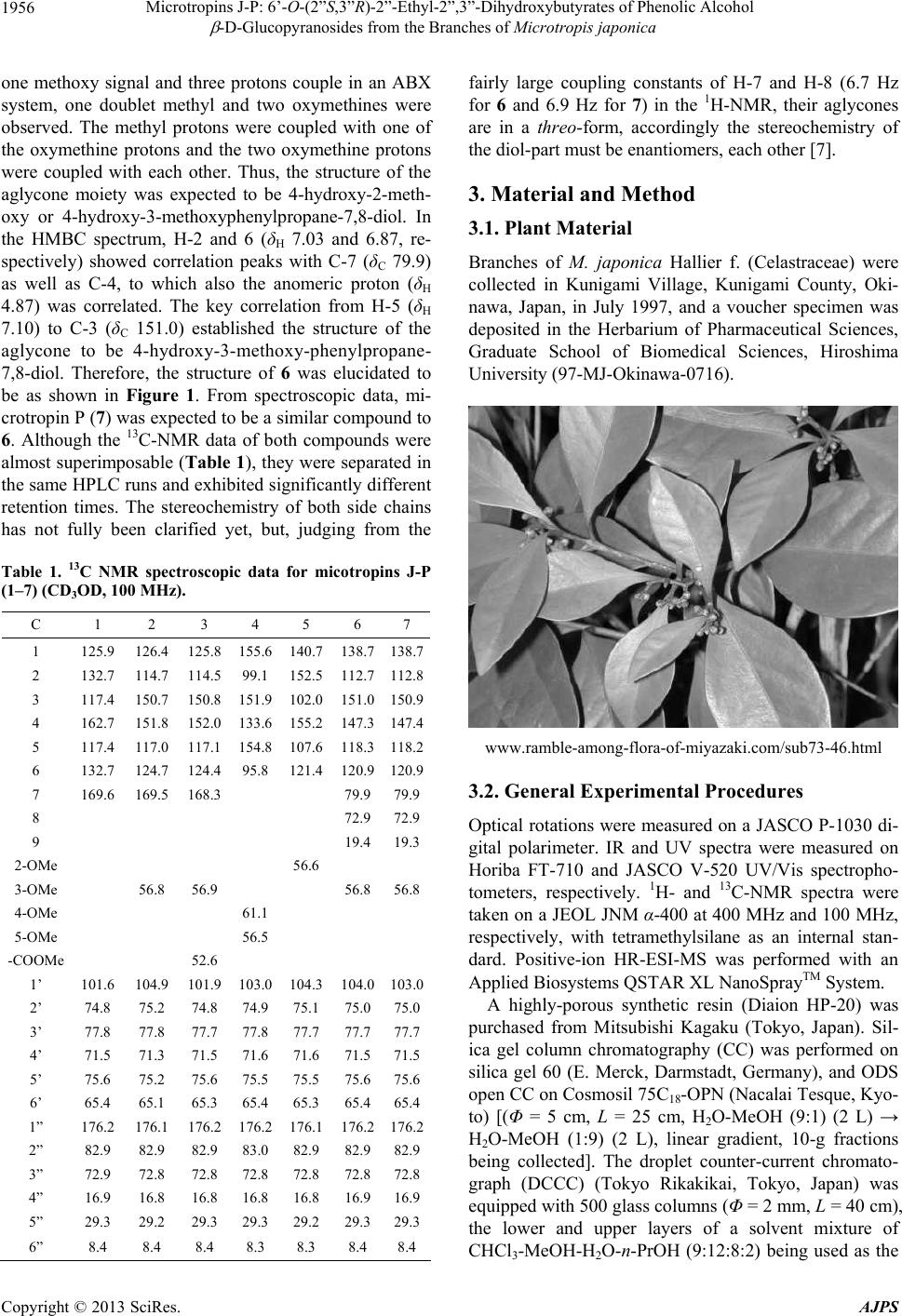 Microtropins J-P: 6’-O-(2”S,3”R)-2”-Ethyl-2”,3”-Dihydroxybutyrates of Phenolic Alcohol -D-Glucopyranosides from the Branches of Microtropis japonica 1956 one methoxy signal and three protons couple in an ABX system, one doublet methyl and two oxymethines were observed. The methyl protons were coupled with one of the oxymethine protons and the two oxymethine protons were coupled with each other. Thus, the structure of the aglycone moiety was expected to be 4-hydroxy-2-meth- oxy or 4-hydroxy-3-methoxyphenylpropane-7,8-diol. In the HMBC spectrum, H-2 and 6 (δH 7.03 and 6.87, re- spectively) showed correlation peaks with C-7 (δC 79.9) as well as C-4, to which also the anomeric proton (δH 4.87) was correlated. The key correlation from H-5 (δH 7.10) to C-3 (δC 151.0) established the structure of the aglycone to be 4-hydroxy-3-methoxy-phenylpropane- 7,8-diol. Therefore, the structure of 6 was elucidated to be as shown in Figure 1. From spectroscopic data, mi- crotropin P (7) was expected to be a similar compound to 6. Although the 13C-NMR data of both compounds were almost superimposable (Table 1), they were separated in the same HPLC runs and exhibited significantly different retention times. The stereochemistry of both side chains has not fully been clarified yet, but, judging from the Table 1. 13C NMR spectroscopic data for micotropins J-P (1‒7) (CD3OD, 100 MHz). C 1 2 3 4 5 6 7 1 125.9 126.4 125.8155.6 140.7 138.7138.7 2 132.7 114.7 114.599.1 152.5 112.7112.8 3 117.4 150.7 150.8151.9 102.0 151.0150.9 4 162.7 151.8 152.0133.6 155.2 147.3147.4 5 117.4 117.0 117.1154.8 107.6 118.3118.2 6 132.7 124.7 124.495.8 121.4 120.9120.9 7 169.6 169.5 168.3 79.979.9 8 72.972.9 9 19.419.3 2-OMe 56.6 3-OMe 56.8 56.9 56.856.8 4-OMe 61.1 5-OMe 56.5 -COOMe 52.6 1’ 101.6 104.9 101.9103.0 104.3 104.0103.0 2’ 74.8 75.2 74.8 74.9 75.1 75.075.0 3’ 77.8 77.8 77.7 77.8 77.7 77.777.7 4’ 71.5 71.3 71.5 71.6 71.6 71.5 71.5 5’ 75.6 75.2 75.6 75.5 75.5 75.675.6 6’ 65.4 65.1 65.3 65.4 65.3 65.465.4 1” 176.2 176.1 176.2176.2 176.1 176.2176.2 2” 82.9 82.9 82.9 83.0 82.9 82.982.9 3” 72.9 72.8 72.8 72.8 72.8 72.872.8 4” 16.9 16.8 16.8 16.8 16.8 16.916.9 5” 29.3 29.2 29.3 29.3 29.2 29.329.3 6” 8.4 8.4 8.4 8.3 8.3 8.4 8.4 fairly large coupling constants of H-7 and H-8 (6.7 Hz for 6 and 6.9 Hz for 7) in the 1H-NMR, their aglycones are in a threo-form, accordingly the stereochemistry of the diol-part must be enantiomers, each other [7]. 3. Material and Method 3.1. Plant Material Branches of M. japonica Hallier f. (Celastraceae) were collected in Kunigami Village, Kunigami County, Oki- nawa, Japan, in July 1997, and a voucher specimen was deposited in the Herbarium of Pharmaceutical Sciences, Graduate School of Biomedical Sciences, Hiroshima University (97-MJ-Okinawa-0716). www.ramble-among-flora-of-miyazaki.com/sub73-46.html 3.2. General Experimental Procedures Optical rotations were measured on a JASCO P-1030 di- gital polarimeter. IR and UV spectra were measured on Horiba FT-710 and JASCO V-520 UV/Vis spectropho- tometers, respectively. 1H- and 13C-NMR spectra were taken on a JEOL JNM α-400 at 400 MHz and 100 MHz, respectively, with tetramethylsilane as an internal stan- dard. Positive-ion HR-ESI-MS was performed with an Applied Biosystems QSTAR XL NanoSprayTM System. A highly-porous synthetic resin (Diaion HP-20) was purchased from Mitsubishi Kagaku (Tokyo, Japan). Sil- ica gel column chromatography (CC) was performed on silica gel 60 (E. Merck, Darmstadt, Germany), and ODS open CC on Cosmosil 75C18-OPN (Nacalai Tesque, Kyo- to) [(Φ = 5 cm, L = 25 cm, H2O-MeOH (9:1) (2 L) → H2O-MeOH (1:9) (2 L), linear gradient, 10-g fractions being collected]. The droplet counter-current chromato- graph (DCCC) (Tokyo Rikakikai, Tokyo, Japan) was equipped with 500 glass columns (Φ = 2 mm, L = 40 cm), the lower and upper layers of a solvent mixture of CHCl3-MeOH-H2O-n-PrOH (9:12:8:2) being used as the Copyright © 2013 SciRes. AJPS  Microtropins J-P: 6’-O-(2”S,3”R)-2”-Ethyl-2”,3”-Dihydroxybutyrates of Phenolic Alcohol -D-Glucopyranosides from the Branches of Microtropis japonica 1957 stationary and mobile phases, respectively. Five-gram fractions were collected and numbered according to their order of elution with the mobile phase. HPLC was per- formed on an ODS column (Inertsil; ODS-3, GL Science, Tokyo, Japan; Φ = 6 mm, L = 250 mm, 1.6 mL/min), and the eluate was monitored with a UV detector at 254 nm, and a refractive index monitor. 3.3. Extraction and Isolation Air-dried branches of M. japonica (13.0 kg) were ex- tracted three times with MeOH (30 L × 3) at room tem- perature for one week and then concentrated to 3 L in vacuo. The concentrated extract was washed with n-he- xane (3 L, 53.8 g), and then the MeOH layer was con- centrated to a gummy mass. The latter was suspended in water (3 L) and then extracted with EtOAc (3 L) to give 103 g of an EtOAc-soluble fraction. The aqueous layer was extracted with 1-BuOH (3 L) to give a 1-BuOH-so- luble fraction (40.9 g), and the remaining water-layer was concentrated to furnish 107 g of a water-soluble fraction. The 1-BuOH-soluble fraction (39.9 g) was sub- jected to Diaion HP-20 CC (Φ = 50 mm, L = 40 cm), us- ing H2O-MeOH (4:1, 2 L), (3:2, 2 L), (2:3, 2 L), and (1:4, 2 L), and MeOH (2 L), 500 mL-fractions being collected. The residue (7.53 g out of 8.73 g) in fractions 7 - 10 was subjected to silica gel (250 g) CC with increasing amounts of MeOH in CHCl3 [CHCl3 (3 L), and CHCl3-MeOH (49:1, 1.5 L), (24:1, 1.5 L), (23:2, 1.5 L), (9:1, 1.5 L), (17:3, 1.5 L), (4:1, 1.5 L), (3:1, 1.5 L), and (7:3, 1.5 L)], CHCl3-MeOH-H2O (35:15:2, 1.5 L), and MeOH (1.5 L), 300 mL-fractions being collected. The residue (0.962 g) in fractions 23 - 29 was separated by ODS open CC and the residue (108 mg) in fractions 81 - 87 was applied to DCCC. The residue (4.03 mg) in fractions 51 - 61 was purified by HPLC (H2O-MeOH, 3:1) to give 2.4 mg of 5 from the peak at 21 min. The residue (64.0 mg) in frac- tions 99 - 105 obtained on ODS open CC was subjected to DCCC and the residue (6.7 mg) in fractions 84 - 93 was purified by HPLC (H2O-MeOH, 13:7) to give 2.8 mg of 4 from the peak at 14 min. The residue (2.91 g out of 3.10 g) in fractions 30 - 38 obtained on silica gel CC was purified by ODS open CC to give three fractions. The residue (306 mg) in the second lot of fractions, 81 - 93, was separated by DCCC and the residue (45.5 mg) in fractions 33 - 35 was then purified by HPLC (H2O- MeOH, 33:7) to give 3.96 mg of 7 and 10.1 mg of 6 from the peaks at 46 min and 50 min, respectively. The residue (1.67 g) in fractions 94 - 124 obtained on ODS open CC was subjected to DCCC to give 1.21 g of 2 in fractions 63 - 86, 21.6 mg of 8 in fractions 142 - 164, and 10.1 mg of 3 in fractions 165 - 183. The residue (1.17 g out of 1.27 g) in fractions 39 - 47 obtained on silica gel CC was separated by ODS open CC and the residue (351 mg) in fractions 121 - 144 was applied to DCCC. The residue (86.6 mg out of 279 mg) in fractions 30 - 46 was purified by HPLC (H2O-MeOH, 4:1) to yield 14.2 mg of 1 and 21.0 mg of 2 from the peaks at 50 min and 65 min, respectively. The residue (5.68 g out of 6.22 g) in fractions 11 - 14 obtained on Diaion HP-20 CC was subjected to silica gel CC (180 g) with increasing amounts of MeOH in CHCl3 [CHCl3 (2 L), and CHCl3-MeOH (49:1, 1 L), (24:1, 1 L), (23:2, 1 L), (9:1, 1 L), (17:3, 1 L), (4:1, 1 L), (3:1, 1 L), and (7:3, 1 L)], CHCl3-MeOH-H2O (35:15:2, 1 L), and MeOH (1 L), 200 mL-fractions being collected. The re- sidue (1.30 g out of 1.41 g) in fractions 27 - 34 was se- parated by ODS open CC to give 13.6 mg of 9 in frac- tions 10 - 22. 3.4. Microtropin J (1) Amorphous powder, [ ]D 24 ‒59.8 (c 0.92, MeOH); IR max (film): 3380, 2934, 1730, 1705, 1606, 1511, 1454, 1239, 1073, 1016 cm‒1; UV max (MeOH): 245 (4.10), 209 (3.90) nm (log ); 1H-NMR (CD3OD, 400 MHz) δ: 7.98 (2H, d, J = 8.7 Hz, H-2 and 6), 7.14 (2H, d, J = 8.7 Hz, H-3 and 5), 5.03 (1H, d, J = 7.7 Hz, H-1’), 4.65 (1H, dd, J = 11.9, 2.1 Hz, H-6’a), 4.21 (1H, dd, J = 11.9, 6.3 Hz, H-6’b), 3.89 (1H, q, J = 6.4 Hz, H-3”), 3.72 (1H, ddd, J = 9.5, 6.3, 2.1 Hz, H-5’), 3.51 - 3.40 (3H, m, H-2’, 3’ and 4’), 1.73 (1H, dq, J = 14.8, 7.4 Hz, H-5”a), 1.55 (1H, dq, J = 14.8, 7.4 Hz, H-5”b), 1.15 (3H, d, J = 6.4 Hz, H3-4”), 0.84 (3H, dd, J = 7.4, 7.4 Hz, H3-6”); 13C-NMR (CD3OD, 100 MHz): Table 1; HR-ESI-MS (positive-ion mode) m/z: 453.1367 [M+Na]+ (calcd for C19H26O11Na, 453.1367). 3.5. Microtropin K (2) Amorphous powder, [ ]D 24 ‒50.6 (c 1.20, MeOH); IR max (KBr): 3410, 2978, 1735, 1706, 1602, 1513, 1462, 1270, 1073, 1022; UV max (MeOH): 285 (3.58), 250 (4.00), 214 (4.15) nm (log ); 1H-NMR (CD3OD, 400 MHz) δ: 7.626 (1H, d, J = 8.8 Hz, H-5), 7.625 (1H, s, H-2), 7.19 (1H, d, J = 8.8 Hz, H-6), 5.03 (1H, d, J = 7.3 Hz, H-1’), 4.64 (1H, dd, J = 11.9, 2.1 H-6’a), 4.19 (1H, dd, J = 11.9, 6.4 Hz, H-6’b), 3.90 (3H, s, -OCH3), 3.87 (1H, q, J = 6.4 Hz, H-3”), 3.69 (1H, ddd, J = 9.7, 6.4, 2.1 Hz, H-5’), 3.57 - 3.42 (3H, m, H-2’, 3’ and 4’), 1.70 (1H, dq, J = 14.6, 7.4 Hz, H-5”a), 1.53 (1H, dq, J = 14.6, 7.4 Hz, H-5”b), 1.14 (3H, d, J = 6.4 Hz, H3-4”), 0.82 (3H, dd, J = 7.4, 7.4 Hz, H3-6”); 13C-NMR (CD3OD, 100 MHz): Table 1; HR-ESI-MS (positive-ion mode) m/z: 483.1474 [M+Na]+ (calcd for C20H28O12Na, 483.1472). 3.6. Microtropin L (3) Amorphous powder, [ ]D 23 ‒31.6 (c 0.67, MeOH); IR Copyright © 2013 SciRes. AJPS  Microtropins J-P: 6’-O-(2”S,3”R)-2”-Ethyl-2”,3”-Dihydroxybutyrates of Phenolic Alcohol -D-Glucopyranosides from the Branches of Microtropis japonica 1958 max (film): 3392, 2938, 1733, 1716, 1512, 1458, 1274, 1115, 1073, 1028 cm‒1; UV max (MeOH): 335 (2.75), 287 (3.60), 248 (3.80), 220 (3.84) nm (log ); 1H-NMR (CD3OD, 400 MHz) δ: 7.62 (1H, d, J = 8.6 Hz, H-6), 7.61 (1H, s, H-2), 7.20 (1H, d, J = 8.6 Hz, H-5), 5.03 (1H, d, J = 7.3 Hz, H-1’), 4.62 (1H, dd, J = 11.9, 2.2 Hz, H-6’a), 4.19 (1H, dd, J = 11.9, 5.2 Hz, H-6’b), 3.90 (3H, s, -OCH3), 3.89 (3H, s, -COOCH3), 3.87 (1H, m, H-3”), 3.69 (1H, ddd, J = 9.5, 5.2, 2.2 Hz, H-5’), 3.56 - 3.22 (3H, m, H-2’, 3’ and 4’), 1.71 (1H, dq, J = 13.7, 7.4 Hz, H-5”a), 1.53 (1H, dq, J = 13.7, 7.4 Hz, H-5”b), 1.13 (3H, d, J = 6.6 Hz, H3-4”), 0.82 (3H, dd, J = 7.4, 7.4 Hz, H3-6”); 13C-NMR (CD3OD, 100 MHz): Table 1; HR-ESI-MS (positive-ion mode) m/z: 497.1630 [M+Na]+ (calcd for C21H30O12Na, 497.1630). 3.7. Microtropin M (4) Amorphous powder; [ ]D 25 ‒96.7 (c 0.18, MeOH); IR max (film): 3393, 2939, 1736, 1508, 1457, 1231, 1170, 1102, 1075, 1017 cm‒1; UV max (MeOH): 275 (3.18), 216 (3.79) nm (log ); 1H-NMR (CD3OD, 400 MHz) δ: 6.32 (1H, d, J = 2.7 Hz, H-2), 6.27 (1H, d, J = 2.7 Hz, H-6), 4.77 (1H, d, J = 7.5 Hz, H-1’), 4.64 (1H, dd, J = 11.9, 2.2 Hz, H-6’a), 4.21 (1H, dd, J = 11.9, 6.2 Hz, H-6’b), 3.95 (1H, q, J = 6.4 Hz, H-3”), 3.80 (3H, s, 5-OCH3), 3.73 (3H, s, 4-OCH3), 3.62 (1H, ddd, J = 9.5, 6.2, 2.2 Hz, H-5’), 3.48 - 3.36 (3H, m, H-2’, 3’ and 4’), 1.75 (1H, dq, J = 13.9, 7.4 Hz, H-5”a), 1.52 (1H, dq, J = 13.9, 7.4 Hz, H-5”b), 1.16 (3H, d, J = 6.4 Hz, H3-4”), 0.84 (3H, dd, J = 7.4, 7.4 Hz, H3-6”); 13C-NMR (CD3OD, 100 MHz): Table 1; HR-ESI-MS (positive-ion mode) m/z: 485.1633 [M+Na]+ (calcd for C20H30O12Na, 485.1629). 3.8. Microtropin N (5) Amorphous powder; [ ]D 25 ‒11.5 (c 0.16, MeOH); IR max (film): 3362, 2938, 1736, 1512, 1457, 1233, 1213, 1167, 1072, 1025 cm‒1; UV max (MeOH): 279 (3.30), 213 (3.71) nm (log ); 1H-NMR (CD3OD, 400 MHz) δ: 6.96 (1H, d, J = 8.6 Hz, H-6), 6.46 (1H, d, J = 2.7 Hz, H-3), 6.28 (1H, dd, J = 8.6, 2.7 Hz, H-5), 4.67 (1H, d, J = 7.7 Hz, H-1’), 4.63 (1H, dd, J = 11.8, 2.2 Hz, H-6’a), 4.15 (1H, dd, J = 11.8, 6.2 Hz, H-6’b), 3.88 (1H, q, J = 6.5 Hz, H-3”), 3.80 (3H, s, -OCH3), 3.50 (1H, ddd, J = 9.5, 6.2, 2.2 Hz, H-5’), 3.44 - 3.31 (3H, m, H-2’, 3’ and 4’), 1.66 (1H, dq, J = 13.9, 7.4 Hz, H-5”a), 1.50 (1H, dq, J = 13.9, 7.4 Hz, H-5”b), 1.15 (3H, d, J = 6.5 Hz, H3-4”), 0.80 (3H, dd, J = 7.4, 7.4 Hz, H3-6”); 13C-NMR (CD3OD, 100 MHz): Table 1; HR-ESI-MS (positive-ion mode) m/z: 455.1524 [M+Na]+ (calcd for C19H28O11Na, 455.1523). 3.9. Microtropin O (6) Amorphous powder; [ ]D 25 ‒32.1 (c 0.34, MeOH); IR max (film): 3366, 2932, 1736, 1512, 1457, 1265, 1241, 1167, 1073, 1038 cm‒1; UV max (MeOH): 271 (3.35), 224 (3.82) nm (log ); 1H-NMR (CD3OD, 400 MHz) δ: 7.10 (1H, dd, J = 8.3, 0.4 Hz, H-5), 7.03 (1H, d, J = 1.9 Hz, H-2), 6.87 (1H, ddd, J = 8.3, 1.9, 0.4 Hz, H-6), 4.87 (1H, d, J = 7.5 Hz, H-1’), 4.63 (1H, dd, J = 11.9, 2.1 Hz, H-6’a), 4.31 (1H, d, J = 6.7 Hz, H-7), 4.18 (1H, dd, J = 11.9, 5.6 Hz, H-6’b), 3.89 (1H, q, J = 6.4 Hz, H-3”), 3.87 (3H, s, -OCH3), 3.79 (1H, dq, J = 6.7, 6.4 Hz, H-8), 3.61 (1H, ddd, J = 9.5, 5.6, 2.1 Hz, H-5’), 3.50 - 3.38 (3H, m, H-2’, 3’ and 4’), 1.71 (1H, dq, J = 14.9, 7.4 Hz, H-5”a), 1.53 (1H, dq, J = 14.9, 7.4 Hz, H-5”b), 1.15 (3H, d, J = 6.4 Hz, H3-4”), 0.98 (3H, d, J = 6.4 Hz, H3-9), 0.83 (3H, dd, J = 7.4, 7.4 Hz, H-6”); 13C-NMR (CD3OD, 100 MHz): Table 1; HR-ESI-MS (positive-ion mode) m/z: 513.1947 [M+Na]+ (calcd for C22H34O12Na, 513.1942). 3.10. Microtropin P (7) Amorphous powder; [ ]D 26 ‒49.0 (c 0.13, MeOH); IR max (film): 3362, 2933, 1736, 1512, 1457, 1266, 1231, 1165, 1073, 1025 cm‒1; UV max (MeOH): 273 (3.44), 223 (3.94) nm (log ); 1H-NMR (CD3OD, 400 MHz) δ: 7.10 (1H, d, J = 8.4 Hz, H-5), 7.03 (1H, d, J = 1.9 Hz, H-2), 6.87 (1H, dd, J = 8.4, 1.9 Hz, H-6), 4.87 (1H, d, J = 7.5 Hz, H-1’), 4.62 (1H, dd, J = 11.8, 2.2 Hz, H-6’a), 4.31 (1H, d, J = 6.9 Hz, H-7), 4.18 (1H, dd, J = 11.8, 6.1 Hz, H-6’b), 3.90 (1H, q, J = 6.4 Hz, H-3”), 3.87 (3H, s, -OCH3), 3.79 (1H, dq, J = 6.9, 6.4 Hz, H-8), 3.61 (1H, ddd, J = 9.5, 6.1, 2.2 Hz, H-5’), 3.52 - 3.30 (3H, m, H-2’, 3’ and 4’), 1.70 (1H, dq, J = 14.9, 7.5 Hz, H-5”a), 1.54 (1H, dq, J = 14.9, 7.5 Hz, H-5”b), 1.15 (3H, d, J = 6.4 Hz, H3-4”), 0.98 (3H, d, J = 6.4 Hz, H3-9), 0.83 (3H, dd, J = 7.5, 7.5 Hz, H-6”); 13C-NMR (CD3OD, 100 MHz): Table 1; HR-ESI-MS (positive-ion mode) m/z: 513.1944 [M+Na]+ (calcd for C22H34O12Na, 513.1942). 3.11. Sugar Analysis About 500 g of each of microtropins J-P (1-7) was hy- drolyzed with 1M HCl (0.2 mL) at 90˚C for 2 h. The reaction mixtures were partitioned with an equal amount of EtOAc (0.2 mL), and then the water layers were ana- lyzed with a chiral detector (JASCO OR-2090 plus) on an amino column [Asahipak NH2P-50 4E, CH3CN-H2O (3:1), 1 mL/min]. The hydrolyzates gave a peak for D- glucose at 8.8 min with each positive optical rotation sign. The peaks were identified by co-chromatography with authentic samples. 3.12. Mild Alkaline Hydrolysis of 2 Microtropin K (2) (19.9 mg) in 450 L of MeOH was Copyright © 2013 SciRes. AJPS 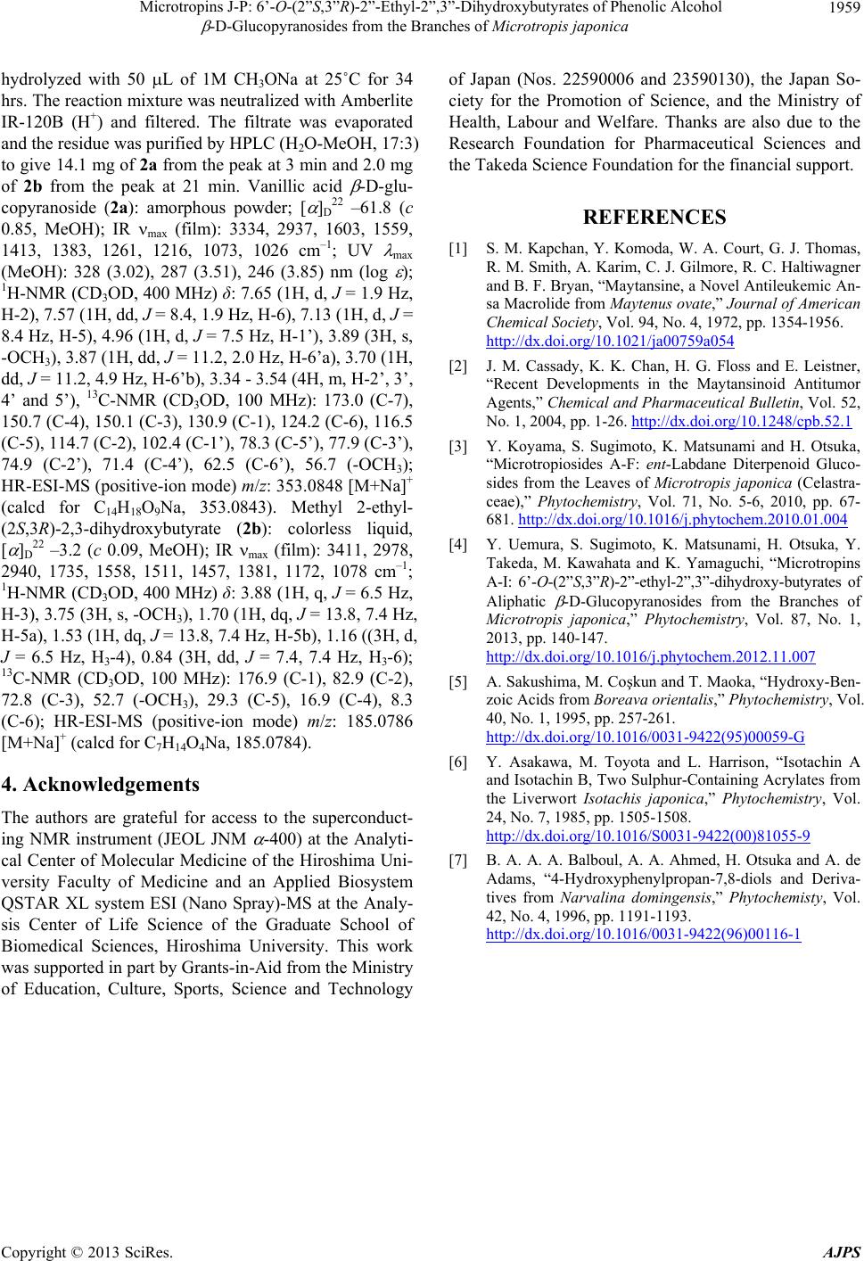 Microtropins J-P: 6’-O-(2”S,3”R)-2”-Ethyl-2”,3”-Dihydroxybutyrates of Phenolic Alcohol -D-Glucopyranosides from the Branches of Microtropis japonica Copyright © 2013 SciRes. AJPS 1959 hydrolyzed with 50 L of 1M CH3ONa at 25˚C for 34 hrs. The reaction mixture was neutralized with Amberlite IR-120B (H+) and filtered. The filtrate was evaporated and the residue was purified by HPLC (H2O-MeOH, 17:3) to give 14.1 mg of 2a from the peak at 3 min and 2.0 mg of 2b from the peak at 21 min. Vanillic acid -D-glu- copyranoside (2a): amorphous powder; [ ]D 22 ‒61.8 (c 0.85, MeOH); IR max (film): 3334, 2937, 1603, 1559, 1413, 1383, 1261, 1216, 1073, 1026 cm‒1; UV max (MeOH): 328 (3.02), 287 (3.51), 246 (3.85) nm (log ); 1H-NMR (CD3OD, 400 MHz) δ: 7.65 (1H, d, J = 1.9 Hz, H-2), 7.57 (1H, dd, J = 8.4, 1.9 Hz, H-6), 7.13 (1H, d, J = 8.4 Hz, H-5), 4.96 (1H, d, J = 7.5 Hz, H-1’), 3.89 (3H, s, -OCH3), 3.87 (1H, dd, J = 11.2, 2.0 Hz, H-6’a), 3.70 (1H, dd, J = 11.2, 4.9 Hz, H-6’b), 3.34 - 3.54 (4H, m, H-2’, 3’, 4’ and 5’), 13C-NMR (CD3OD, 100 MHz): 173.0 (C-7), 150.7 (C-4), 150.1 (C-3), 130.9 (C-1), 124.2 (C-6), 116.5 (C-5), 114.7 (C-2), 102.4 (C-1’), 78.3 (C-5’), 77.9 (C-3’), 74.9 (C-2’), 71.4 (C-4’), 62.5 (C-6’), 56.7 (-OCH3); HR-ESI-MS (positive-ion mode) m/z: 353.0848 [M+Na]+ (calcd for C14H18O9Na, 353.0843). Methyl 2-ethyl- (2S,3R)-2,3-dihydroxybutyrate (2b): colorless liquid, [ ]D 22 ‒3.2 (c 0.09, MeOH); IR max (film): 3411, 2978, 2940, 1735, 1558, 1511, 1457, 1381, 1172, 1078 cm‒1; 1H-NMR (CD3OD, 400 MHz) δ: 3.88 (1H, q, J = 6.5 Hz, H-3), 3.75 (3H, s, -OCH3), 1.70 (1H, dq, J = 13.8, 7.4 Hz, H-5a), 1.53 (1H, dq, J = 13.8, 7.4 Hz, H-5b), 1.16 ((3H, d, J = 6.5 Hz, H3-4), 0.84 (3H, dd, J = 7.4, 7.4 Hz, H3-6); 13C-NMR (CD3OD, 100 MHz): 176.9 (C-1), 82.9 (C-2), 72.8 (C-3), 52.7 (-OCH3), 29.3 (C-5), 16.9 (C-4), 8.3 (C-6); HR-ESI-MS (positive-ion mode) m/z: 185.0786 [M+Na]+ (calcd for C7H14O4Na, 185.0784). 4. Acknowledgements The authors are grateful for access to the superconduct- ing NMR instrument (JEOL JNM -400) at the Analyti- cal Center of Molecular Medicine of the Hiroshima Uni- versity Faculty of Medicine and an Applied Biosystem QSTAR XL system ESI (Nano Spray)-MS at the Analy- sis Center of Life Science of the Graduate School of Biomedical Sciences, Hiroshima University. This work was supported in part by Grants-in-Aid from the Ministry of Education, Culture, Sports, Science and Technology of Japan (Nos. 22590006 and 23590130), the Japan So- ciety for the Promotion of Science, and the Ministry of Health, Labour and Welfare. Thanks are also due to the Research Foundation for Pharmaceutical Sciences and the Takeda Science Foundation for the financial support. REFERENCES [1] S. M. Kapchan, Y. Komoda, W. A. Court, G. J. Thomas, R. M. Smith, A. Karim, C. J. Gilmore, R. C. Haltiwagner and B. F. Bryan, “Maytansine, a Novel Antileukemic An- sa Macrolide from Maytenus ovate,” Journal of American Chemical Society, Vol. 94, No. 4, 1972, pp. 1354-1956. http://dx.doi.org/10.1021/ja00759a054 [2] J. M. Cassady, K. K. Chan, H. G. Floss and E. Leistner, “Recent Developments in the Maytansinoid Antitumor Agents,” Chemical and Pharmaceutical Bulletin, Vol. 52, No. 1, 2004, pp. 1-26. http://dx.doi.org/10.1248/cpb.52.1 [3] Y. Koyama, S. Sugimoto, K. Matsunami and H. Otsuka, “Microtropiosides A-F: ent-Labdane Diterpenoid Gluco- sides from the Leaves of Microtropis japonica (Celastra- ceae),” Phytochemistry, Vol. 71, No. 5-6, 2010, pp. 67- 681. http://dx.doi.org/10.1016/j.phytochem.2010.01.004 [4] Y. Uemura, S. Sugimoto, K. Matsunami, H. Otsuka, Y. Takeda, M. Kawahata and K. Yamaguchi, “Microtropins A-I: 6’-O-(2”S,3”R)-2”-ethyl-2”,3”-dihydroxy-butyrates of Aliphatic -D-Glucopyranosides from the Branches of Microtropis japonica,” Phytochemistry, Vol. 87, No. 1, 2013, pp. 140-147. http://dx.doi.org/10.1016/j.phytochem.2012.11.007 [5] A. Sakushima, M. Coşkun and T. Maoka, “Hydroxy-Ben- zoic Acids from Boreava orientalis,” Phytochemistry, Vol. 40, No. 1, 1995, pp. 257-261. http://dx.doi.org/10.1016/0031-9422(95)00059-G [6] Y. Asakawa, M. Toyota and L. Harrison, “Isotachin A and Isotachin B, Two Sulphur-Containing Acrylates from the Liverwort Isotachis japonica,” Phytochemistry, Vol. 24, No. 7, 1985, pp. 1505-1508. http://dx.doi.org/10.1016/S0031-9422(00)81055-9 [7] B. A. A. A. Balboul, A. A. Ahmed, H. Otsuka and A. de Adams, “4-Hydroxyphenylpropan-7,8-diols and Deriva- tives from Narvalina domingensis,” Phytochemisty, Vol. 42, No. 4, 1996, pp. 1191-1193. http://dx.doi.org/10.1016/0031-9422(96)00116-1
|