Vol.3, No.1, 13-19 (2011) Health
doi:10.4236/health.2011.31003
Copyright © 2011 SciRes. Openly accessible at http://www.scirp.org/journal/HEALTH/
Platelet cytochrome c-oxidase activity in patients with
acute schizophrenia in the course of their treatment with
risperidone
Gulnur Sh. Burbaeva1, Irina S. Boksha1, Marina S. Turishcheva1, Olga K. Savushkina1,
Alan G. Beniashvili2, Georgiy E. Rupchev2, Margarit a A. M orozova2
1Laboratory of Neurochemistry, Mental Health Research Centre, Russian Academy of Medical Sciences, Moscow, Russia;
*Corresponding Author: neurochem06@mail.ru
2Laboratory of Psychopharmacology at the Clinical Department Mental Health Research Centre, Russian Academy of Medical Sci-
ences, Moscow, Russia; margmorozova@gmail.com
Received 5 October 2011; revised 15 October 2011; accepted 19 October 2011
ABSTRACT
Objectives: Testing a hypothesis, that platelet
cytochrome c-oxidase (COX) activities in pa-
tients with paranoid schizophrenia, acute epi-
sode, may be li nked to d ynamics of their cl in ical
patterns and quality of cognitive functioning
under antipsychotic treatment. Methods: Psy-
chopathological (PANSS, NSA-16) and cognitive
assessments; platelet COX enzymatic activity
determination, post-hoc nonparametric statis-
tical analysis. Results: Psychopathological and
cognitive assessments were done and blood
was sampled in p atients before (at baseline) and
af ter treatment with risperidone. Following regu-
larities were found after the treatment of pa-
tients: Significant elevation of COX, w herein the
higher was COX at baseline, the more prom inent
was dec rease in PANSSneg and NSA rates; sig-
nificant negative correlation between COX and
executive time in cognitive tests. When the pa-
tient group was divided by median of COX at
baseline into two subgroups (greater or equal
median, and <m edian), signi ficantly larger amount
of patients with PANSSpos decrease by >20% was
assigned to the fir st subgr oup; signi ficantly larger
amount of patients with PANSSneg by <20% was
assigned to the second group. Conclusions:
Therapy with risperidone seems to be more ef-
fective for patients with higher COX activity at
baseline, but this fact requires further study.
Keywords: Schizophrenia; Risperidone;
Biochemical Markers; Acute Phase Treatment;
Atypical Antipsychotics
1. INTRODUCTION
Many evidences have been obtained for energy me-
tabolism impairment in brain of patients with mental
disorders [1-6]. The decrease in rates of glucose oxida-
tion and oxygen consumption in some brain areas of
demented patients with Alzheimer’s disease [1,2] or
schizophrenia [4,7] has been demonstrated by MRT and
PET methods. Significant decrease in cytochrome c-
oxidase activity (COXthe complex IV of mitochon-
drial respiratory chain) has been observed in several
autopsied brain structures obtained from patients with
schizophrenia as compared with control [4,8]. This fact
suggests for defect of oxidation phosphorylation in
brain of patients with s chizophr enia , puta tively resulting
in energy metabolism impairment [9].
Blood platelets are often used for biochemical model-
ing of processes occurring in nervous tissue, wherein
platelet mitochondria possess the complete respiratory
electron transport chain, including all its enzymatic
complexes, sufficiently active for in vitro biochemical
studies with clinical aims [10,12,13]. Attempts have
been made to evaluate platelet COX activity in mental
disorders, such as dementia (due to Alzheimer ’s disease),
and mild cognitive impairment (MCI) [14-16], cognitive
decline in Parkinson disease [17], and schizophrenia.
Platelet COX activity in the groups with dementia and
MCI was found lower than that in controls, wherein the
patients with MCI “occupied” positions between the
control group and demented patients [14-16]. Taken to-
gether this data has enabled to put forward an assump-
tion on probable diagnostic validity of COX as a biolog-
ical marker of Alzheimer’s disease [14]. Another group
of investigators explored the diagnostic aspect of platelet
respiratory chain activity and found elevation of respira-
tory complex I activity in patients with schizophrenia,
G. S. Burbaeva et al. / Health 3 (2011) 13-19
Copyright © 2011 SciRes. Openly accessible at http://www.scirp.org/jo urnal/HEALTH/
14
whereas no difference was found in COX activity be-
tween the samples from patients with schizophrenia and
controls [13].
In the present study we tested a hypothesis, which
assumed that platelet COX activities in patients with
acute schizophrenia might be linked to the dynamics
of their clinical patterns and quality of their cognitive
functioning under the antipsychotic treatment. The
study included following tasks: comparative estima-
tion of COX activity in patients with schizophrenia
and in control group; estimation of COX activity
changes under antipsychotic treatment; evaluation of
links between COX and clinical and neurocognitive
assessments before treatment course (“at baseline”)
and after the treatment. Besides, on the basis of the
tasks solved, we attempted to evaluate a possibility of
COX usage for prognosis of antipsychotic treatment
efficacy.
2. MATERIALS AND METHODS
2.1. Subjects and Methods of the Study
After receiving approval for the study from the Ethics
Committee of MHRC RAMS, 27 patients (men, 20-56
years old; median 33 years, 25%-, and 75%-quartiles are
25 and 44 years, respectively) with acute schizophrenia,
paranoid type (DSM-IV 295.30), have been included
into the study and assigned to the antipsychotic treat-
ment with risperidone (4-6 mg per day).
Psychopathological assessments by PANSS, negative
symptom scale NSA-16, and neurocognitive assessments
(battery of neurocognitive tests evaluating various as-
pects of memory, attention, executive functioning) were
regarded at baseline and after treatment course with ris-
peridone for 2 months, blood was sampled at the same
time ranges for COX activity assay.
Neurocognitive assessment tests:
1) Working memory: Wechsler memory scale, subtest
V: series A; series B; sum A and B.
2) Associative memory: Wechsler memory scale, sub-
test VII;
3) Psychomotor speed: WAIS test, subtest VII: Sym-
bol coding.
4) Verbal memory: Text No1 reconstruction, and Text
No2 reconstruction.
5) Visua l-spatial memory: Benton visual in tention test
(BVRT), Fifth Educati on .
6) Attention: Schulte tables and Bourdon test (pen-
cil-and-paper test), Continues attention task (CAT),
(Computerised testing).
7) Executive functions: Tower of London (TOL dx);
Computerised Wisconsin Card Sort Task Version 4
(WCST).
The control group consisted of 25 volunteers (men,
17-43 years old; median 25 years, 25%- and 75%-quartiles
are 20, and 27 years, respectively)healthy persons with
no history of mental or ne urologica l disorders.
There was found significant age difference between
the patient and control groups (p < 0.00033), however,
no influences of age on COX activity in controls or pa-
tients were found (Spearmen R < 0.22, p > 0.32), there-
fore we admitted this control group.
Isolation of platelets and extraction method for COX
activity determination .
Each blood sample was processed in 2 hours after
sampling. Blood plasma prepared from 10 ml of venous
blood taken with citrate buffer containing dextrose, pH
5.7 (1:5, v/v), by centrifugation at 1000 rpm for 10 min
at room temperature (Beckman J-6 centrifuge), was
carefully collected for following centrifugation at 10,000
rpm for 20 min at 5˚C (Beckman J-21 centrifuge , JA20.1
rotor). The supernatant was removed, and the pellet was
washed with the citrate buffer by centrifugation, super-
natant removed, and the pellet re-suspended in 62.5 mM
Tris -HCl buffer, pH 6.8, then frozen in liquid nitrogen
and stored at 70˚C prior the analysis of COX activity.
Just before the measurement the sample was defrosted,
and n-dodec yl-be ta-D-maltoside was added to its final
concentration of 1%, then incubated for 10 min at 4˚C,
centrifuged (10 min 10,000 g at 4˚C), and COX activity
was measured in supernatant.
COX enzymatic activity was determined by kinetic
method using spectrophotometer (λ = 550 nm) [18,19].
Specific activity of COX was calculated (U/mg), taken
the total protein measured by Lowry et al. [20].
Internal s t andard
Platelet sample collection accumulated in th e Laboratory
of Neurochemistry RAMS includes the samples from
healthy volunteers used for the internal standard prepara-
tion. Protein extracts prepared from these samples ac-
cording with the described above procedure are used as
the internal standard (a gauge) in comparative COX es-
timations in each experiment.
2.2. St atistica l Analysis
Clinical data, demography, and biochemical data of
the patients co mposed a da tabase. Nonpar ametric statistics
(Spearmen correlations, Wilcoxon Matched Pairs Test,
Mann-Whitney U-test, Chi-square distribution with Yates
correction factor using Statistica 6.0 software) was em-
ployed for assessment of changes in the target parame-
ters and links between them.
3. RESULTS
1) Comparison of COX baseline activity in group of
G. S. Burbaeva et al. / Health 3 (2011) 13-19
Copyright © 2011 SciRes. Openly accessible at http://www.scirp.org/journal/HEALTH/
15
Table 1. Result of statistical COX activity data processing in patient group (before the treatment course) and in controls by
Mann-Whitney U-Test.
Rank Sum Rank Sum U Z
Patients with schizophrenia
N
Controls
N
p-level
COX 462 916 844.642 27 25 0.000001
patients with schizophrenia (before the beginning of
treatment course with risperidone) and in control group.
Comparison of COX baseline activity in group of pa-
tients with schizophrenia and in control group (by
Mann-Whitney U-test) has revealed, that platelet COX
activity is significantly d ecreased in the patient group (p
= 0.000001, Table 1).
The distributions of observation numbers of various
COX activities in the control group and in the group of
patients before the treatment course are shown in the
Figures 1(a) and (b). Comparison of these diagrams
demonstrates, that the number of lower COX activity
observations is higher in the patient group than in the
control group.
2) COX comparison in patient group before and after
treatment course.
Comparison of COX activity before and after the
treatment course by non-parametric statistics (Wilcoxon
Matched Pairs Test) has demonstrated its significant
change (Table 2), wherein the increase in COX activity
was observed in 20 patients, and its decrease was ob-
served in 7 patients. Although COX has increased in
most patients after the treatment course, the patient
group has been found significantly different still from
the control group (p = 0.00015).
The increase in COX activity after the treatment
course is illustrated with the figur e showing diagrams of
COX activity distribution in the patient group (COX
activity vs. observations’ number) before and after the
treatment course (diagrams b and c, respectively). It is
obviously from the comparison of the diagrams that the
number of patients with higher COX activities increases
after the treatment course.
3) Search for links between COX activity and altera-
tions in clinical and cognition assessments under the
treatment course with risperidone.
Since the decreased COX activity has been found in
patients with schizophrenia in comparison with controls,
we have supposed this parameter has to be reflecting the
severity of psychosis and cognitive dysfunction in the
patients. Search for links between COX activity and
these assessments has provided no direct support for this
hypothesis, however, baseline COX activity has proved
to have prognostic value due to its link with PANSSneg
magnitude of alteration under the treatment course (R =
0.45, p < 0.02): the higher was baseline COX activity,
the more prominent was the decrease in deficit rates.
Similar link was found with magnitude of NSA de-
crease after the treatment course (R = 0.48, p < 0.05).
The patients with decreasing COX activity after the
treatment course (N = 7) displayed negative significant
correlations between COX activity and PANSS total and
PANSS psychopathological scores at baselines (R =
0.85, p < 0.01, R = 0.82, p < 0.02, respectively), i.e.,
the higher is COX at baseline, the less is psychotic
symptoms’ score.
The Figure 1(b) shows, that most patients demon-
strate COX baseline activities near the median in the
group (60 U/mg), however, prominent deviations from
median value are seen as well. Hence, the group of
patients was divided into two subgroups by COX base-
line activity “
median” (subgroup 1) and “<median”
(subgroup 2) (no significant difference in age between
them).
Significantly higher number of patients with decreasing
PANSS positive by 20% and more has been detected in
the subgroup 1, and the subgroup 2 contained signify-
cantly higher number of patients with PANSS negative
score decreasin g by less than 20% after the treatment (Χ2
= 9.53, Yates corrected = 7.6, p < 0,01, and Χ2 = 6.52,
Yates corrected = 4.53, p < 0.025, respectively). These
findings mean, that patients with higher COX baseline
activities demonstrate more prominent reduction of psy-
chotic symptoms than those with lower COX baseline
activities.
Patients with COX baseline activity
median (sub-
group 1) have demonstrated also significant improvement
in results of neurocognitive testing: Psychomotor speed
(Symbol coding), Total Executive Time of TOL, Middle
Total Executive Time of TOL, and Psychomotor Speed.
No significant changes in results of clinical assessments
were found in patients with COX baseline activity < me-
dian (subgroup 2), however, a significant improvement was
observ ed in results of neurocognitive testing (Working
memory a nd Verbal m emory reconstruction).
The negative significant correlation was found in the
total patient group between COX activity and time spent
Table 2. Result of comparison of COX activity in the patient
group before (at baseline) and after the treatment course by
Wilcoxon Matched Pairs Test.
Valid
N T Z p-level
COX baseline & COX after
the treatment 27 65.500 2.414 0.016
G. S. Burbaeva et al. / Health 3 (2011) 13-19
Copyright © 2011 SciRes. Openly accessible at http://www.scirp.org/jo urnal/HEALTH/
16
(a)
(b)
(c)
Figure 1. Observation numbers plotted versus various COX activities
(U/mg) in control group (a), in patient group before treatment course (b),
and in the patient group after the treatment course with risperidone (c).
for planning tests after the treatment course (R = 0.44,
p < 0.03). This link means that the higher is COX activi-
ty, the easier patient fulfills these tests after the treatment.
4. CONCLUSIONS
Platelet COX activity is significantly decreased in the
studied group of patients with schizophrenia at baseline
(before the antipsychotic treatment course) in comparison
with control grou p.
Significantly higher number of patients with signifi-
cantly better dynamics of clinical parameters is revealed
in the subgroup of patients with COX baseline activities
G. S. Burbaeva et al. / Health 3 (2011) 13-19
Copyright © 2011 SciRes. Openly accessible at http://www.scirp.org/journal/HEALTH/
17
median. This is fairly for psychotic symptoms (more
than 20% reduction of PANSS total score), and for men-
tal deficit (significant reduction of NSA scores). Some
results of neurocognitive tests have improved after the
treatment therewith.
One would suppose, that the COX baseline levels
median in acute st at e of pa tients with schizophrenia coul d
be a sign of favorable dynamics of psychotic e pi s ode , a n d,
possibly, positive response to antipsychotic treatment,
although note, that the last statement requires an addi-
tional study.
5. DISCUSSION
In the present work, activity of enzymatic complex IV
(cytochrome c-oxidase, COX) determined in platelet
fraction enriched with mitochondrial membranes has
proved to be sufficient for quantitative comparative stu-
dies in all inspected subjects. The platelet COX activity
has been found therewith significantly decreased in the
group of patients with paranoid schizophrenia (DSM
-IV-TR 295.3), in comparison with control group. Re-
view of Dr. Kato [21] pointed out some inconsistency in
results obtained by different researchers, who studied
enzymatic activity of mitochondrial electron transport
chain and expression levels of genes encoding its com-
plexes in schizophrenia on various populations and var-
ious tissues from patients. Recent data is not uniform as
well [13,22]. So, significantly increased activity and
gene expression of complex I (NADH: ubiquinone oxi-
doreductase) in blood of patients with schizophrenia has
been reported [13], whereas decreas ed NDUFV2 exp res-
sion levels have been detected in other study on patients
with schizophrenia from Caucasian population [22].
The data indicating that the activity of complex I is
higher in patients than in controls has enabled the au-
thors [13] to propose complex I as diagnostic peripheral
marker for sc hizophrenia . As for COX, the authors st ated
the absence of significant differences in its activity be-
tween patients and controls, but the experimental data on
individual COX activity was not published making im-
possible a comparison between these results and data of
the present work. However, one could suppose from our
data, that “normalizing” platelet enzymatic activities by
COX (proposed by Ben-Shachar and Klein) would result
in overestimations of these activities in patients with
schizophrenia.
Some disagreement in data on influence of neurolep-
tics on the activity of respiratory complexes is seen in
literature as well, as noted earlier by Kato [ 21]. The pa-
per of Ben-Shachar and Klein [13] reports on higher
activity of complex I in patients treated w ith neurolep tics
versu s u ntreated patients. Other research groups reported
on inhib itio n of complex I activity with neuroleptics
(more prominent with typical, such as haloperidol, chlor-
promazine, less prominent with atypical, such as rispe-
ridone), whereas COX activity was either not influenced,
or affected. These results were obtained in vitro [23], in
model experiments with animals [24], and on mononuc-
lears in peripheral blood of patients with schizophrenia
[25].
In the present study, significant elevation of COX ac-
tivity was observed in the most patients after treatment
course with risperidone, possibly it is associated with
improvement of thei r cli nical stat e. So, our spe cial attention
was paid to the treatment efficiency (clinical response)
evaluation in the patients. In our previous study, when
searching for biochemical predictor of pharmacological
therapy efficiency we have taken the decrease in PANSS
total for positive treatment effect [26]. In the present
work, we additionally regard PANSS positive, PANSS
negative, PANSS psychopathological, NSA, and neuro-
cognitive tests.
The initially higher COX activity has been found to
account for favorable prognosis in respect to whole clin-
ical dynamics’ pattern, especially to psychotic symptoms.
This may be an indication, that higher level of energy
metabolism is a good “background” for improvement in
mental state of patients with schizophrenia, and, vice
versa, lower levela b ad one. The initially higher COX
activities ( median in total group) are associated with
favorable prognosis for improvement of cognitive test
results (WAIS subtest 7). Taking into account that this
sub-test is the most complex by the number of intellec-
tual functions included and is associated with various
properties of attention, perception, visual- motor coor-
dination, rate of new skills’ formation, and ability of
visual-motor stimuli integration [27], the successful im-
provement in the results of this test can be considered as
a sign of cognitive function enhancement.
We recognize that the design of the present study does
not allow unambiguous stating, that favorable clinical
dynamics is associated with the therapy employed only,
but we surely do admit this, taking into account numer-
ous risperidone studies supporting its high antipsychotic
activity [28]. Hence, one can admit, that the found link
between the baseline COX activity and positi ve res pon s e
to the treatment of patients in acute state is not casual:
possibly, these patients have yet a “potential” to restore,
whereas the patients with low COX level possibly need
in additional therapy supporting energy metabolism.
Thus, the measurement of single parameter, such as
platelet COX activity, related to platelet mitochondrial
respiratory chain could have some prognostic value for
prediction of individual efficacy of medication with ris-
peridone in patients with schizophrenia (although, ob-
viously, inspection of larger groups of patients is neces-
G. S. Burbaeva et al. / Health 3 (2011) 13-19
Copyright © 2011 SciRes. Openly accessible at http://www.scirp.org/jo urnal/HEALTH/
18
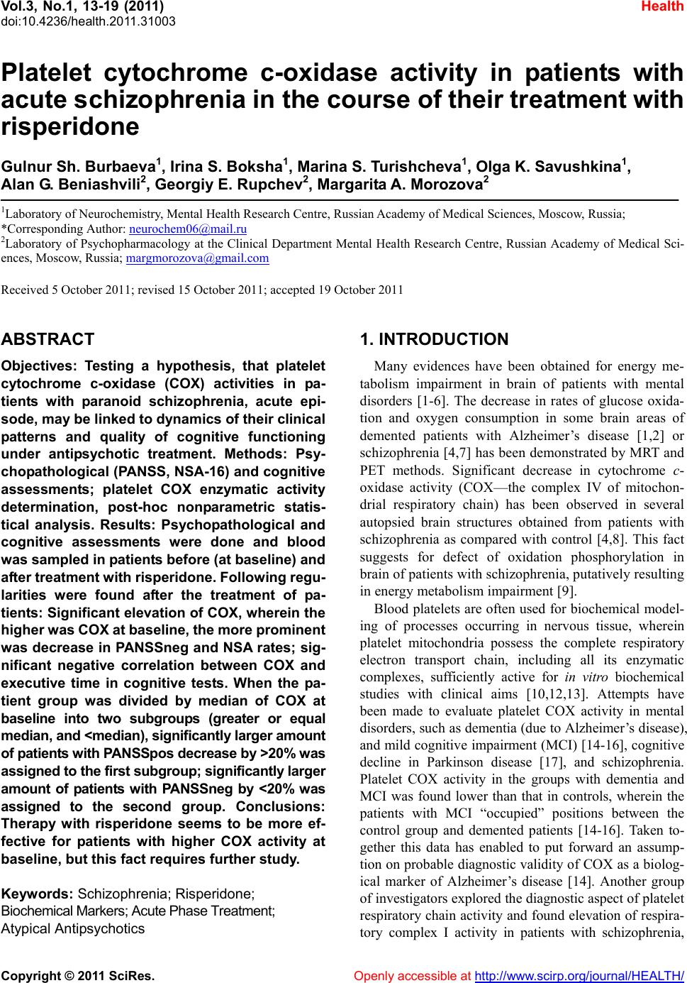
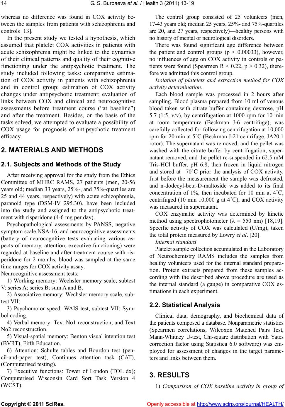
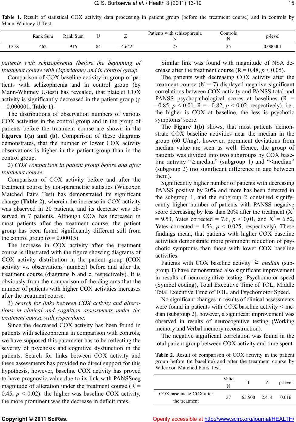
sary for accomplishment individual prognosis, and an
additional study is needed on dependence of COX activ-
ity on various factors).
REFERENCES
[1] Mohanakrishnan, P., Fowler, A.H., Vonsattel, J.P., Husain,
M.M., Jolles, P.R., Liem, P. and Komoroski, R.A. (1995)
An in vitro 1H nuclear magnetic resonance study of the
tempoparietal cortex of Alzheimer brains. Experimental
Brain Research, 102, 503-510.
doi:10.1007/BF00230654
[2] Kennedy, A.M., Frackowiak, R.S., Newman, S.K., Bloom-
field, P.M., Seaward, J., Roques, P., Lewington, G., Cun-
ningham, V.J. and Rossor, M.N. (1995) Deficits in cere-
bral glucose metabolism demonstrated by positron emis-
sion tomography in individuals at risk of familial Alz-
heimer’s disease. Neuroscience Letters, 186, 17-20.
doi:10.1016/0304-3940(95)11270-7
[3] Volz, H.R., Riehemann, S., Maurer, I., Smesny, S.,
Sommer, M., Rzanny, R., Holstein, W., Czekalla, J. and
Sauer, H. (2000) Reduced phosphodiesters and high-
energy phosphates in the frontal lobe of schizophrenic
patients: A 31P chemical shift spectroscopic-imaging
study. Biological Psychiatry, 47, 954-961.
doi:10.1016/S0006-3223(00)00235-3
[4] Maurer, I., Zierz, S. and Moller, H.-J. (2001) Evidence
for a mitochondrial oxidative phosphorylation defect in
brains from patients with schizophrenia. Schizophrenia
Research, 48, 125-136.
doi:10.1016/S0920-9964(00)00075-X
[5] Fukuzako, H. (2001) Neurochemical investigation of the
schizophrenic brain by in vivo phosphorus magnetic re-
sonance spectroscopy. The World Journal of Biological
Psychiatry, 2, 70-82. doi:10.3109/15622970109027496
[6] Jou, S.-H., Chiu, N.-Y. and Liu, C.-S. (2009) Mitochon-
drial dysfunction and psychiatric disorders. Chang Gung
Medical Journal, 32, 370-379.
[7] Buchsbaum, M.S. and Hazlett, E.A. (1998) Positron
emission tomography studies of abnormal glucose meta-
bolism in schizophrenia. Schizophrenia Bulletin, 24,
343-364.
[8] Cavelier, L., Jazin, E.E., Eriksson, I., Prince, J., Bave, U.,
Oreland, L. and Gyllensten, U. (1995) Decreased cy-
tochrome c-oxidase activity and lack of age-related ac-
cumulation of mitochondrial DNA de letions in the brains
of schizophrenics. Genomics, 29, 217-224.
doi:10.1006/geno.1995.1234
[9] Shao, L., Martin, M.V., Watson, S.J., Schatzb erg, A., A k i l,
H., Myers, R.M., Jones, E.G., Bunney, W.E. and Vawter,
M.P. (2008) Mitochondrial involvement in psychiatric
disorders. Annals of Medicine, 40, 281-295.
[10] Mann, V.M., Cooper, J.M., Krige, D., Daniel, S.E., Scha-
pira, A.H.V. and Marsden, C.D. (1992) Brain, skeletal
muscle and platelet homogenate mitochondrial function
in Рarkinson’s disease. Brain, 115, 333-342.
[11] Dror, N., Klein, E., Karry, R., Sheinkman, A., Kirsh, Z.,
Mazor, M., Tzukerman, M. and Ben-Shachar D. (2002)
State-dependent alterations in mitochondrial complex I
activity in platelets: A potential peripheral marker for
schizophrenia. Molecular Psychiatry, 7, 995-1001.
[12] Böhm, M., Papezova, H., Hansikova, H., Wenchich, L. and
Zeman, J. (2007) Activities of respiratory chain complexes
in isolated platelets in females with anorexia nervosa.
International Journal of Eating Disorders, 40, 659-663.
doi:10.1002/eat.20403
[13] Ben-Shachar, D. and Klein, E. (2008) Methods and kits
for diagnosis of schizophrenia. US Patent, 7, 442-496.
[14] Valla, J., Schneider, L., Niedzielko, T., Coon, K.D., Caselli,
R., Sabbagh, M. N., Ahern, G. L., Baxter, L., Alexander, G.,
Walker, D.G. and Reiman, E.M. (2006) Impaired platelet
mitochondrial activity in Alzheimer’s disease and mild
cognitive impair ment. Mitochondrion, 6, 323-330.
[15] Burbaeva, G.Sh., Boksha, I.S., Turishcheva, M.S., Teresh-
kina, E.B., Savushkina, O.K., Starodubtseva, L.I., Fedo-
rova, Ya.B. and Gavrilova, S.I. (2008) Cytochrome c-
oxidase and glutamine synthetase-like protein in blood
platelets as candidates for the role of early markers of
Alzheimer’s disease. Proceedings of the IV-th Scientific
and Practical Conference “Alzheimer ’s disease and cog-
nitive impairments in old age: Advances in neurobiology
and therapy”, 89-97 [Russian].
[16] Boksha, I., Burbaeva, G., Savushkina, O., Tereshkina , E.,
Turishcheva, M., Starodubtseva, L. and Vorobyeva, E.
(2009) Blood proteins as markers and predictors of cog-
nitive deficit in mental pathologies. Abstracts, 3rd World
Congress Gene-2009, Foshan, China.
http://www.bitlifesciences.com/wcg2009/Program.asp
[17] Benecke, R., Strümper , P. and Weiss, H. (1993) Electron
transfer complexes I and IV of platelets are abnormal in
Parkinson's disease but normal in Parkinson-plus syn-
dromes. Brain 116, 1451-1463.
doi:10.1093/brain/116.6.1451
[18] Parker, W.D. Jr and Parks, J.K. (1995) Cytochrome c-
oxidase in Alzheimer’s disease brain: purification and
characterization. Neurology, 45, 482-486.
[19] Cardoso, S.M., Proenca, M.T., Santos, S., Santana, I . and
Oliveira, C.R. (2004) Cytochrome c-oxidase is decreased
in Alzheimer’s disease platelets. Neurobiology of Aging,
25, 105-110. doi:10.1016/S0197-4580(03)00033-2
[20] Lowry, O.H., Rosebrough, N.J., Farr, A.L. and Randall,
R.J. (1951) Protein measurement with the folin phenol
reagent. The Journal of Biological Chemistry, 193, 265-
275.
[21] Kato, T. (2001) The other, forgotten genome: Mitochon-
drial DNA and mental disorders. Molecular Psychiatry, 6,
625-633. doi:10.1038/sj.mp.4000926
[22] Washizuka, S., Iwamoto, K., Kakiuchi, C., Bundo, M.
and Kato, T. (2009) Expression of mitochondrial com-
plex I subunit gene NDUFV2 in the lymphoblastoid cells
derived from patients with bipolar disorder and schi-
zophrenia. Neuroscience Research, 63, 199-204.
doi:10.1016/j.neures.2008.12.004
[23] Maurer, I. and Moller, H.J. (1997) Inhibition of complex
I by neuroleptics in normal human brain cortex parallels
the extrapyramid al toxicity of neurol eptics. Molecular and
Cellular Biochem is try, 174, 255-259.
doi:10.1023/A:1006872911332
[24] Streck, E.L., Rezin, G.T., Barbosa, L.M., Assis, L.C.,
Grandi, E. and Quevedo, J. (2007) Effect of antipsychot-
ics on succinate dehydrogenase and cytochrome oxidase
activities in rat brain. Naunyn-Schmiedeberg’s Archives
G. S. Burbaeva et al. / Health 3 (2011) 13-19
Copyright © 2011 SciRes. Openly accessible at http://www.scirp.org/journal/HEALTH/
19
of Pharmacology, 376, 127-133.
doi:10.1007/s00210-007-0178-2
[25] Casademont, J., Garrabou, G., Miró, Ò., López, S., Pons,
A., Bernardo, M. and Cardellach, F. (2007) Neuroleptic
treatment effect on mitochondrial electron transport chain:
Peripheral blood mononuclear cells analysis in psychotic
patients. Journal of Clinical Psychopharmacology, 27,
284-288. doi:10.1097/JCP.0b013e318054753e
[26] Burbaeva, G.Sh., Boksha, I.S., Tereshkina, E.B., Savush-
kina, O.K., Turishcheva, M.S., Starodubtseva, L.I., Bru-
sov, O.S. and Morozova, M.A. (2006) Effect of olanza-
pine treatment on platelet glutamine synthetase-like pro-
tein and glutamate dehydrogenase immunoreactivity in
schizophrenia. The World Journal of Biological Psychia-
try, 7, 75-81. doi:10.1080/15622970510029957
[27] Wechsler, D. (1981) Wechsler adult intelligence scale-
revised,San Antonio, TX: Psychological Corporation.
[28] Marder, S.R., Davis, J.M. and Chouinard, G. (1997) The
effects of risperidone on the five dimensions of schi-
zophrenia derived by factor analysis: combined results of
the North American trials. The Journal of Clinical Psy-
chiatry, 58, 538-546. doi:10.4088/JCP.v58n1205