Modern Plastic Surgery, 2013, 3, 120-122
http://dx.doi.org/10.4236/mps.2013.34024 Published Online October 2013 (http://www.scirp.org/journal/mps)
Transdermal Nitroglycerine Patch: An Optional Device to
Reduce Flap Venous Congestion? A Case Report
Sara Di Lorenzo, Bartolo Corradino, Adriana Cordova
Department of Surgi cal Oncology, Pla stic Surgery Unit, University of Palermo, Palermo, Italy.
Email: saradilorenz@gmail.com, dilsister@libero.it
Received August 1st, 2013; revised September 1st, 2013; accepted September 8th, 2013
Copyright © 2013 Sara Di Lorenzo et al. This is an open access article distributed under the Creative Commons Attribution License,
which permits unrestricted use, distribution, and reproduction in any medium, provided the original work is properly cited.
ABSTRACT
Sometimes in free flap there is a venous congestion without an obstruction of the venous anastomosis or other organic
causes of reduction venous drainage (haematoma, seroma compressing the pedicle). In these cases the authors suggest
the application of nitrog lycerine patch in the cong ested area of the fla p few hours befor e the surgical ex ploration of the
anastomosis. If there is a fast improvement of the clinical feature of the flap, the surgical exploration could be avoided.
The authors underline that applying the nitroglycerin patch should not be regarded in any way as a therapy of a free flap
venous thrombosis but only as an useful device, an option to be taken only when the surgeon is undecided whether to
revisit the anastomosis or not.
Keywords: Free Flap Failure; Venous Congestion; Vascular Pedicle Thrombosis; Microsurgical Complications
1. Introduction
The majority of surgical complications after tissue trans-
fer surgery are related to vascular thrombosis, which
usually occur within 3 days of surgery, but sometimes
thrombosis can occur later.
There are many techniques available for evaluation of
flap viability. The most common technique is clinical
observation and Doppler vascular pedicle monitoring.
The clinical observation is very useful when the flap has
a skin island.
Venous thrombosis occurs more frequently because of
its low-flow, low-pressure nature, and because it may
evolve over se ver al h ours.
A venous congestion usually results in oedema and
darkening of the skin colour.
During early venous obstruction a needle stick will
cause a rapid bleeding of dark blood. The capacity to
understand and id entify the early signs of flap suff erance
is important in order to decide if surgical explorations,
and an additional pharmacological therapy or something
else are necessary.
If a flap’s vascular integrity is in question, if there is a
doubt of venous thrombo sis at the anastomosis, a prompt
surgical exploration is mandatory [1].
Surgical exploration is still mandatory when there are
haematoma and seroma that could make a compression
on the vascular pedicle.
Sometimes there is a venous congestion of the flap
without an obstruction of the venous vessel at the anas-
tomotic site. In the surgical exploration sometimes there
is no problem at the anastomotic site or other organic
causes of reduction venous drainage.
This kind of insufficient venous drainage may cause a
slow partial flap loss that may require further surgical
steps, multiple debridements to remove necrotic tissue
and prevent further complications as infections, and to
improve the outcome.
Authors expose their experience with a nitroglycerine
plaster applied on flap in order to reduce the venous
congestion of a free DIEP flap. It begun in the 5th day
after surgery.
2. Clinical Case
The patient is a 38-year-old female undergone a right
mastectomy for a ductal carcinoma and a quadrantec-
tomy on the left breast for a dysp lasia.
A delayed free Diep Flap reconstruction of the right
breast was performed using the internal mammary ves-
sels, as recipient vessels. The ischemia time was less than
2 hours.
The flap seemed well perfused.
The patient didn’t smoke and hadn’t risk factor or sys-
temic disease.
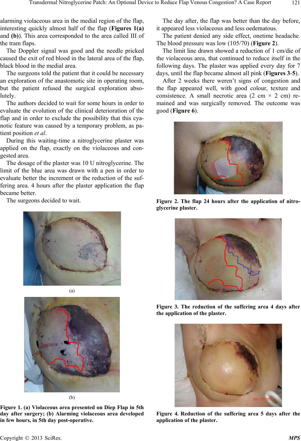
On 5th day after surgery the patient presented an
Copyright © 2013 SciRes. MPS
Transdermal Nitroglycerine Patch: An Optional Device to Reduce Flap Venous Congestion? A Case Report 121
alarming violaceous area in the medial region of the flap,
interesting quickly almost half of the flap (Figures 1(a)
and (b)). This area corresponded to the area called III of
the tram flaps.
The Doppler signal was good and the needle pricked
caused the exit o f red blood in the lateral area o f the flap,
black blood in the medial area.
The surgeons told the patient that it could be necessary
an exploration of th e anastomotic site in operating room,
but the patient refused the surgical exploration abso-
lutely.
The authors decided to wait for some hours in order to
evaluate the evolution of the clinical deterioration of the
flap and in order to exclude the possibility that this cya-
notic feature was caused by a temporary problem, as pa-
tient position et al.
During this waiting-time a nitroglycerine plaster was
applied on the flap, exactly on the violaceous and con-
gested area.
The dosage of the plaster was 10 U nitroglycerine. The
limit of the blue area was drawn with a pen in order to
evaluate better the increment or the reduction of the suf-
fering area. 4 hours after the plaster application the flap
became better.
The surgeons decided to wait.
(a)
(b)
Figure 1. (a) Violaceous area presented on Diep Flap in 5th
day after surgery; (b) Alarming violaceous area developed
in few hours, in 5th day post-operative.
The day after, the flap was better than the day before,
it appeared less violaceous and less oedematous.
The patient denied any side effect, onetime headache.
The blood pressure was low (105/70) (Figure 2).
The limit line drawn showed a reduction of 1 cm/die of
the violaceous area, that continued to reduce itself in the
following days. The plaster was applied every day for 7
days, until the flap became almost all pink (Figures 3-5).
After 2 weeks there weren’t signs of congestion and
the flap appeared well, with good colour, texture and
consistence. A small necrotic area (2 cm × 2 cm) re-
mained and was surgically removed. The outcome was
good (Figure 6).
Figure 2. The flap 24 hours after the application of nitro-
glycerine plaster.
Figure 3. The reduction of the suffering area 4 days after
the application of the plaster.
Figure 4. Reduction of the suffering area 5 days after the
application of the plaster.
Copyright © 2013 SciRes. MPS
Transdermal Nitroglycerine Patch: An Optional Device to Reduce Flap Venous Congestion? A Case Report
Copyright © 2013 SciRes. MPS
122
Figure 5. The reduction of the suffering area 6 days after
the application of the plaster.
Figure 6. The outcome after 2 weeks.
3. Conclusion
Despite the variety of anticoagulation protocols, the
studies about the prophylactic anticoagulation therapy
and the almost perfect reconstructive techniques used by
microsurgeons, the reported failure rate among free flap
ranges from 4 % to 10%. The free flap suffering or fail-
ure is a complication that often requires, after a rapid
recognition of the clinical aspect of the flap, the surgical
re-exploration in order to remove haematomas, seroma
over the pedicle or to perform a new anastomoses [2].
Sometime the venous cong estion of the flap is caused by
a temporary problem (spasm) that could not require a
surgical revision of the anastomosis. The authors expose
their experience with a nitroglycerine plaster applied on
flap in order to reduce the venous congestion of a free
DIEP flap. It begun in the 5th day after surgery. The use
of Nitro-derived drugs in the treatment of angina pectoris
is well known. The nitrates caused a d ilatation of the ve-
nous compartment and then a dilatation of arterial com-
partment included coronary arteries. Organic nitrates as
nitroglycerine, are the most employed drugs in cardi-
ological clinical practice. Nitroglycerine is largely given
as a plaster whit transdermal absorption. The mechanism
of action of organic nitrates is well known. They act on
the endothelial layer inducing the production of NO (ni-
tric oxide). This gas, physiologically produced by endo-
thelial cells, white blood cells, muscular cells and brain
cells, caused vasodilatation and reduced the platelet ag-
gregation. NO releasing of endothelial cells (induced by
nitroglycerine) causes an increment of level of GMPc
inside the smooth muscular cells of the vessels, so the
muscular smooth cell relaxation finally causes the vaso-
dilatation of the vessels. The authors expose their ex-
perience; it’s suggested that during the period, the sur-
geon is still evaluating if a reexploration of the flap or of
the anastomotic site is necessary, and applying a nytro-
glicerin patch on the suffering flap without local or sys-
temic side effects is possible. If there is a fast improve-
ment of the clinical feature of the flap, the surgical ex-
ploration could be avoided.
REFERENCES
[1] B. C. D. Evans and G. R. D. Evans, “Microvascular Sur-
gery,” Plastic & Reconstructive Surgery, Vol. 119, No. 2,
2007, pp. 18e-30e.
http://dx.doi.org/10.1097/01.prs.0000246717.81077.e4
[2] M. Askari, C. Fisher, F. G. Weniger, S. Bidic and A. W.
P. Lee, “Anticoagulation Therapy in Microsurgery: A
Review,” Journal of Hand Surgery, Vol. 31, No. 5, 2006,
pp. 836-846.