World Journal of Cardiovascular Diseases, 2013, 3, 1-4 WJCD
http://dx.doi.org/10.4236/wjcd.2013.36A001 Published Online September 2013 (http://www.scirp.org/journal/wjcd/)
Surgical-interventional hybrid orchestra consisting of
Potts shunt, transcatheter tricuspid valve repair by
Edwards-valve in a 26-year-old patient with
pulmonary hypertension and right
ventricular failure
Sabine Recla1, Blanka Steinbrenner1, Jennifer Schreier2, Stephan Fichtlscherer2, Dorle Schmidt1,
Christian Apitz1, Matthias Müller1, Juergen Bauer1, Hakan Akintuerk1, Dietmar Schranz1*
1Hessiches Kinderherzzentrum, Universitätskliniken Giessen, Frankfurt, Germany
2Medizinische Klinik III, Kardiologie Universitätsklinikum, Frankfurt, Germany
Email: *dietmar.schranz@paediat.med.uni-giessen.de
Received 30 June 2013; revised 5 August 2013; accepted 21 August 2013
Copyright © 2013 Sabine Recla et al. This is an open access article distributed under the Creative Commons Attribution License,
which permits unrestricted use, distribution, and reproduction in any medium, provided the original work is properly cited.
ABSTRACT
Reported here is a 26-year-old patient with corrected
congenital interrupted aortic arch admitted with
massive hemoptysis and severe suprasystemic pul-
monary hypertension provided him for heart-lung
transplantation. Initial closure of a small aorto-pul-
monary collateral remained ineffective. Persistent life-
threatening hemoptysis led to the decision for a modi-
fied Potts shunt with the aim of decompressing the
right ventricle and avoiding pulmonary-hypertensive
crisis. The hemoptysis did not recur. However, the
patient’s oxygen transport co ndition deteriorated and
necessitated an orchestra of interventional-surgical
approaches from re-coarctation stenting, Potts shunt
flow reducing to surgical and transcatheter tricuspid
valve repair with transient ECMO before the patient
could be discharged home.
Keywords: Pulmonary Hypertension; Potts Shunt;
Edwards Tricuspid Valve; Hybrid-Procedure
1. INTRODUCTION
Pulmonary hypertension (PH) with hemoptysis and
suprasystemic pressures is associated with poor progno-
sis [1-3]. Decrease of cardiac output is not only related to
impaired right ventricular (RV) function, but also a con-
sequence of impaired LV filling due to right-to-left ven-
tricular interaction [4]. As a novel approach for palli-
ating children with suprasystemic idiopathic pulmonary
arterial hypertension (PAH), surgical Potts shunt has
been reported as an alternative treatment to lung trans-
plantation [5,6].
2. CASE REPORT
A 26-year-old young man (body weight 52 kg) was em-
ergently referred with massive hemoptysis. He was
known to have suprasystemic pre-capillary PH secondary
to a complex congenital heart malformation (IAA-Type
B). Reconstruction of the aortic arch by a modified
Waldhausen technique combined with a pulmonary ar-
tery banding was performed in infancy. VSD closure and
pulmonary artery de-banding followed at the age of 2
years. 15 years later aortic arch was re-reconstructed by
PTFE-patch enlargement. He was known to have pre-
capillary pulmonary hypertension, which had been treated
by a calcium-antagonist, with the later addition of silde-
nafil and bosentan.
At admission life-threatening hemoptysis was asso-
ciated with right ventricular dilatation (143 ml/m2, MRI)
severe tricuspid valve insufficiency and significant cya-
nosis necessitating continuous supplemental oxygen
therapy. Heart catheterization was performed but only
one small collateral vessel could be detected and closed
by coils, but hemoptysis persisted. The suprasystemic
pulmonary arterial pressure (PAP) was confirmed with
PAP to systemic artery pressure (SAP) ratio of 1.2 and
1.5 after contrast medium injection which was used to
delineate the relationship between the left pulmonary
*Corresponding a uthor.
OPEN ACCESS
S. Recla et al. / World Journal of Cardiovascular Diseases 3 (2013) 1-4
2
artery (LPA) and descending aorta (Figure 1). Nitric
oxide test-inhalation (20 ppm) did not show any signifi-
cant effect on the PAH (PAP 102/63/76 mmHg; corre-
sponding PCWP of 16 mmHg, systemic arterial pressure
(SAP) in the ascending aorta of 95/61/73 mmHg, de-
scending aorta (DAO) of 84/58/66 mmHG). After de-
tailed discussion between the heart team, the patient and
his family led to the decision for a surgical Potts shunt
rather then listing the patient for combined lung- or heart-
lung transplantation. Based on the data from France [6],
a 13 mm PTFE-graft tube was surgically connected from
the left-PA to the DAO (Figure 2). The left-intercostal
surgical approach was technically uneventful. Hemopty-
sis was not observed again. However, the combination of
a post-operative left-lower lobe atelectasis, newly created
right-to-left shunt through the PTFE-tube and extreme
rarefied pulmonary vascular bed resulted in an extreme
lower body oxygen saturation of 28% distal to the Potts
shunt with an associated inferior caval vein (IVC) oxy-
gen saturation of 13%, but without an increased sys-
temic lactate level. The superior caval vein oxygen satu-
ration declined to 31% along with to a low left ventricular
cardiac output. The MRI blood-flow measurements in the
pulmonary artery demonstrated 61 ml/beat, 25 ml in the
ascending, and 12 ml in the descending aorta pre-, and
45 ml/beat post-Potts shunt, respectively. A reduced left
ventricular preload with consecutive low left ventricular
cardiac output was considered as a co-factor for the ex-
treme lower body desaturation. Therefore, the slightly re-
Figure 1. Demonstrates simultaneous injection of contrast me-
dium in the descending aorta (DAO) and left pulmonary artery
(LPA) to evaluate the morphology prior to Potts shunt surgery.
Measured LPA diameter was 24 mm, and DAO 16 mm, respec-
tively.
Figure 2. Depicts the surgical performed PTFE-graft after
connecting the LPA to the DAO.
coarctated descending aortic arch was stented by an An-
dra 39 mm XL stent mounted on a 20 × 45 mm Balloon-
in-Balloon (PFM). However, afterload reduction by di-
minishing the gradient across the aortic arch less than 5
mmHg improved only slightly the oxygen transport pa-
rameters. Therefore, we decided for flow reducetion
through the Potts shunt. A premounted 29 × 14 mm
Atrium graft stent (Advanta V12) advanced from a ve-
nous access was implanted in a diabolo shape by a slow
flared balloon inflation from out-side in until a narrowed
part of 7 mm in the middle of the graft-stent was
achieved. Thereafter measurements of oxygen transport
parameters improved. The additional trial to create an
interatrial communication for improving the left ven-
tricular preload by atrioseptostomy using the Brocken-
brough technique and gradual static balloon dilatation of
atrial septum resulted in ischemic ECG changes and
hemodynamic instability. Immediate device closure
(Amplatzer ASD-12 mm, AGA) was performed to treat
the hypoxemic reaction of the upper body part in par-
ticular of the coronary perfusion. The patient recovered
slowly but he remained bed-dependent as a NYHA func-
tional class of IV. Despite some clinical improvement the
tricuspid regurgitation increased progressively. Again,
surgical tricuspid valve reconstruction was considered as
an alternative to listing for tricuspid repair together with
lung transplantation. Surprisingly, the surgical approach
was well tolerated despite the need for an open-heart
surgery, However, the reconstructed tricuspid valve util-
izing a Carpentier-Edwards 30 mm band, remained suffi-
cient for just 2 - 3 weeks. Due to progressive grad III
regurgitation again, an off label use of a transcatheter 29
mm Edwards-Sapien Heart valve was considered to
Copyright © 2013 SciRes. OPEN ACCESS
S. Recla et al. / World Journal of Cardiovascular Diseases 3 (2013) 1-4 3
avoiding an additional open-heart surgery. Transcatheter
valve implantation was performed under general anes-
thesia with fluoroscopic and 3D-TEE guidance. An 18 F
Cook sheath was placed in femoral vein, and after sizing
of the Carpentier-band, a 39 mm Andra XXL stent
mounted on 30 × 60 mm balloon catheter (Balt) was po-
sitioned in the tricuspid-annulus and slight diabolo shape
created by expanding to 30 mm at both stent-ends fol-
lowed by using a 30 × 40 mm nucleus balloon (PFM).
The already prepared Edwards-valve was immediately
implanted within the previous Andra-stent and in the
metal band-marked tricuspid valve annulus. However,
the abrupt afterload increase after placement of the com-
petent Edwards-valve caused the right ventricular func-
tion to deteriorate. Balloon dilatation of the Atrium stent
within the Potts shunt to almost 9 mm diameter and In-
creasing dosages of catecholamines in addition to the
baseline inotropic therapy with milrinone and levo-
simendan failed to stabilize his circulation. Therefore,
the patient was put on ECMO (Extra-Corporal Mem-
brane Oxygenation) by utilizing the percutaneous femo-
ral vein (22 Fr sheath) and artery (16 Fr sheath) access.
Bi-ventricular and in particular right ventricular function
recovered over 5-days of ECMO therapy, and 7 days
ventilatory support. The “hybrid orchestra” of interven-
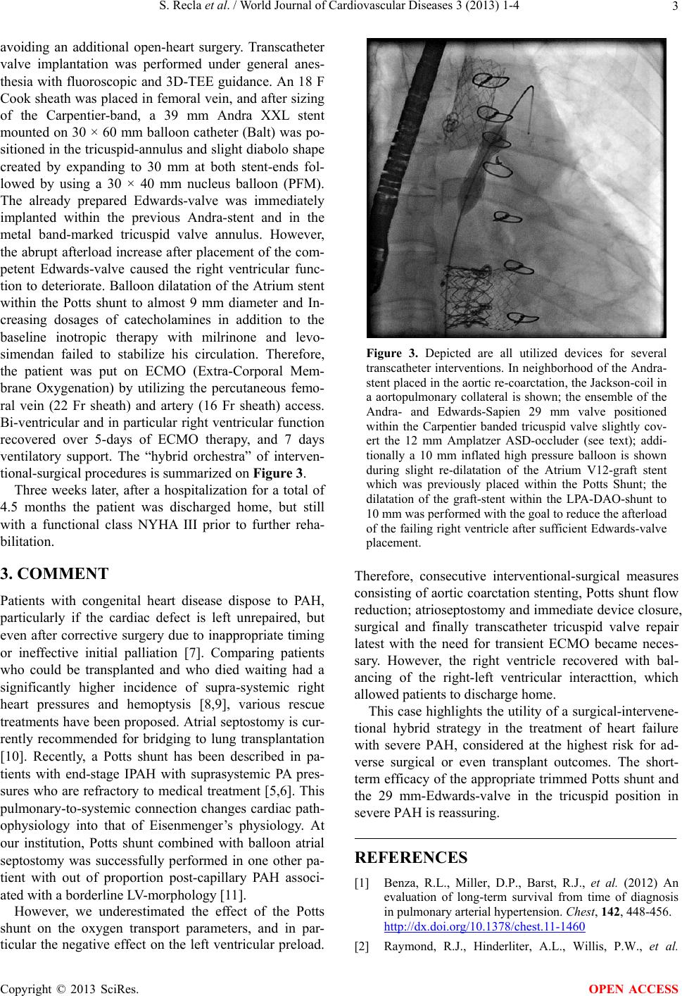
tional-surgical procedures is summarized on Figur e 3.
Three weeks later, after a hospitalization for a total of
4.5 months the patient was discharged home, but still
with a functional class NYHA III prior to further reha-
bilitation.
3. COMMENT
Patients with congenital heart disease dispose to PAH,
particularly if the cardiac defect is left unrepaired, but
even after corrective surgery due to inappropriate timing
or ineffective initial palliation [7]. Comparing patients
who could be transplanted and who died waiting had a
significantly higher incidence of supra-systemic right
heart pressures and hemoptysis [8,9], various rescue
treatments have been proposed. Atrial septostomy is cur-
rently recommended for bridging to lung transplantation
[10]. Recently, a Potts shunt has been described in pa-
tients with end-stage IPAH with suprasystemic PA pres-
sures who are refractory to medical treatment [5,6]. This
pulmonary-to-systemic connection changes cardiac path-
ophysiology into that of Eisenmenger’s physiology. At
our institution, Potts shunt combined with balloon atrial
septostomy was successfully performed in one other pa-
tient with out of proportion post-capillary PAH associ-
ated with a borderline LV-morpholo gy [11].
However, we underestimated the effect of the Potts
shunt on the oxygen transport parameters, and in par-
ticular the negative effect on the left ventricular preload.
Figure 3. Depicted are all utilized devices for several
transcatheter interventions. In neighborhood of the Andra-
stent placed in the aortic re-coarctation, the Jackson-coil in
a aortopulmonary collateral is shown; the ensemble of the
Andra- and Edwards-Sapien 29 mm valve positioned
within the Carpentier banded tricuspid valve slightly cov-
ert the 12 mm Amplatzer ASD-occluder (see text); addi-
tionally a 10 mm inflated high pressure balloon is shown
during slight re-dilatation of the Atrium V12-graft stent
which was previously placed within the Potts Shunt; the
dilatation of the graft-stent within the LPA-DAO-shunt to
10 mm was performed with the goal to reduce the afterload
of the failing right ventricle after sufficient Edwards-valve
placement.
Therefore, consecutive interventional-surgical measures
consisting of aortic coarctation stenting, Potts shunt flow
reduction; atrioseptostomy and immediate device closure,
surgical and finally transcatheter tricuspid valve repair
latest with the need for transient ECMO became neces-
sary. However, the right ventricle recovered with bal-
ancing of the right-left ventricular interacttion, which
allowed patients to discharge home.
This case highlights the utility o f a surgical-intervene-
tional hybrid strategy in the treatment of heart failure
with severe PAH, considered at the highest risk for ad-
verse surgical or even transplant outcomes. The short-
term efficacy of the appropriate trimmed Potts shunt and
the 29 mm-Edwards-valve in the tricuspid position in
severe PAH is reassuring.
REFERENCES
[1] Benza, R.L., Miller, D.P., Barst, R.J., et al. (2012) An
evaluation of long-term survival from time of diagnosis
in pulmonary arterial hypertension. Chest, 142, 448-456.
http://dx.doi.org/10.1378/chest.11-1460
[2] Raymond, R.J., Hinderliter, A.L., Willis, P.W., et al.
Copyright © 2013 SciRes. OPEN ACCESS
S. Recla et al. / World Journal of Cardiovascular Diseases 3 (2013) 1-4
Copyright © 2013 SciRes. OPEN ACCESS
4
(2002) Echocardiographic predictors of adverse outcomes
in primary pulmonary hypertension. Journal of the Ameri-
can College of Cardiology, 39, 1214-1219.
http://dx.doi.org/10.1016/S0735-1097(02)01744-8
[3] Zylkowska, J., Kurzyna, M., Pietura, R., et al. (2011)
Recurrent hemoptysis: An emerging life-threatening com-
plication in idiophathic pulmonary arterial hypertension.
Chest, 139, 690-693.
http://dx.doi.org/10.1378/chest.10-1134
[4] Gan, C.T., Lankhaar, J.W., Marcus, J.T., et al. (2006)
Impaired left ventricular filling due to right-to-left ven-
tricular interaction in patients with pulmonary arterial
hypertension. American Journal of Physiology—Heart
and Circulatory Physiology, 290, H1528-H1533.
[5] Blanc, J., Vouhe, P. and Bonnet, D. (2004) Potts shunt in
patients with pulmonary hypertension. The New England
Journal of Medicine, 350, 623.
http://dx.doi.org/10.1056/NEJM200402053500623
[6] Baruteau, A.E., Serraf, A., Lévy, M., et al. (2012) Potts
shunt in children with idiopathic pulmonary arterial hy-
pertension: Long-term results. The Annals of Thoracic
Surgery, 94, 817-824.
http://dx.doi.org/10.1016/j.athoracsur. 2012.03.099
[7] Beghetti, M. and Tissot, C. (2010) Pulmonary hyperten-
sion in congenital shunts. Revista Española de Cardi-
ología, 63, 1179-1193.
http://dx.doi.org/10.1016/S0300-8932(10)70250-5
[8] Cantu, J., Wang, D. and Safdar, Z. (2012) Clinical impli-
cations of haemoptysis in patients with pulmonary arterial
hypertension. International Journal of Clinical Practice,
177, 5-12. http://dx.doi.org/10.1111/ijcp.12004
[9] Goldstein, B.S., Sweet, S.C., Mao, J., Huddleston, C.B.
and Grady, R.M. (2011) Lung transplantation in children
with idiopathic pulmonary arterial hypertension: An 18-
year experience. The Journal of Heart and Lung Trans-
plantation, 30, 1148-1152.
http://dx.doi.org/10.1016/j.healun.2011.04.009
[10] Micheletti, A., Hislop, A.A., Lammers, A., et al. (2006)
Role of atrial septostomy in the treatment of children with
pulmonary arterial hypertension. Heart, 92, 969-972.
http://dx.doi.org/10.1136/hrt.2005.077669
[11] Latus, H., Apitz, C., Schmidt, D., Jux, C., Müller, M.,
Bauer, J., Akintuerk, H., Schneider, M. and Schranz, D.
(2013) Potts shunt and atrioseptostomy in pulmonary hy-
pertension due to left ventricular disease. The Annals of
Thoracic Surgery, 96, 317-319.
http://dx.doi.org/10.1016/j.athoracsur. 2012.10.069