Vol.1, No.1, 1-3 (2013) Open Journal of Emergency Medicine
Presentation of multiple midface fractures combined
with intracranial injury treated conservatively
Talal Rane1, Firas Nasser2, Samir Taha2*
1Department of Oral & Maxillofacial Surgery, Al-Ahli Hospital, Doha, Qatar
2Department of Oral and Maxillofacial Surgery, Hamad Medical Corporation, Doha, Qatar; *Corresponding Author: staha3@hmc.org.qa
Received 17 July 2013; revised 19 August 2013; accepted 1 September 2013
Copyright © 2013 Talal Rane et al. This is an open access article distributed under the Creative Commons Attribution License, which
permits unrestricted use, distribution, and reproduction in any medium, provided the original work is properly cited.
ABSTRACT
Midface injuries are normally the result of high
forces impacted on the fac e , that can bypass the
power of the connection buttresses, which are
thick and strong, base of skull is also a rigid and
tough structure which requires significant forces
to break. In our case, multiple midface and cra-
nial base fractures are presented, which reflect
injury caused by high forces and norma lly, these
result in instability in the facial skeleton, or
cause CSF rhinorrhea, or sharp spicules to en-
danger the orbit or the anterior cranial fossa, or
the presence of any other indication of surgery.
It is unusual to find such an extent of facial
trauma and such a multiplication of fractures on
the midface and cranial base levels, yet they
were st able enough an d without a re al indication
to require surgical interference.
Keywords: Midface Fracture; Upp er Face Fracture;
Conservative Management
1. INTRODUCTION
Combined intracranial injury with midface and upper
face multiple fractures signify severe force involved [1-
4], published work on forces calculated to produce a
fracture of each craniofacial bone structure is well docu-
mented [1,2], normally, this results in severe injuries to
the face [1-3]. Hodgson, V.R. et al. presented detailed
work on the tolerance of facial bones to impact [3], and
indicated that impacts produce fracture near 200 pounds,
but that is closely related to the length of the pulse of
impact measured in milliseconds.
A lot of work is presented in the literature about treat-
ment [5-8], sequencing of which bone to fix first [6,7],
timing of treatment [6,7].
We present a case of facial trauma that resulted in
multiple midface and upper face fractures combined with
a base-of-skull fracture, but all were undisplaced and not
mobile therefore, did not require any intervention, and
were managed conservatively, together with the intracra-
nial hemorrhage being minor, that did not require surgi-
cal treatment as well.
2. CASE REPORT
A (46) years old year old male was brought to the A &
E Department of Hamad General Hospital on 12 May
2009, he was lowering a swimming pool basin down into
a pre-dug site in a house garden, when the edge of the
basin hit him on the face.
Patient was conscious, oriented with GCS 15/15, his
BP was 146/82 and Oxygen saturation of 98%, airway
was stable and neck is clear of injury.
Craniofacial examination revealed bilateral periorbital
ecchymosis, active oronasal bleeding, and oro-facial lac-
erations.
Pupils were round, equal and brisk reactive, directly
and consensually, visual acu ity intact with mild p roptosis
of right eye, no obvious restriction of ocular movements.
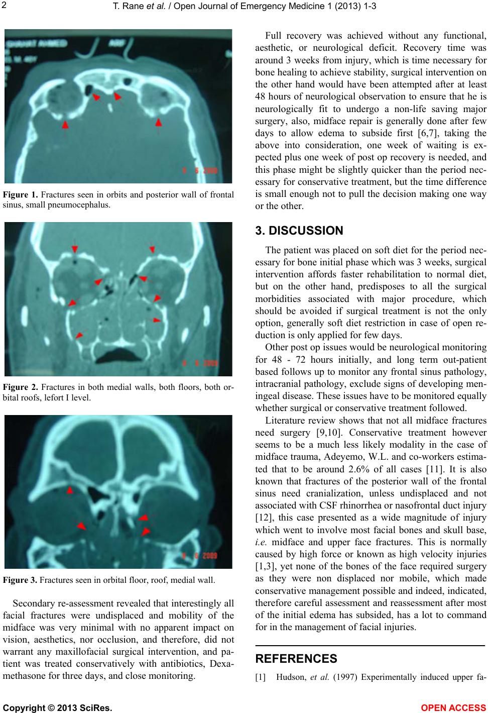
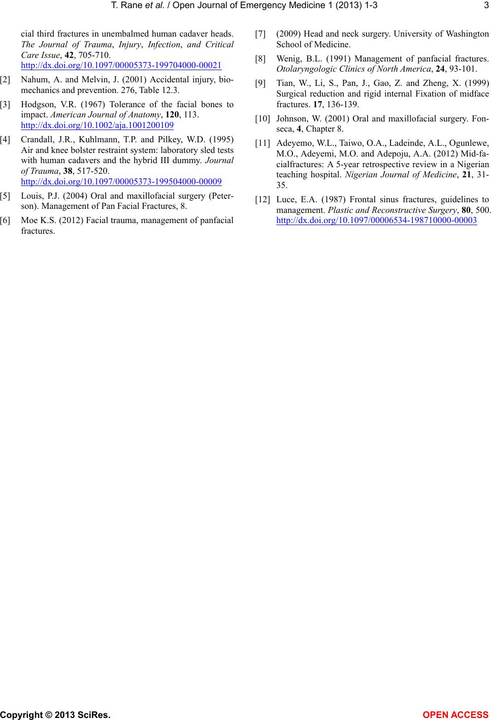
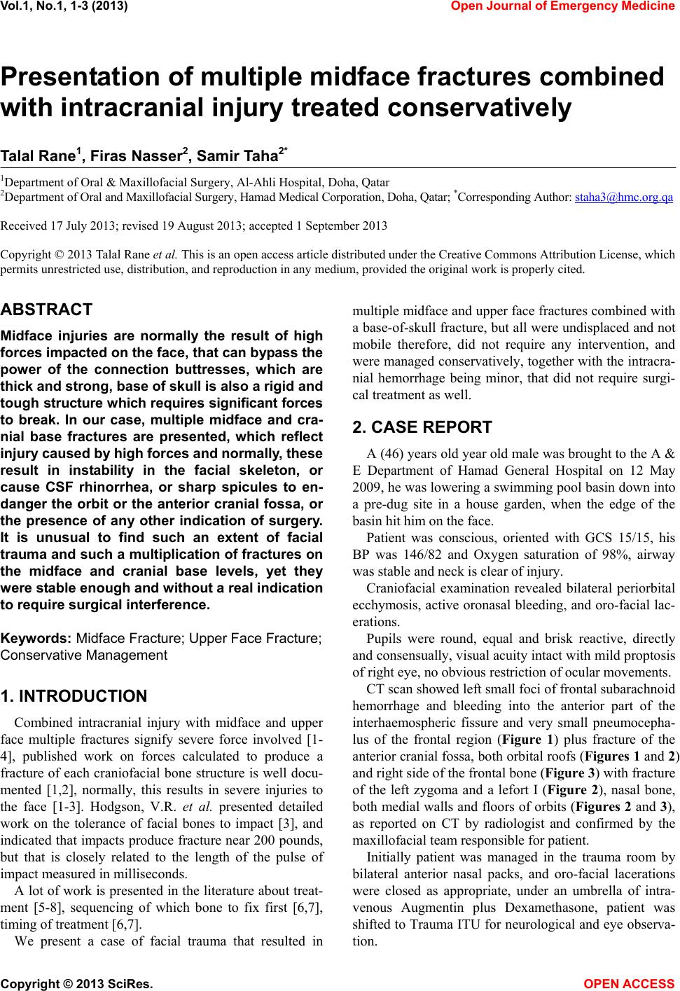
CT scan showed left small foci of frontal subarachno id
hemorrhage and bleeding into the anterior part of the
interhaemospheric fissure and very small pneumocepha-
lus of the frontal region (Figure 1) plus fracture of the
anterior cranial fossa, both orbital roofs (Figures 1 and 2)
and right side of the frontal bone (Figure 3) with fracture
of the left zygoma and a lefort I (Figure 2), nasal bone,
both medial walls and floors of orbits (Figures 2 and 3),
as reported on CT by radiologist and confirmed by the
maxillofacial team responsible for patient.
Initially patient was managed in the trauma room by
bilateral anterior nasal packs, and oro-facial lacerations
were closed as appropriate, under an umbrella of intra-
venous Augmentin plus Dexamethasone, patient was
shifted to Trauma ITU for neurological and eye observa-
tion.
Copyright © 2013 SciRes. OPEN AC CESS
T. Rane et al. / Open Journal of Emergency Medicine 1 (2 013) 1-3
2
Figure 1. Fractures seen in orbits and posterior wall of frontal
sinus, small pneumocephalus.
Figure 2. Fractures in both medial walls, both floors, both or-
bital roofs, lefort I level.
Figure 3. Fractures seen in orbital floor, roof, medial wall.
Secondary re-assessment revealed that interestingly all
facial fractures were undisplaced and mobility of the
midface was very minimal with no apparent impact on
vision, aesthetics, nor occlusion, and therefore, did not
warrant any maxillofacial surgical intervention, and pa-
tient was treated conservatively with antibiotics, Dexa-
methasone for three days, and close monitoring.
Full recovery was achieved without any functional,
aesthetic, or neurological deficit. Recovery time was
around 3 weeks from injury, which is time necessary for
bone healing to achieve stability, surgical interv ention on
the other hand would have been attempted after at least
48 hours of neurological observation to ensure that he is
neurologically fit to undergo a non-life saving major
surgery, also, midface repair is generally done after few
days to allow edema to subside first [6,7], taking the
above into consideration, one week of waiting is ex-
pected plus one week of post op recovery is needed, and
this phase might be slightly quicker than the period nec-
essary for conservative treatment, but the time difference
is small enough not to pull the d ecision making one way
or the other.
3. DISCUSSION
The patient was placed on soft diet for the period nec-
essary for bone initial phase which was 3 weeks, surgical
intervention affords faster rehabilitation to normal diet,
but on the other hand, predisposes to all the surgical
morbidities associated with major procedure, which
should be avoided if surgical treatment is not the only
option, generally soft diet restriction in case of open re-
duction is only applied for few days.
Other post op issues would be neurological monitoring
for 48 - 72 hours initially, and long term out-patient
based follows up to monitor any frontal sinus pathology,
intracranial pathology, exclude signs of developing men-
ingeal disease. These issues have to be monitored equally
whether surgical or conservative treatment followed.
Literature review shows that not all midface fractures
need surgery [9,10]. Conservative treatment however
seems to be a much less likely modality in the case of
midface trauma, Adeyemo, W.L. and co-workers estima-
ted that to be around 2.6% of all cases [11]. It is also
known that fractures of the posterior wall of the frontal
sinus need cranialization, unless undisplaced and not
associated with CSF rhinorrhea or nasofrontal duct injury
[12], this case presented as a wide magnitude of injury
which went to involve most facial bones and skull base,
i.e. midface and upper face fractures. This is normally
caused by high force or known as high velocity injuries
[1,3], yet none of the bones of the face required surgery
as they were non displaced nor mobile, which made
conservative management possible and indeed, indicated,
therefore careful assessment and reassessment after most
of the initial edema has subsided, has a lot to command
for in the management of facial injuries.
REFERENCES
[1] Hudson, et al. (1997) Experimentally induced upper fa-
Copyright © 2013 SciRes. OPEN AC CESS
T. Rane et al. / Open Journal of Emergency Medicine 1 (2 013) 1-3
Copyright © 2013 SciRes. OPEN AC CESS
3
cial third fractures in unembalmed human cadaver heads.
The Journal of Trauma, Injury, Infection, and Critical
Care Issue, 42, 705-710.
http://dx.doi.org/10.1097/00005373-199704000-00021
[2] Nahum, A. and Melvin, J. (2001) Accidental injury, bio-
mechanics and prevention. 276, Table 12.3.
[3] Hodgson, V.R. (1967) Tolerance of the facial bones to
impact. American Journal of Anatomy, 120, 113.
http://dx.doi.org/10.1002/aja.1001200109
[4] Crandall, J.R., Kuhlmann, T.P. and Pilkey, W.D. (1995)
Air and knee bolster restraint system: laboratory sled tests
with human cadavers and the hybrid III dummy. Journal
of Trauma, 38, 517-520.
http://dx.doi.org/10.1097/00005373-199504000-00009
[5] Louis, P.J. (2004) Oral and maxillofacial surgery (Peter-
son). Management of Pan Facia l Fractures, 8.
[6] Moe K.S. (2012) Facial trauma, management of panfacial
fractures.
[7] (2009) Head and neck surgery. University of Washington
School of Medicine.
[8] Wenig, B.L. (1991) Management of panfacial fractures.
Otolaryngologic Clinics of North America, 24, 93-101.
[9] Tian, W., Li, S., Pan, J., Gao, Z. and Zheng, X. (1999)
Surgical reduction and rigid internal Fixation of midface
fractures. 17, 136-139.
[10] Johnson, W. (2001) Oral and maxillofacial surgery. Fon-
seca, 4, Chapter 8.
[11] Adeyemo, W.L., Taiwo, O.A., Ladeinde, A.L., Ogunlewe,
M.O., Adeyemi, M.O. and Adepoju, A.A. (2012) Mid-fa-
cialfractures: A 5-year retrospective review in a Nigerian
teaching hospital. Nigerian Journal of Medicine, 21, 31-
35.
[12] Luce, E.A. (1987) Frontal sinus fractures, guidelines to
management. Plastic and Reconstructive Surgery, 80, 500.
http://dx.doi.org/10.1097/00006534-198710000-00003