Journal of Cancer Therapy, 2013, 4, 1236-1241
http://dx.doi.org/10.4236/jct.2013.47144 Published Online September 2013 (http://www.scirp.org/journal/jct)
Combination Therapy of Capecitabine with
Cyclophosphamide as a Second-Line Treatment after
Failure of Paclitaxel plus Bevacizumab Treatment in a
Human Triple Negative Breast Cancer Xenograft Model
Mieko Yanagisawa, Keigo Yorozu, Mitsue Kurasawa, Yoichiro Moriya*, Naoki Harada
Product Research Department, Chugai Pharmaceutical Co., Ltd., Kamakura, Japan.
Email: *moriyayui@chugai-pharm.co.jp
Received July 25th, 2013; revised August 21st, 2013; accepted August 26th, 2013
Copyright © 2013 Mieko Yanagisawa et al. This is an open access article distributed under the Creative Commons Attribution Li-
cense, which permits unrestricted use, distribution, and reproduction in any medium, provided the original work is properly cited.
ABSTRACT
We examined the antitumor efficacy of the capecitabine (CAPE) plus cyclophosphamide (CPA) combination as a
2nd-line therapy after paclitaxel (PTX) plus bevacizumab (BEV) treatment in a xenograft model of human triple nega-
tive breast cancer (TNBC) cell line, MX-1. After tumor growth was confirmed, PTX (20 mg/kg; i.v.) + BEV (5 mg/kg;
i.p.) treatment was started (Day 1). Each agent was administered once a week for 5 weeks and tumor regression was
observed for at least the first 3 weeks. For 2nd-line treatment, we selected mice in which the tumor volume had increased
from day 29 to day 36 and was within 130 - 250 mm3 on day 36. After randomization of mice selected on day 36, CPA
(10 mg/kg; p.o.) and CAPE (539 mg/kg; p.o.) were administered daily for 14 days (days 36 - 49), followed by cessation
of the drugs for 1 week. The tumor growth on day 57 was significantly suppressed in the CPA, CAPE and CAPE +
CPA groups as compared with the control group (p < 0.05). Furthermore, the antitumor activity on day 57 of CAPE +
CPA was significantly stronger than that of CPA or CAPE alone (p < 0.05). The thymidine phosphorylase (TP) level in
tumor tissue was evaluated by immunohistochemistry on day 50, and was significantly higher in the CPA group than
those in the control group (p < 0.05). Upregulation of TP in tumor tissues by CPA treatment would increase the 5-FU
level in tumor tissues treated with CAPE. This would explain the possible mechanism that made CAPE + CPA superior
to CAPE alone in the 2nd-line treatment. Our preclinical results suggest that the CAPE + CPA combination therapy may
be effective as 2nd-line therapy after disease progression in PTX + BEV 1st-line treatment for TNBC patients.
Keywords: Triple Negative Breast Cancer; Capecitabine; Cyclophosphamide; Bevacizumab; Paclitaxel;
Xenograft Model
1. Introduction
Bevacizumab (BEV) is a genetically engineered human-
ized monoclonal antibody derived from murine anti-hu-
man vascular endothelial growth factor (VEGF) mono-
clonal antibody A4.6.1 [1,2]. It binds specifically to hu-
man VEGF, thereby blocking the binding of VEGF to
VEGF receptors expressed on vascular endothelial cells.
By blocking the biological activity of VEGF [3], anti-
human VEGF antibodies such as BEV inhibit neovascu-
larization in tumor tissues and thus suppress tumor
growth [1,4-9]. Paclitaxel (PTX) binds to β-tubulin and
stabilizes microtubules, which represses the dynamic in-
stability of spindle microtubules and results in blocking
the cell cycle at the metaphase-to-anaphase transition [10].
In clinical, BEV in combination with PTX (PTX +
BEV) significantly prolonged progression-free survival
as compared with paclitaxel alone in the 1st-line treat-
ment of metastatic breast cancers [11]. However, which
treatment modality is effective as a 2nd-line therapy after
progressive disease of PTX + BEV treatment is controver-
sial. On the other hand, combination therapy of capecit-
abine (N4-pentyloxycarbonyl-5’-deoxy-5-fluorocytidine,
CAPE) plus cyclophosphamide (CPA) is considered to
be effective for patients with HER2-negative metastatic
breast cancer who have been treated with anthracyclines
[12]. CAPE is an oral fluoropyrimidine drug widely used
for breast cancers which generates the active substance
5-FU in tumors by a three-step cascade of enzymes lo-
*Corresponding author.
Copyright © 2013 SciRes. JCT
Combination Therapy of Capecitabine with Cyclophosphamide as a Second-Line Treatment after Failure of Paclitaxel
plus Bevacizumab Treatment in a Human Triple Negative Breast Cancer Xenograft Model
1237
cated in the liver and tumors. The final step is the con-
version of 5’-DFUR, an intermediate metabolite, to 5-FU
by thymidine phosphorylase (TP), which is highly ex-
pressed in tumors. Therefore, in CAPE treatment, tumor
tissues that have higher expression levels of TP would be
expected to have higher levels of 5-FU. Indeed, it has
been reported that the antitumor activity of CAPE did
correlate with TP levels in tumor in xenograft models,
whereas that of 5-FU did not [13,14]. Some antitumor
modalities, such as CPA, taxanes, oxaliplatin, erlotinib,
and radiation, have been reported to increase the levels of
TP in tumors in xenograft models and to show signifi-
cantly more potent antitumor activity in combination
with CAPE than each agent or treatment as a monother-
apy [15-20].
In this study, we examined the antitumor efficacy of
the CAPE + CPA combination as a 2nd-line therapy after
disease progression in PTX + BEV 1st-line treatment in a
xenograft model. For this purpose, we used a MX-1 hu-
man triple negative breast cancer (TNBC) cell xenograft
model because, as we have previously reported, treat-
ment with the PTX + BEV combination in this model
showed higher antitumor activity than PTX or BEV
alone [8] and, thanks to CPA upregulation of TP, CAPE
+ CPA showed a significant antitumor activity as a 1st-
line treatment [15].
2. Materials and Methods
2.1. Antitumor Drugs and Reagents
BEV and CAPE were obtained from F. Hoffmann-La
Roche, Ltd. (Basle, Switzerland). Human IgG (HuIgG)
was purchased from MP Biomedicals, LLC (Solon, OH,
USA). BEV and HuIgG were diluted with saline and
were administered intraperitoneally (i.p.). PTX was com-
mercially obtained from Wako Pure Chemical Industries,
Ltd. (Osaka, Japan). PTX was dissolved in Cremophor
EL-ethanol solution (1:1) and diluted 1:10 with saline
just before intravenous (i.v.) administration. Cremophor
EL-ethanol solution (1:1) diluted 1:10 with saline was
administered as the PTX vehicle. Cremophor EL was
purchased from Sigma-Aldrich Corp. (St. Louis, MO,
USA). CPA, which was purchased from Shionogi & Co.,
Ltd. (Osaka, Japan), was diluted with distilled water
(DW) and administered orally (p.o.). DW was adminis-
tered as the CPA vehicle. CAPE was suspended in 40
mmoles/L citrate buffer (pH 6.0) containing 5% gum
arabic as the vehicle and given p.o. The 40 mmoles/L
citrate buffer (pH 6.0) containing 5% gum arabic was
administered as the CAPE vehicle.
2.2. Animals
Five-week-old female BALB-nu/nu (CAnN.Cg-Foxn1 <
nu > /CrlCrlj nu/nu) mice were obtained from Charles
River Laboratories Japan, Inc. (Kanagawa, Japan). All
mice were housed in a pathogen-free environment under
controlled conditions (temperature 20˚C - 26˚C, humidity
30% - 70%, light/dark cycle 12 hours/12 hours). Chlo-
rinated water and irradiated food (CE-2; Clea Japan, Inc.,
Tokyo, Japan) were provided ad libitum. All mice were
allowed to acclimatize and recover from shipping-related
stress for 11 days prior to the study. The health of the
mice was monitored by daily observation. The protocol
was reviewed by the Institutional Animal Care and Use
Committee of Chugai Pharmaceutical Co., Ltd. and all
mouse experiments were performed in accordance with
the Guidelines for the Accommodation and Care of La-
boratory Animals promulgated in Chugai Pharmaceutical
Co., Ltd.
2.3. MX-1 Human Breast Cancer Xenograft
Model
The MX-1 human breast cancer cell line was kindly pro-
vided by Dr T. Tashiro (Cancer Chemotherapy Center,
Japanese Foundation for Cancer Research, Tokyo, Japan).
A piece of minced MX-1 tumor (approx. 10 mm3) was
inoculated subcutaneously into the right flank region of
each mouse.
2.4. 1st-Line Treatment in the MX-1 Model
Nineteen days after the MX-1 inoculation, mice bearing a
tumor of 200 - 800 mm3 in volume were selected and
were randomly allocated (day 1) to control (5 mice) or
PTX + BEV treatment group (158 mice). As a 1st-line
treatment, PTX (20 mg/kg, i.v.) with BEV (5 mg/kg, i.p.)
was administered weekly for 5 weeks starting from day 1.
HuIgG (5 mg/kg) and PTX vehicle were administered in
the control group.
2.5. 2nd-Line Treatment and the Evaluation of
Antitumor Efficacy
For 2nd-line treatment, mice bearing a tumor that was 130
- 250 mm3 on day 36 and that had increased between day
29 and day 36 were selected. The selected mice were
randomized on day 36 into 4 groups (control [2ndL],
CAPE, CPA, and CAPE + CPA; 6 mice per group) for
the evaluation of antitumor efficacy, and 2 groups (con-
trol [2ndL], CPA; 6 mice per group) for TP analysis. CPA
at 10 mg/kg and CAPE at 539 mg/kg (MTD) [13,14]
were given p.o. daily for 14 days (day 36 to 49), fol-
lowed by cessation of the drugs for 1 week. The antitu-
mor efficacy was evaluated by tumor volume (TV) and
the percentage of tumor growth inhibition (TGI%) on
day 57. The TV was estimated using the equation V =
ab2/2, where a and b are the length and width of the tu-
Copyright © 2013 SciRes. JCT
Combination Therapy of Capecitabine with Cyclophosphamide as a Second-Line Treatment after Failure of Paclitaxel
plus Bevacizumab Treatment in a Human Triple Negative Breast Cancer Xenograft Model
1238
mor, respectively. TGI% was calculated as follows:
TGI% = [1 (mean change in TV in each group treated
with antitumor drugs/mean change in TV in control
group)] × 100. TV and body weight were monitored
twice a week starting from the first day of the treatment.
2.6. Immunohistochemistry (IHC) of TP in
Tumor Tissues from 2nd-Line Treatment
After mice from the 1st-line treatment had been randomly
allocated to CPA and control groups (6 mice per group).
CPA or CPA vehicle (DW) was given daily for 14 days
(day 36 to 49) as the 2nd-line treatment. The tumors were
excised on day 50, and 4 μm-thick sections were pre-
pared from paraffin-embedded formalin-fixed tissues.
IHC of TP was performed using anti-TP antibody (Anti-
TYMP, rabbit monoclonal antibody; SIGMA Life Sci-
ence, MO, USA) and peroxidase-labeled polymer-horse-
radish peroxidase (HRP) conjugated goat anti-rabbit im-
munoglobulins (Envision + System-HRP-DAB; Dako,
Tokyo).
IHC was evaluated by scoring the positive staining
area and positive staining strength in each mouse in CPA
post-PTX+BEV or control group post-PTX+BEV. Scores
are as follows: , negative; ±, very slightly positive; +,
slightly positive; ++, moderately positive; +++, markedly
positive. In order to perform a statistical analysis, the
IHC scores , ±, +, ++ and +++ were quantified as 0, 1, 2,
3 and 4, respectively.
2.7. Statistical Analysis
Statistical analysis of TV and IHC scores was performed
using the Wilcoxon test (SAS preclinical package, SAS
Institute, Inc., Tokyo, Japan). Differences were consid-
ered to be significant at p < 0.05.
3. Results
3.1. Antitumor Activity of 1st-Line and 2nd-Line
Treatment
During 1st-line treatment, an obvious antitumor effect
was observed in the PTX + BEV group, as was seen in
the previous study [8]. As for the 2nd-line treatment, the
average TV in each group on the starting day of 2nd-line
treatment (day 36) was 182 - 184 mm3. On day 57, the
TV (mean ± SD) of each group was as follows: control
[2ndL] group, 2721 ± 772 mm3; CAPE group, 1325 ± 294
mm3; CPA group, 1665 ± 314 mm3; and CAPE + CPA
group, 214 ± 42 mm3. TGI% on day 57 was 55% in
CAPE group, 42% in CPA group, and 99% in CAPE +
CPA group. The TV of the CPA, CAPE, and CAPE +
CPA groups was significantly lower compared to that of
the control [2ndL] group (p < 0.05, Figure 1). It is note-
worthy that the antitumor activity of the CAPE + CPA
group was significantly higher than that of the CPA or
CAPE groups (p < 0.05, Figure 1).
3.2. IHC of TP in Tumor Tissue
The results of IHC on TP in tumor tissues obtained on
day 50 are shown in Figure 2 and Table 1. The score of
positive staining area in the CPA group was significantly
higher than that of the control [2ndL] group (p < 0.05).
The score of positive staining strength of the CPA group
was also significantly higher than that of the control
[2ndL] group (p < 0.05).
4. Discussion
In a phase III trial (E2100), BEV in combination with
PTX significantly prolonged progression-free survival
and increased the objective response rate compared with
PTX alone in patients with metastatic breast cancer [11].
On the other hand, combination therapy of CAPE + CPA
is considered to be effective for HER2-negative metas-
tatic breast cancer [12]. In preclinical study, using an
MX-1 xenograft model, it has been reported that antitu-
Figure 1. Antitumor activity of CAPE in combination with
CPA as 2nd-line treatment, after 1st-line treatment with PTX
and BEV. PTX at 20 mg/kg (i.v.) and BEV at 5 mg/kg (i.p.)
were administered weekly for 5 weeks starting from day 1.
For 2nd-line treatment, mice bearing a tumor of 130 - 250
mm3 in volume on day 36 that had increased between day
29 and day 36 were selected. The mice were randomized
into 4 groups of 6 mice each on day 36 as follows; control
[2ndL], CPA, CAPE, CAPE + CPA. CPA at 10 mg/kg and
CAPE at 539 mg/kg were orally administered daily for 14
days followed by cessation of the drugs for 1 week. control
(open circles), PTX + BEV (blocked squares), control [2ndL]
(open diamonds), CPA (open triangles), CAPE (blocked
diamonds), CAPE + CPA (blocked triangles). Data points
represent TV average + SD. (a) p < 0.05 vs control [2ndL]
group; (b) p < 0.05 vs CPA group; (c) p < 0.05 vs CAPE
roup by Wilcoxon test. g
Copyright © 2013 SciRes. JCT
Combination Therapy of Capecitabine with Cyclophosphamide as a Second-Line Treatment after Failure of Paclitaxel
plus Bevacizumab Treatment in a Human Triple Negative Breast Cancer Xenograft Model
Copyright © 2013 SciRes. JCT
1239
Table 1. IHC score of TP.
Group Control [2nd L] group CPA group
Mouse No. 1 2 3 4 5 6 1 2 3 4 5 6
Positive staining area* + +++ ++ ++ ++ ++ +++ +++ +++ +++ +++ +++
Positive staining strength* ++ ++ ++ ++ ++ + ++ +++ +++ ++ +++ +++
IHC was evaluated by scoring the positive staining area and positive staining strength of each mouse in the CPA and control [2ndL] groups. Scores are repre-
sented as: +, slightly positive; ++, moderately positive; +++, markedly positive. The statistical analysis was performed after quantification of the scores as
described in Materials and Methods. *p < 0.05 CPA vs control group by Wilcoxon test.
Figure 2. IHC of TP in tumor tissues from mice treated
with CPA or vehicle as the 2nd-line treatment. Mice were
treated as described in Figure 1. The selected mice were
randomized into control [2ndL] and CPA groups of 6 mice
each on day 36. CPA or DW as a vehicle was given daily for
14 days. The tumors were collected on day 50 for IHC of
TP.
mor activity of PTX + BEV was stronger than that of
PTX alone or BEV alone [8]. It has also been reported
that CPA upregulated TP in tumors and the CAPE +
CPA combination showed a synergistic antitumor activ-
ity as a 1st-line treatment in the MX-1 xenograft model
[15]. These clinical and preclinical findings prompted us
to examine the antitumor efficacy of the CAPE + CPA
combination as a 2nd-line therapy after PTX + BEV 1st-
line treatment in an MX-1 TNBC xenograft model.
During the 1st-line treatment, an obvious antitumor ef-
fect was observed in the PTX + BEV group in a similar
way as previously reported [8]. However, when the 1st-
line treatment was prolonged, tumors started to grow. At
present, it is unclear why the once-regressed tumors
tended to grow even though the treatment was still con-
tinued. One explanation may be that the tumors acquired
resistance to PTX and/or BEV. The PTX resistance may
be caused in part by molecules responsible for multidrug
resistance, because it has been reported that the degree of
expression in P-glycoprotein/P-gp or multidrug resis-
tance-associated protein 3/MRP3 affects the resistance to
various cancer drugs, including taxanes [21,22]. As for
the resistance to antiangiogenic therapy, the following
mechanisms have been proposed: upregulation of alter-
native pro-angiogenic signaling pathways that include
fibroblast growth factor, PlGF, ephrin, angiopoietin, or
the Notch ligand/receptor system; recruitment of bone
marrow-derived cells that secrete numerous angiogenic
factors; and increased pericyte coverage of tumor blood
vessels to support vasculature [23,24]. However, because
there is at present no specifically defined marker for
BEV resistance [25,26], we speculate that the tumor re-
growth during the 1st-line treatment (PTX + BEV) in our
experiment may be caused by one or more of the above
mechanisms.
In the 2nd-line treatment, CAPE or CPA as a single
agent showed a significant antitumor activity even
though the 2nd-line treatment had been started when tu-
mors were in the growing phase (day 36). This implies
that resistant mechanisms affecting the antitumor effi-
cacy of CAPE or CPA were not induced during 1st-line
treatment in our model. As explained above, the final
step in the conversion of CAPE to 5-FU is governed by
TP, which is highly expressed in tumors, and the correla-
tion of CAPE antitumor activity with tumor levels of TP
has been shown [13,14]. Even though BEV reportedly
induced no significant increase in TP levels in 2 human
Combination Therapy of Capecitabine with Cyclophosphamide as a Second-Line Treatment after Failure of Paclitaxel
plus Bevacizumab Treatment in a Human Triple Negative Breast Cancer Xenograft Model
1240
colorectal cancer xenograft models [7], PTX has been
reported to increase the level of TP in xenografted tu-
mors [16] and, therefore,, the TP level in tumor would be
increased by PTX + BEV treatment in the 1st-line treat-
ment in our study. However, because antitumor activity
gradually receded during the 1st-line treatment, the amount
of TP induced by PTX might also decrease, if the anti-
tumor activity was attenuated by PTX resistance. To cla-
rify the above hypothesis, the change over time in tumor
TP levels in 1st-line treatment should be examined. In the
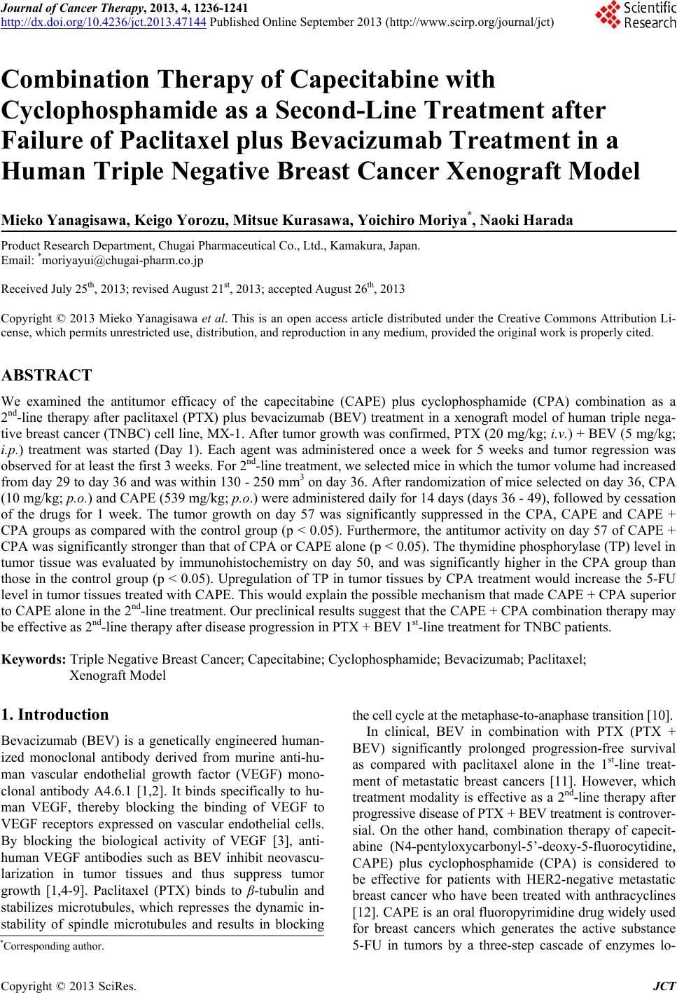
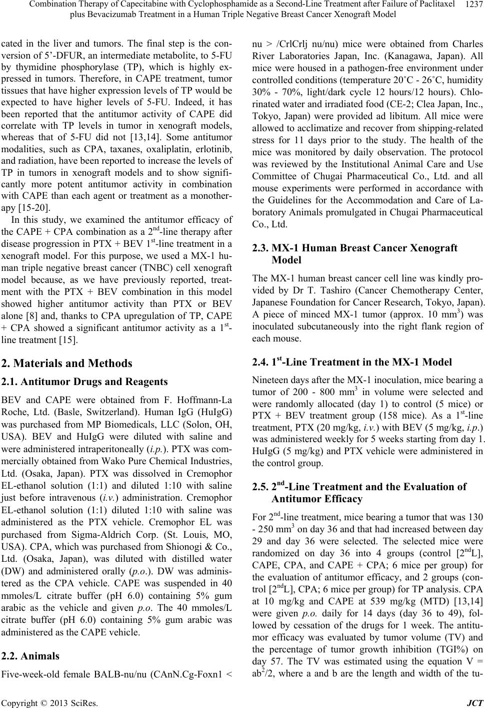
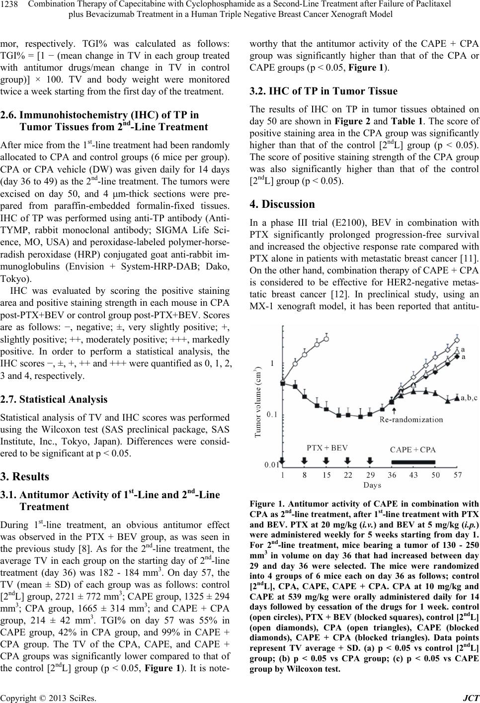
2nd-line treatment, CAPE + CPA combination showed an
extremely high antitumor activity compared to CAPE or
CPA monotherapy. Because the TP level in tumor was
upregulated by CPA treatment, the superior antitumor
effect of CAPE + CPA combination compared to CAPE
monotherapy may be attributed to the increased 5-FU
level in tumor tissue caused by facilitated conversion
from CAPE by TP. These results are similar to that in the
1st-line therapy reported previously [15].
Our preclinical results suggest that the CAPE + CPA
combination therapy may be effective as a 2nd-line ther-
apy for progressive disease after PTX + BEV 1st-line
treatment in TNBC patients.
5. Acknowledgements
We thank Dr. Kazushige Mori for helpful discussion and
comments regarding the manuscript.
REFERENCES
[1] L. G. Presta, H. Chen, S. J. O’Connor, V. Chisholm, Y. G.
Meng, et al., “Humanization of an Anti-Vascular Endo-
thelial Growth Factor Monoclonal Antibody for the Ther-
apy of Solid Tumors and Other Disorders,” Cancer Re-
search, Vol. 57, No. 20, 1997, pp. 4593-4599.
[2] K. J. Kim, B. Li, K. Houck, J. Winer and N. Ferrara, “The
Vascular Endothelial Growth Factor Proteins: Identifica-
tion of Biologically Relevant Regions by Neutralizing
Monoclonal Antibodies,” Growth Factors, Vol. 7, No.1,
1992, pp. 53-64. doi:10.3109/08977199209023937
[3] Y. Wang, D. Fei, M. Vanderlaan and A. Song, “Biologi-
cal Activity of Bevacizumab, a Humanized Anti-Vegf
Antibody in Vitro,” Angiogenesis, Vol. 7, No. 4, 2004, pp.
335-345. doi:10.1007/s10456-004-8272-2
[4] K. J. Kim, B. Li, J. Winer, M. Armanini, N. Gillett, et al.,
“Inhibition of Vascular Endothelial Growth Factor-In-
duced Angiogenesis Suppresses Tumour Growth in Vivo,”
Nature, Vol. 362, No. 6423, 1993, pp. 841-844.
doi:10.1038/362841a0
[5] H. P. Gerber and N. Ferrara, “Pharmacology and Phar-
macodynamics of Bevacizumab as Monotherapy or in
Combination with Cytotoxic Therapy in Preclinical Stud-
ies,” Cancer Research, Vol. 65, No. 3, 2005, pp. 671-680.
[6] P. V. Dickson, J. B. Hamner, T. L. Sims, C. H. Fraga, C.
Y. Ng, et al., “Bevacizumab-Induced Transient Remod-
eling of the Vasculature in Neuroblastoma Xenografts
Results in Improved Delivery and Efficacy of Systemi-
cally Administered Chemotherapy,” Clinical Cancer Re-
search, Vol. 13, No. 13, 2007, pp. 3942-3950.
doi:10.1158/1078-0432.CCR-07-0278
[7] M. Yanagisawa, K. Fujimoto-Ouchi, K. Yorozu, Y. Ya-
mashita and K. Mori, “Antitumor Activity of Bevacizu-
mab in Combination with Capecitabine and Oxaliplatin in
Human Colorectal Cancer Xenograft Models,” Oncology
Reports, Vol. 22, No. 2, 2009, pp. 241-247.
[8] M. Yanagisawa, K. Yorozu, M. Kurasawa, K. Nakano, K.
Furugaki, et al., “Bevacizumab Improves the Delivery
and Efficacy of Paclitaxel,” Anticancer Drugs, Vol. 21,
No. 7, 2010, pp. 687-694.
[9] Y. Yamashita-Kashima, K. Fujimoto-Ouchi, K. Yorozu,
M. Kurasawa, M. Yanagisawa, et al., “Biomarkers for
Antitumor Activity of Bevacizumab in Gastric Cancer
Models,” BMC Cancer, Vol. 12, 2012, p. 37.
doi:10.1186/1471-2407-12-37
[10] S. B. Horwitz, “Mechanism of Action of Taxol,” Trends
in Pharmacological Sciences, Vol. 13, No. 4, 1992, pp.
134-136. doi:10.1016/0165-6147(92)90048-B
[11] K. Miller, M. Wang, J. Gralow, M. Dickler, M. Cobleigh,
et al., “Paclitaxel plus Bevacizumab versus Paclitaxel
Alone for Metastatic Breast Cancer,” New England Jour-
nal of Medicine, Vol. 357, No. 26, 2007, pp. 2666-2676.
doi:10.1056/NEJMoa072113
[12] M. Tanaka, Y. Takamatsu, K. Anan, S. Ohno, R. Nishi-
mura, et al., “Oral Combination Chemotherapy with Ca-
pecitabine and Cyclophosphamide in Patients with Me-
tastatic Breast Cancer: A Phase II Study,” Anti-Cancer
Drugs, Vol. 21, No. 4, 2010, pp. 453-458.
doi:10.1097/CAD.0b013e328336acb1
[13] H. Yasuno, M. Kurasawa, M. Yanagisawa, Y. Sato, N.
Harada, et al., “Predictive Markers of Capecitabine Sen-
sitivity Identified from the Expression Profile of Py-
rimidine Nucleoside-Metabolizing Enzymes,” Oncology
Reports, Vol. 29, No. 2, 2013, pp. 451-458.
[14] T. Ishikawa, F. Sekiguchi, Y. Fukase, N. Sawada and H.
Ishitsuka, “Positive Correlation between the Efficacy of
Capecitabine and Doxifluridine and the Ratio of Thy-
midine Phosphorylase to Dihydropyrimidine Dehydro-
genase Activities in Tumors in Human Cancer Xeno-
grafts,” Cancer Research, Vol. 58, No. 4, 1998, pp. 685-
690.
[15] M. Endo, N. Shinbori, Y. Fukase, N. Sawada, T. Ishikawa,
et al., “Induction of Thymidine Phosphorylase Expression
and Enhancement of Efficacy of Capecitabine or 5’-De-
oxy-5-Fluorouridine by Cyclophosphamide in Mammary
Tumor Models,” International Journal of Cancer, Vol. 83,
No. 1, 1999, pp. 127-134.
doi:10.1002/(SICI)1097-0215(19990924)83:1<127::AID-
IJC22>3.0.CO;2-6
[16] N. Sawada, T. Ishikawa, Y. Fukase, M. Nishida, T. Yo-
shikubo, et al., “Induction of Thymidine Phosphorylase
Activity and Enhancement of Capecitabine Efficacy by
Taxol/Taxotere in Human Cancer Xenografts,” Clinical
Copyright © 2013 SciRes. JCT
Combination Therapy of Capecitabine with Cyclophosphamide as a Second-Line Treatment after Failure of Paclitaxel
plus Bevacizumab Treatment in a Human Triple Negative Breast Cancer Xenograft Model
Copyright © 2013 SciRes. JCT
1241
Cancer Research, Vol. 4, No. 4, 1998, pp. 1013-1019.
[17] J. Cassidy, J. Tabernero, C. Twelves, R. Brunet, C. Butts,
et al., “Xelox (Capecitabine plus Oxaliplatin): Active
First-Line Therapy for Patients with Metastatic Colorectal
Cancer,” Journal of Clinical Oncology, Vol. 22, No. 11,
2004, pp. 2084-2091. doi:10.1200/JCO.2004.11.069
[18] N. Sawada, K. Kondoh and K. Mori, “Enhancement of
Capecitabine Efficacy by Oxaliplatin in Human Colorec-
tal and Gastric Cancer Xenografts,” Oncology Reports,
Vol. 18, No. 4, 2007, pp. 775-778.
[19] K. Fujimoto-Ouchi, M. Yanagisawa, F. Sekiguchi and Y.
Tanaka, “Antitumor Activity of Erlotinib in Combination
with Capecitabine in Human Tumor Xenograft Models,”
Cancer Chemotherapy and Pharmacology, Vol. 57, No. 5,
2006, pp. 693-702. doi:10.1007/s00280-005-0079-3
[20] N. Sawada, T. Ishikawa, F. Sekiguchi, Y. Tanaka and H.
Ishitsuka, “X-Ray Irradiation Induces Thymidine Phos-
phorylase and Enhances the Efficacy of Capecitabine
(Xeloda) in Human Cancer Xenografts,” Clinical Cancer
Research, Vol. 5, 1999, pp. 2948-2953.
[21] E. Mechetner, A. Kyshtoobayeva, S. Zonis, H. Kim, R.
Stroup, et al., “Levels of Multidrug Resistance (mdr1)
p-Glycoprotein Expression by Human Breast Cancer Cor-
relate with in Vitro Resistance to Taxol and Doxorubi-
cin,” Clinical Cancer Research, Vol. 4, 1998, pp. 389-
398.
[22] J. P. de Hoon, J. Veeck, B. E. Vriens, T. G. Calon, M. van
Engeland, et al., “Taxane Resistance in Breast Cancer: A
Closed Her2 Circuit?” Biochimica et Biophysica Acta,
Vol. 1825, No. 2, 2012, pp. 197-206.
[23] A. K. Koutras, I. Starakis, U. Kyriakopoulou, P. Katsaou-
nis, A. Nikolakopoulos, et al., “Targeted Therapy in Co-
lorectal Cancer: Current Status and Future Challenges,”
Current Medicinal Chemistry, Vol. 18, No. 11, 2011, pp.
1599-1612. doi:10.2174/092986711795471338
[24] S. Tejpar, H. Prenen and M. Mazzone, “Overcoming
Resistance to Antiangiogenic Therapies,” Oncologist, Vol.
17, No. 8, 2012, pp. 1039-1050.
doi:10.1634/theoncologist.2012-0068
[25] N. Ferrara and R. S. Kerbel, “Angiogenesis as a Thera-
peutic Target,” Nature, Vol. 438, No. 7070, 2005, pp.
967-974. doi:10.1038/nature04483
[26] M. F. Mulcahy, “Bevacizumab in the Therapy for Re-
fractory Metastatic Colorectal Cancer,” Biologics, Vol. 2,
No. 1, 2008, pp. 53-59.