 Open Journal of Urology, 2013, 3, 210-216 http://dx.doi.org/10.4236/oju.2013.35039 Published Online September 2013 (http://www.scirp.org/journal/oju) Characteristics of Prostate Cancers Missed by Biopsies: Evaluation of Cumulative Tumor Volume Missed According to Cancer True Prevalence* Nicolas B. Delongchamps1#, Gustavo de la Roza2, Paul Perrin3, Michaël Peyromaure1, Gabriel P. Haas4 1Department of Urology, Cochin Hospital, Paris Descartes University, Paris, France 2Department of Pathology, Upstate Medical University, Syracuse, USA 3Department of Urology, Lyon South Hospital, Lyon, France 4Astellas Pharma Global Development, Inc., Deerfield, USA Email: #nicolasbdl@hotmail.com Received May 14, 2013; revised June 16, 2013; accepted June 22, 2013 Copyright © 2013 Nicolas B. Delongchamps et al. This is an open access article distributed under the Creative Commons Attribution License, which permits unrestricted use, distribution, and reproduction in any medium, provided the original work is properly cited. ABSTRACT Purpose: To characterize missed prostate tumors and their cumulative volume with various biopsy regimens to deter- mine optimal biopsy schemes. Methods: We performed 6, 12 and 18-core needle biopsies on 165 and 36-core biopsies on 47 autopsy prostates, respectively. The 6-core biopsy included 6 cores from the mid peripheral zone (MPZ), the 12-core biopsy included 6 cores from the MPZ and lateral PZ (LPZ), and the 18-core biopsies included 6 cores from the MPZ, LPZ and central zone (CZ). The 36-core biopsies included 12 cores in each of these 3 areas. We analyzed the sensitivity of biopsies at each site and evaluated the cumulative volume of cancers and tumor foci missed. Results: Whole-mount analysis identified 59 cancers, 110 tumor foci, and a total cumulative tumor volume of 43 cm3. The per- centage of tumor foci and corresponding cumulative volume missed with 6, 12, 18 and 36-core biopsies were of 79% and 58%, 64% and 48%, 57% and 26%, and 42% and 17%, respectively (p < 0.05). 12-core biopsies from the MPZ and LPZ performed best for clinically significant cancers detection. However, increasing the number of cores over the 6-core biopsy cutoff increased solely the detection of tumor foci < 0.5 cm3. Conclusion: Twelve biopsies from the MPZ and LPZ detected most of the clinically significant cancers while missing most of the tumor foci. These missed tumors represented only a small amount of the overall cancer volume. Keywords: Prostate Cancer; Prevalence; Biopsy; Clinical Significance 1. Introduction The trend among clinicians has been performed more prostate biopsies to detect more prostate cancers. Sextant biopsies have been all but abandoned, and most studies recommend extended biopsy protocols of 12 cores [1-3]. Although these efforts have undoubtedly led to increased detection of cancer, they have also led to the over diag- nosis of small and well-differentiated tumors, designated as clinically insignificant [4,5]. Even undetected, they may not be of immediate threat, and should they be de- tected, not to be managed with radical and possibly mor- bid treatment. This issue recently came under increased scrutiny as a consequence of the recent recommendations by the Task Force for Preventive Medicine [6]. Notwith- standing the evidence that PSA screening followed by early stage treatment may reduce mortality [7-9], they concluded that the use of PSA was harmful because it caused increased morbidity and over treatment of indo- lent tumors. While recommendations from the Task Force for Pre- ventive Medicine focused on the adverse effects of PSA screening, they did not address the role of prostate biopsy as a staging tool. Also, we have to be concerned not only with the ability of the biopsy to detect cancer, but also with the potential characteristics of cancers missed. We previously reported cancer detection rates obtained with different ex-vivo biopsy protocols performed on autop- sied prostates [10]. Based on the cancer true prevalence, we found that 12 biopsies targeting the mid peripheral *Funding: National Institute on Aging (AG021389 to G.P.H.); National Cancer Institute (CA097751 to G.P.H.); National Institutes of Health (2U42RR006042-13). This study or its publication did not receive financial support from Astellas Pharma Global Development, Inc. #Corresponding author. C opyright © 2013 SciRes. OJU 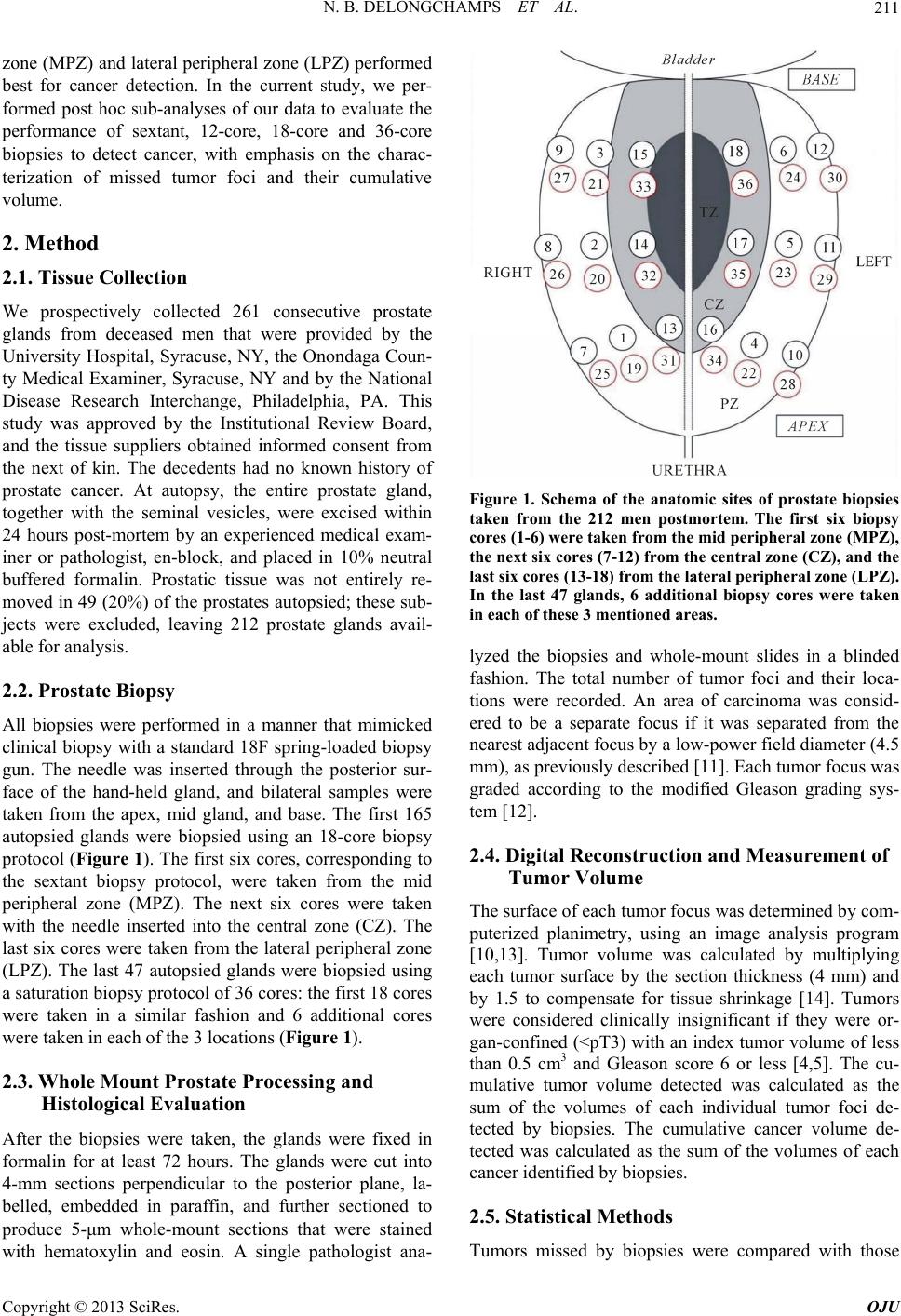 N. B. DELONGCHAMPS ET AL. 211 zone (MPZ) and lateral peripheral zone (LPZ) performed best for cancer detection. In the current study, we per- formed post hoc sub-analyses of our data to evaluate the performance of sextant, 12-core, 18-core and 36-core biopsies to detect cancer, with emphasis on the charac- terization of missed tumor foci and their cumulative volume. 2. Method 2.1. Tissue Collection We prospectively collected 261 consecutive prostate glands from deceased men that were provided by the University Hospital, Syracuse, NY, the Onondaga Coun- ty Medical Examiner, Syracuse, NY and by the National Disease Research Interchange, Philadelphia, PA. This study was approved by the Institutional Review Board, and the tissue suppliers obtained informed consent from the next of kin. The decedents had no known history of prostate cancer. At autopsy, the entire prostate gland, together with the seminal vesicles, were excised within 24 hours post-mortem by an experienced medical exam- iner or pathologist, en-block, and placed in 10% neutral buffered formalin. Prostatic tissue was not entirely re- moved in 49 (20%) of the prostates autopsied; these sub- jects were excluded, leaving 212 prostate glands avail- able for analysis. 2.2. Prostate Biopsy All biopsies were performed in a manner that mimicked clinical biopsy with a standard 18F spring-loaded biopsy gun. The needle was inserted through the posterior sur- face of the hand-held gland, and bilateral samples were taken from the apex, mid gland, and base. The first 165 autopsied glands were biopsied using an 18-core biopsy protocol (Figure 1). The first six cores, corresponding to the sextant biopsy protocol, were taken from the mid peripheral zone (MPZ). The next six cores were taken with the needle inserted into the central zone (CZ). The last six cores were taken from the lateral peripheral zone (LPZ). The last 47 autopsied glands were biopsied using a saturation biopsy protocol of 36 cores: the first 18 cores were taken in a similar fashion and 6 additional cores were taken in each of the 3 locations (Figure 1). 2.3. Whole Mount Prostate Processing and Histological Evaluation After the biopsies were taken, the glands were fixed in formalin for at least 72 hours. The glands were cut into 4-mm sections perpendicular to the posterior plane, la- belled, embedded in paraffin, and further sectioned to produce 5-μm whole-mount sections that were stained with hematoxylin and eosin. A single pathologist ana- Figure 1. Schema of the anatomic sites of prostate biopsies taken from the 212 men postmortem. The first six biopsy cores (1-6) were taken from the mid peripheral zone (MPZ), the next six cores (7-12) from the central zone (CZ), and the last six cores (13-18) from the lateral peripheral zone (LPZ). In the last 47 glands, 6 additional biopsy cores were taken in each of these 3 mentioned areas. lyzed the biopsies and whole-mount slides in a blinded fashion. The total number of tumor foci and their loca- tions were recorded. An area of carcinoma was consid- ered to be a separate focus if it was separated from the nearest adjacent focus by a low-power field diameter (4.5 mm), as previously described [11]. Each tumor focus was graded according to the modified Gleason grading sys- tem [12]. 2.4. Digital Reconstruction and Measurement of Tumor Volume The surface of each tumor focus was determined by com- puterized planimetry, using an image analysis program [10,13]. Tumor volume was calculated by multiplying each tumor surface by the section thickness (4 mm) and by 1.5 to compensate for tissue shrinkage [14]. Tumors were considered clinically insignificant if they were or- gan-confined (<pT3) with an index tumor volume of less than 0.5 cm3 and Gleason score 6 or less [4,5]. The cu- mulative tumor volume detected was calculated as the sum of the volumes of each individual tumor foci de- tected by biopsies. The cumulative cancer volume de- tected was calculated as the sum of the volumes of each cancer identified by biopsies. 2.5. Statistical Methods Tumors missed by biopsies were compared with those Copyright © 2013 SciRes. OJU 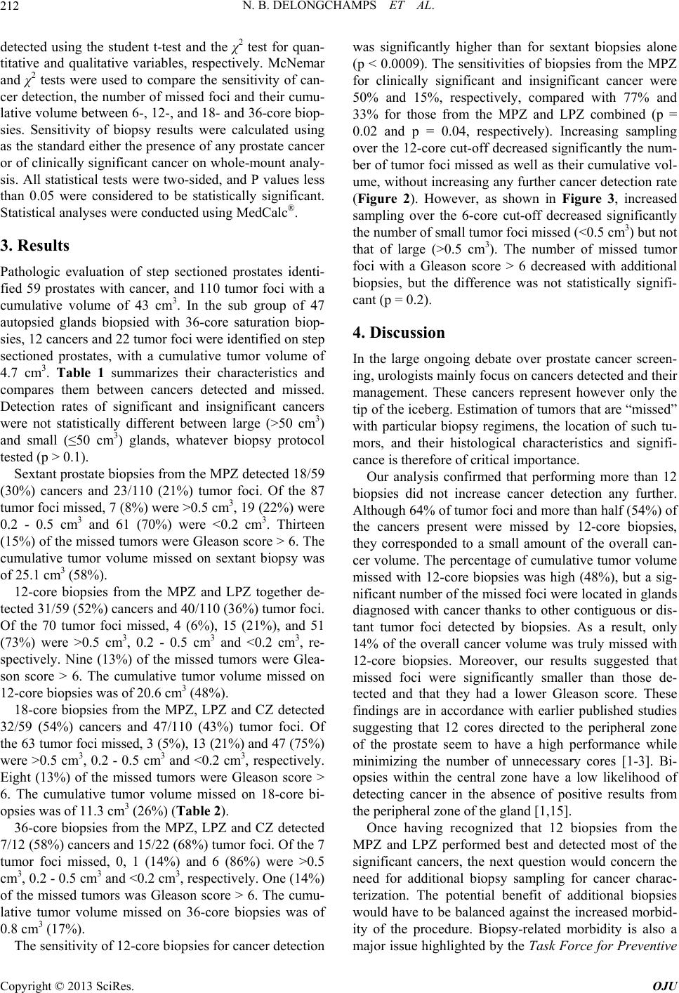 N. B. DELONGCHAMPS ET AL. 212 detected using the student t-test and the χ2 test for quan- titative and qualitative variables, respectively. McNemar and χ2 tests were used to compare the sensitivity of can- cer detection, the number of missed foci and their cumu- lative volume between 6-, 12-, and 18- and 36-core biop- sies. Sensitivity of biopsy results were calculated using as the standard either the presence of any prostate cancer or of clinically significant cancer on whole-mount analy- sis. All statistical tests were two-sided, and P values less than 0.05 were considered to be statistically significant. Statistical analyses were conducted using MedCalc®. 3. Results Pathologic evaluation of step sectioned prostates identi- fied 59 prostates with cancer, and 110 tumor foci with a cumulative volume of 43 cm3. In the sub group of 47 autopsied glands biopsied with 36-core saturation biop- sies, 12 cancers and 22 tumor foci were identified on step sectioned prostates, with a cumulative tumor volume of 4.7 cm3. Table 1 summarizes their characteristics and compares them between cancers detected and missed. Detection rates of significant and insignificant cancers were not statistically different between large (>50 cm3) and small (≤50 cm3) glands, whatever biopsy protocol tested (p > 0.1). Sextant prostate biopsies from the MPZ detected 18/59 (30%) cancers and 23/110 (21%) tumor foci. Of the 87 tumor foci missed, 7 (8%) were >0.5 cm3, 19 (22%) were 0.2 - 0.5 cm3 and 61 (70%) were <0.2 cm3. Thirteen (15%) of the missed tumors were Gleason score > 6. The cumulative tumor volume missed on sextant biopsy was of 25.1 cm3 (58%). 12-core biopsies from the MPZ and LPZ together de- tected 31/59 (52%) cancers and 40/110 (36%) tumor foci. Of the 70 tumor foci missed, 4 (6%), 15 (21%), and 51 (73%) were >0.5 cm3, 0.2 - 0.5 cm3 and <0.2 cm3, re- spectively. Nine (13%) of the missed tumors were Glea- son score > 6. The cumulative tumor volume missed on 12-core biopsies was of 20.6 cm3 (48%). 18-core biopsies from the MPZ, LPZ and CZ detected 32/59 (54%) cancers and 47/110 (43%) tumor foci. Of the 63 tumor foci missed, 3 (5%), 13 (21%) and 47 (75%) were >0.5 cm3, 0.2 - 0.5 cm3 and <0.2 cm3, respectively. Eight (13%) of the missed tumors were Gleason score > 6. The cumulative tumor volume missed on 18-core bi- opsies was of 11.3 cm3 (26%) (Table 2). 36-core biopsies from the MPZ, LPZ and CZ detected 7/12 (58%) cancers and 15/22 (68%) tumor foci. Of the 7 tumor foci missed, 0, 1 (14%) and 6 (86%) were >0.5 cm3, 0.2 - 0.5 cm3 and <0.2 cm3, respectively. One (14%) of the missed tumors was Gleason score > 6. The cumu- lative tumor volume missed on 36-core biopsies was of 0.8 cm3 (17%). The sensitivity of 12-core biopsies for cancer detection was significantly higher than for sextant biopsies alone (p < 0.0009). The sensitivities of biopsies from the MPZ for clinically significant and insignificant cancer were 50% and 15%, respectively, compared with 77% and 33% for those from the MPZ and LPZ combined (p = 0.02 and p = 0.04, respectively). Increasing sampling over the 12-core cut-off decreased significantly the num- ber of tumor foci missed as well as their cumulative vol- ume, without increasing any further cancer detection rate (Figure 2). However, as shown in Figure 3, increased sampling over the 6-core cut-off decreased significantly the number of small tumor foci missed (<0.5 cm3) but not that of large (>0.5 cm3). The number of missed tumor foci with a Gleason score > 6 decreased with additional biopsies, but the difference was not statistically signifi- cant (p = 0.2). 4. Discussion In the large ongoing debate over prostate cancer screen- ing, urologists mainly focus on cancers detected and their management. These cancers represent however only the tip of the iceberg. Estimation of tumors that are “missed” with particular biopsy regimens, the location of such tu- mors, and their histological characteristics and signifi- cance is therefore of critical importance. Our analysis confirmed that performing more than 12 biopsies did not increase cancer detection any further. Although 64% of tumor foci and more than half (54%) of the cancers present were missed by 12-core biopsies, they corresponded to a small amount of the overall can- cer volume. The percentage of cumulative tumor volume missed with 12-core biopsies was high (48%), but a sig- nificant number of the missed foci were located in glands diagnosed with cancer thanks to other contiguous or dis- tant tumor foci detected by biopsies. As a result, only 14% of the overall cancer volume was truly missed with 12-core biopsies. Moreover, our results suggested that missed foci were significantly smaller than those de- tected and that they had a lower Gleason score. These findings are in accordance with earlier published studies suggesting that 12 cores directed to the peripheral zone of the prostate seem to have a high performance while minimizing the number of unnecessary cores [1-3]. Bi- opsies within the central zone have a low likelihood of detecting cancer in the absence of positive results from the peripheral zone of the gland [1,15]. Once having recognized that 12 biopsies from the MPZ and LPZ performed best and detected most of the significant cancers, the next question would concern the need for additional biopsy sampling for cancer charac- terization. The potential benefit of additional biopsies would have to be balanced against the increased morbid- ity of the procedure. Biopsy-related morbidity is also a major issue highlighted by the Task Force for Preventive Copyright © 2013 SciRes. OJU 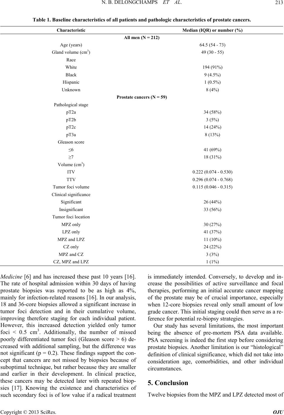 N. B. DELONGCHAMPS ET AL. Copyright © 2013 SciRes. OJU 213 Table 1. Baseline characteristics of all patients and pathologic characteristics of pros t a t e c a ncers. Characteristic Median (IQR) or number (%) All men (N = 212) Age (years) 64.5 (54 - 73) Gland volume (cm3) 49 (30 - 55) Race White 194 (91%) Black 9 (4.5%) Hispanic 1 (0.5%) Unknown 8 (4%) Prostate cancers (N = 59) Pathological stage pT2a 34 (58%) pT2b 3 (5%) pT2c 14 (24%) pT3a 8 (13%) Gleason score ≤6 41 (69%) ≥7 18 (31%) Volume (cm3) ITV 0.222 (0.074 - 0.530) TTV 0.296 (0.074 - 0.768) Tumor foci volume 0.115 (0.046 - 0.315) Clinical significance Significant 26 (44%) Insignificant 33 (56%) Tumor foci location MPZ only 30 (27%) LPZ only 41 (37%) MPZ and LPZ 11 (10%) CZ only 24 (22%) MPZ and CZ 3 (3%) CZ, MPZ and LPZ 1 (1%) Medicine [6] and has increased these past 10 years [16]. The rate of hospital admission within 30 days of having prostate biopsies was reported to be as high as 4%, mainly for infection-related reasons [16]. In our analysis, 18 and 36-core biopsies allowed a significant increase in tumor foci detection and in their cumulative volume, improving therefore staging for each individual patient. However, this increased detection yielded only tumor foci < 0.5 cm3. Additionally, the number of missed poorly differentiated tumor foci (Gleason score > 6) de- creased with additional sampling, but the difference was not significant (p = 0.2). These findings support the con- cept that cancers are not missed by biopsies because of suboptimal technique, but rather because they are smaller and earlier in their development. In clinical practice, these cancers may be detected later with repeated biop- sies [17]. Knowing the existence and characteristics of such secondary foci is of low value if a radical treatment is immediately intended. Conversely, to develop and in- crease the possibilities of active surveillance and focal therapies, performing an initial accurate cancer mapping of the prostate may be of crucial importance, especially when 12-core biopsies reveal only small amount of low grade cancer. This initial staging could then serve as a re- ference for potential re-biopsy strategies. Our study has several limitations, the most important being the absence of pre-mortem PSA data available. PSA screening is indeed the first step before considering prostate biopsies. Another limitation is our “histological” definition of clinical significance, which did not take into consideration age, comorbidities, and other individual circumstances. 5. Conclusion Twelve biopsies from the MPZ and LPZ detected most of  N. B. DELONGCHAMPS ET AL. 214 Table 2. Pathological characteristics of cancers detected and missed with 18-core biopsies. Prostate cancers Detected (n = 32) Missed (n = 27) p value Median (IQR) ITV (cm3) 0.485 (0.265 - 0.845) 0.081 (0.047 - 0.215) 0.02 Median (IQR) TTV (cm3) 0.638 (0.333 - 1.057) 0.087 (0.051 - 0.215) 0.005 Gleason score ≤3 + 3 19 (59%) 21 (78%) ≥3 + 4 13 (41%) 6 (22%) 0.2 Number of foci Unifocal 9 (28%) 21 (78%) Multifocal 23 (72%) 6 (22%) 0.0002 Significance Significant 21 (66%) 5 (18%) Insignificant 11 (34%) 22 (82%) 0.0005 Tumor foci Detected (n = 47) Missed (n = 63) p value Median (IQR) tumor volume (cm3) 0.296 (0.096 - 0.572) 0.081 (0.031 - 0.197) 0.003 Number of tumor foci Volume ≤ 0.2 cm3 20 47 Volume [0.2 - 0.5] cm3 14 13 Volume ≥ 0.5 cm3 13 3 Gleason score ≤3 + 3 30 55 ≥3 + 4 17 8 0.005 Location MPZ 10 (21%) 20 (32%) 0.3 LPZ 17 (37%) 24 (38%) 1 CZ 7 (15%) 17 (27%) 0.2 MPZ + LPZ 9 (19%) 2 (3%) 0.008 MPZ + CZ 3 (6%) 0 MPZ + LPZ + CZ 1 (2%) 0 Figure 2. Rates of missed cancers and individual tumor foci, and corresponding volumes, according to biopsy protocol. Copyright © 2013 SciRes. OJU  N. B. DELONGCHAMPS ET AL. 215 Figure 3. Rates of missed individual tumor foci according to their volume. the clinically significant cancers while missing as much as 48% of cancers. These missed cancers represented however only 13% of the overall volume of cancer. REFERENCES [1] K. Eichler, S. Hempel, J. Wilby, et al., “Diagnostic Value of Systematic Biopsy Methods in the Investigation of Prostate Cancer: A Systematic Review,” Journal of Uro- logy, Vol. 175, No. 5, 2006, pp. 1605-1612. doi:10.1016/S0022-5347(05)00957-2 [2] V. Scattoni, A. Zlotta, R. Montironi, C. Schulman, P. Ri- gatti and F. Montorsi, “Extended and Saturation Prostatic Biopsy in the Diagnosis and Characterisation of Prostate Cancer: A Critical Analysis of the Literature,” European Urology, Vol. 52, No. 5, 2007, pp. 1309-1322. doi:10.1016/j.eururo.2007.08.006 [3] A. Heidenreich, J. Bellmunt, M. Bolla, et al., “EAU Guidelines on Prostate Cancer. Part 1: Screening, Diag- nosis, and Treatment of Clinically Localised Disease,” European Urology, Vol. 59, No. 1, 2011, pp. 61-71. doi:10.1016/j.eururo.2010.10.039 [4] T. A. Stamey F. S. Freiha, J. E. McNeal, et al., “Local- ized Prostate Cancer. Relationship of Tumor Volume to Clinical Significance for Treatment of Prostate Cancer,” Cancer, Vol. 71, Supplement S3, 1993, pp. 933-938. doi:10.1002/1097-0142(19930201)71:3+<933::AID-CNC R2820711408>3.0.CO;2-L [5] J. I. Epstein, P. C. Walsh, M. Carmichael, et al., “Patho- logic and Clinical Findings to Predict Tumor Extent of Nonpalpable (T1c) Prostate Cancer,” JAMA, Vol. 271, No. 5, 1994, pp. 368-374. doi:10.1001/jama.1994.03510290050036 [6] R. Chou, J. M. Croswell, T. Dana, et al., “Screening for Prostate Cancer: A Review of the Evidence for the US Preventive Services Task Force,” Annals of Internal Medicine, Vol. 155, No. 11, 2011, pp. 762-771. doi:10.7326/0003-4819-155-11-201112060-00375 [7] F. H. Schroder, J. Hugosson, M. J. Roobol, et al., “Screen- ing and Prostate-Cancer Mortality in a Randomized Euro- pean Study,” The New England Journal of Medicine, Vol. 360, No. 13, 2009, pp. 1320-1328. doi:10.1056/NEJMoa0810084 [8] F. H. Schröder, J. Hugosson, M. J. Roobol, et al. and ERSPC Investigators, “Prostate-Cancer Mortality at 11 Years of Follow-Up,” The New England Journal of Med- icine, Vol. 366, No. 11, 2012, pp. 981-990. doi:10.1056/NEJMoa1113135 [9] M. Bul and F. H. Schröder, “Screening for Prostate Can- cer—The Controversy Continues, but Can It Be Resol- ved?” Acta Oncologica, Vol. 50, No. S1, 2011, pp. 4-11. doi:10.3109/0284186X.2010.522197 [10] G. P. Haas, N. B. Delongchamps, R. F. Jones, et al., “Needle Biopsies on Autopsy Prostates: Sensitivity of Copyright © 2013 SciRes. OJU  N. B. DELONGCHAMPS ET AL. 216 Cancer Detection Based on True Prevalence,” Journal of the National Cancer Institute, Vol. 99, No. 19, 2007, pp. 1484-1489. doi:10.1093/jnci/djm153 [11] P. Troncoso, R. J. Babaian, J. Y. Ro, et al., “Prostatic Intraepithelial Neoplasia and Invasive Prostatic Adeno- carcinoma in Cystoprostatectomy Specimens,” Urology, Vol. 34, Supplement 6, 1989, pp. 52-56. [12] J. I. Epstein, W. C. Allsbrook Jr., M. B. Amin, et al., “The 2005 International Society of Urological Pathology (ISUP) Concensus Conference on Gleason Grading of Prostatic Carcinoma,” The American Journal of Surgical Pathology, Vol. 29, No. 9, 2005, pp. 1228-1242. doi:10.1097/01.pas.0000173646.99337.b1 [13] M. Noguchi, T. A. Stamey, J. E. McNeal, et al., “As- sessment of Morphometric Measurements of Prostate Carcinoma Volume,” Cancer, Vol. 89, No. 5, 2000, pp. 1056-1064. doi:10.1097/01.pas.0000173646.99337.b1 [14] A. R. Schned, K. J. Wheeler, C. A. Hodorowski, et al., “Tissue-Shrinkage Correction Factor in the Calculation of Prostate Cancer Volume,” The American Journal of Sur- gical Pathology, Vol. 20, No. 12, 1996, pp. 1501-1506. doi:10.1097/00000478-199612000-00009 [15] A. R. Patel, J. S. Jones, J. Rabets, et al., “Parasagittal Biopsies Add Minimal Information in Repeat Saturation Prostate Biopsy,” Urology, Vol. 63, No. 1, 2004, pp. 87- 89. doi:10.1016/j.urology.2003.08.040 [16] R. K. Nam, R. Saskin, Y. Lee, et al., “Increasing Hospital Admission Rates for Urological Complications after Transrectal Ultrasound Guided Prostate Biopsy,” Journal of Urology, Vol. 183, No. 3, 2010, pp. 963-968. doi:10.1016/j.juro.2009.11.043 [17] M. J. Roobol, R. C. N. Van Den Bergh, T. Wolters, et al., “Serum PSA Levels, Number of Prostate Biopsies and Number and Characteristics of Prostate Cancers Detected in Three Consecutive Screening Visits Using a PSA Based Biopsy Indication,” European Urology, Vol. 7, No. 3, 2008, p. 199. doi:10.1016/S1569-9056(08)60509-6 Copyright © 2013 SciRes. OJU
|