Advances in Bioscience and Biotechnology, 2013, 4, 896-899 ABB
http://dx.doi.org/10.4236/abb.2013.49117 Published Online September 2013 (http://www.scirp.org/journal/abb/)
Effect of cytokines and ultraviolet B radiation on the
promoter activity of the metallothionein gene in
keratinocytes
Hiromi Narumi*, Hajime Nakano, Takahide Kaneko, Koji Nakajima, Yasushi Matsuzaki,
Takayuki Aizu, Daisuke Sawamura, Katsumi Hanada
Department of Dermatology, Hirosaki University Graduate School of Medicine, Hirosaki, Japan
Email: *derma@cc.hirosaki-u.ac.jp
Received 18 April 2013; revised 19 May 2013; accepted 10 June 2013
Copyright © 2013 Hiromi Narumi et al. This is an open access article distributed under the Creative Commons Attribution License,
which permits unrestricted use, distribution, and reproduction in any medium, provided the original work is properly cited.
ABSTRACT
Metallothionein (MT) has many functions that are
modulated by several factors, including ultraviolet
(UV) radiation and cytokines. We thought that these
diverse functions of MT might reflect the specific
regulatory mechanisms of its expression. To under-
stand some of the molecular mechanisms underlying
MT expression, we examined the effects of several
cytokines and UVB on the promoter activity of the
MT gene. First, we introduced the MT promoter con-
struct into the HaCaT keratinocytes and treated them
with various concentrations of interleukin-1α (IL-1α)
and IL-6. The addition of IL-1α and IL-6 led to an
increase in the promoter activity of the MT gene.
UVB is known to induce MT expression in epidermal
keratinocytes, and IL-6 is a possible mediator of MT
induction by UV radiation. Therefore, we investi-
gated whether UVB could induce MT promoter ac-
tivity. Our results showed, interestingly, that UVB
radiation has no or little effect on the promoter activ-
ity. This suggested a complex molecular regulation of
the MT gen e .
Keywords: Sunburn; Cytokine; Epidermis; Ultraviolet
Light; Promoter
1. INTRODUCTION
Metallothionein (MT) is a ubiquitously distributed, cys-
teine-rich, low molecular weight protein having a high
binding capacity for metals such as zinc, copper, and
cadmium. It plays a role in zinc homeostasis and detoxi-
fication of heavy metals [1]. Several studies have shown
that MT acts as a reactive oxygen species scavenger [2]
and MT induction has protective effects against oxidative
stresses such as anticancer drugs and ultraviolet (UV)
radiation [3]. MT gene expression is induced not only by
heavy metals but also by various stress-inducing agents
such as UV [4] and X-ray radiation [5]. We found that
β-thujaplicin induced MT expression in keratinocytes,
both in vitro and in vivo, and thereby reduced UVB irra-
diation-induced apoptosis [6]. Furthermore, a number of
cytokines, including interferon-α and β, tumor necrosis
factor-α, interleukin-1 (IL-1), and IL-6, increase MT ex-
pression, suggesting that MT expression also involves
additional functions, including immunomodulation, cell
growth, and cell differentiation [7].
We thought that these diverse functions of MT might
reflect specific regulatory mechanisms of its expression.
We have recently cloned the promoter region of the MT
gene and performed a functional assay [8]. Thus, to un-
derstand some of the molecular mechanism underlying
MT expression, we focused on the effects of several cy-
tokines and UVB radiation on the promoter activity of
the MT gene.
2. MATERIALS AND METHODS
2.1. Cell Culture
The spontaneously transformed human epidermal kerati-
nocyte cell line, HaCaT, (kindly provided by Dr. Husenig)
was cultured in Dulbecco’s modified minimal essential
medium supplemented with 10% fetal calf serum, 1%
l-glutamine, and 1% antibiotic/antimycotic solution. The
cells were maintained at 37˚C in a humidified atmos-
phere containing 5% CO2. The viability of the cells
treated with cytokines and UVB radiation for 24 h was
approximately 95%, as determined by trypan blue exclu-
sion staining.
*Corresponding author.
OPEN ACCESS
H. Narumi et al. / Advances in Bioscience and Biotechnology 4 (2013) 896-899 897
2.2. Plasmid Constructs
The vector p5’MT-CAT, containing the 5’-flanking re-
gion of the MT-IIA gene, was generated by ligating a
HindIII/BamHI fragment of the MT-IIA promoter, span-
ning from 764 to +79, (American Type Culture Collec-
tion, Rockville, MD, USA) to the pBS0CAT reporter
construct [8]. The integrity of the reporter constructs was
confirmed by direct sequencing.
2.3. UVB Radiation Source
As the UVB source, a bank of 7 fluorescent sunlamps
(FL20SE.30; Toshiba Medical Supply, Tokyo, Japan)
emitting rays of 275 - 305 nm and peaking at 305 nm
was used [9]. The radiation dose was measured using a
radiometer (UVR-3036/S; Clinical Supply, Kakamigahara,
Japan).
2.4. Transient Transfection Experiments
Transient transfection was performed using Trans IT trans-
fection reagent (PanVera, Madison, WI, USA). Briefly,
60% confluent HaCaT cells were placed in a 60-mm dish
and then incubated with 0.5 g reporter gene and the
transfection reagent for 6 h. For monitoring the transfec-
tion efficiency, the cells were cotransfected with the
RSV-β-galactosidase expression vector. The cells were
treated with various concentrations of cytokines for 24 h.
The cells were also irradiated with UVB, followed by
further incubation with the medium for 24 h. The treated
cells were rinsed twice with phosphate buffered saline
and then lysed in 200 ml of reporter lysisbuffer (Promega,
Madison, WI, USA). As positive control, we used the
transfected cells that were incubated with 10 M of
cadmium.
2.5. Chloramphenicol Acetyltransferase (CAT)
Assays
The CAT activity, an indicator of the promoter activity,
was determined by incubation with [14C]-chloramphenicol
[8]. The β-galactosidase activity of all the samples were
measured, and each CAT activity value was corrected for
the β-galactosidase activity in the corresponding cell
culture transfected in parallel. CAT activity was quanti-
fied by measuring the amount of [14C]-chloramphenicol
converted to the monoacetylated form. The promoter
activity was expressed as the rate of CAT activity of the
sample to that of the positive control (10 M of cad-
mium).
3. RESULTS
3.1. Effect of IL-1α, IL-6, and IL-10 (Figure 1)
First, we introduced the MT promoter construct into the
HaCaT cells and treated them with various concentra-
tions of IL-6, IL-1, and IL-10. After the addition of
IL-1α, the promoter activity of the MT gene increased in
a dose-dependent manner and reached the maximum at a
concentration of 10 ng/ml. IL-6 induced the promoter
activity at an even lower concentration than IL-1α. On
the other hand, IL-10 did not activate the promoter.
3.2. Effect of UVB (Figure 2)
Next, we examined the effect of UVB radiation on the
promoter activity of the MT gene. We harvested the Ha-
CaT cells 24 h after UVB irradiation. The results showed
(a) (b)
(c)
Figure 1. Effects of IL-1α, IL-6, and IL-10 on the promoter
activity of the MT gene. We introduced the MT promoter con-
struct into the HaCaT cells and treated them with various con-
centrations of IL-1α (a), IL-6 (b), and IL-10 (c). The activity
levels are expressed as mean (SD).
Figure 2. Effects of UVB on the promoter activity of the
MT gene. We introduced the MT promoter construct into
the HaCaT cells and harvested the cells 24 h after UVB
irradiation. The activity levels are expressed as mean (SD).
Copyright © 2013 SciRes. OPEN ACCESS
H. Narumi et al. / Advances in Bioscience and Biotechnology 4 (2013) 896-899
898
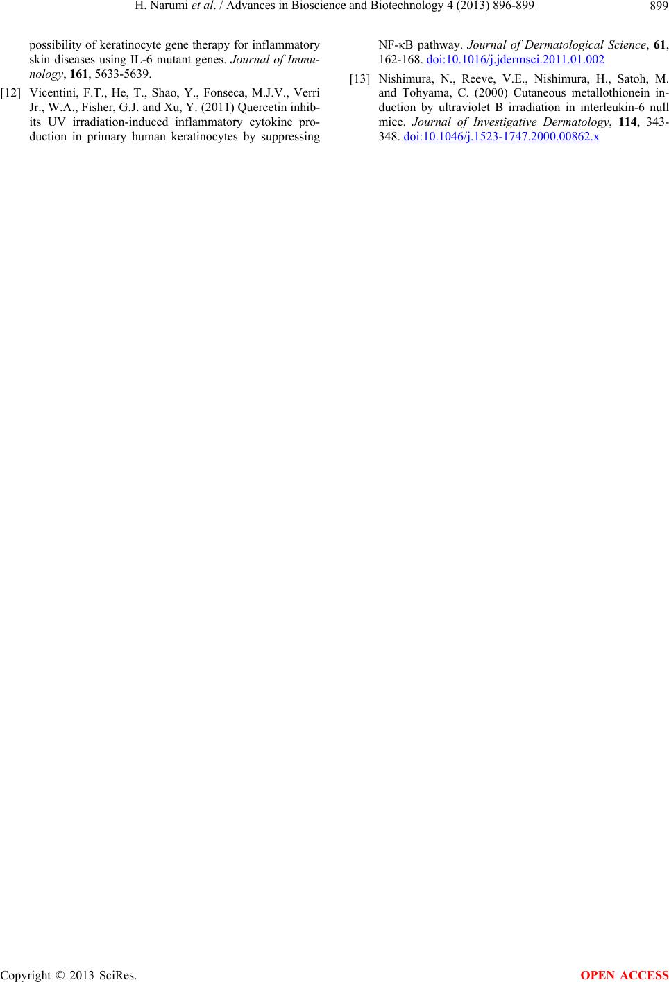
that UVB had no or little enhancing effect on the pro-
moter activity at doses ranging from 5 to 30 mJ/cm2.
Furthermore, we collected the treated cells several times
after irradiation, but we could not observe any strong
induction of the promoter activity.
4. DISCUSSION
IL-1α has many physiological functions in the immune,
metabolic, and hematopoietic systems. Keratinocytes are
a major source of IL-1α. As a proinflammatory cytokine,
IL-1α is involved in inflammatory and allergic skin dis-
eases such as psoriasis and contact dermatitis. It also
plays an important role in many recently defined autoin-
flammatory diseases [10]. IL-6 acts both as a pro- and
anti-inflammatory cytokine. It is also secreted by kerati-
nocytes, and it stimulates the immune response in both
normal and abnormal skin conditions [11]. Furthermore,
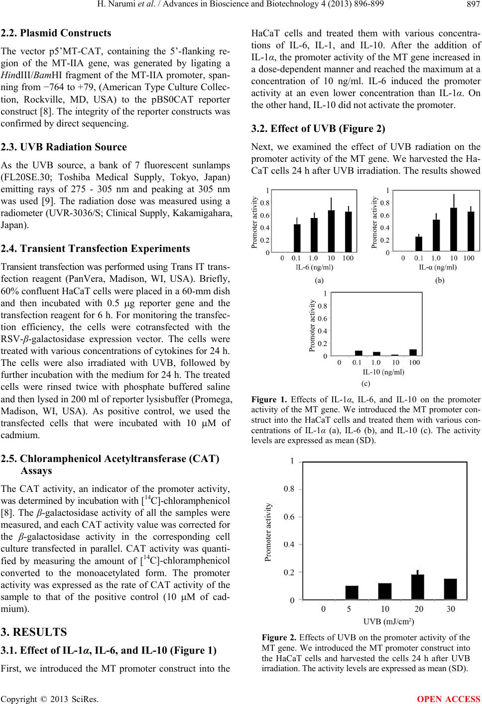
IL-1α and IL-6 are known to increase the MT gene ex-
pression and thereby, its protein expression. In this study,
we first examined the effects of IL-1α and IL-6 on the
MT promoter activity in epidermal keratinocytes. Our
results show that both cytokines induced promoter activ-
ity in a dose-dependent manner. We also investigated the
effect of IL-10 on MT expression, and as expected, we
did not find any significant effect of IL-10 on the pro-
moter activity.
The epidermis is the outermost layer of the skin. Kerati-
nocytes form a majority, i.e., about 95%, of the epider-
mis’ cells and are major targets of solar radiation. UVB
benefits humans by catalyzing the production of vitamin
D, but it also causes sunburn, photoaging, and skin can-
cers. UVB is responsible for the production of many cy-
tokines in the keratinocytes [12]. Furthermore, UVB is
known to induce MT expression in the epidermal kerati-
nocytes, and IL-6 is a possible mediator of MT induction
by UV radiation [13]. Therefore, we investigated whether
UVB radiation could induce MT promoter activity. Our
results showed, interestingly, that UVB radiation has no
or little effects on the promoter activity, although IL-6
clearly induced the promoter activity of the MT gene in
our study. This discrepancy implies non-transcriptional
mechanisms such as an increase in mRNA stability, sug-
gesting complex molecular regulation of the MT gene.
5. ACKNOWLEDGEMENTS
The authors would like to thank Ms Yuka Toyomaki, Mrs Yukiko
Tamura, Mrs Yuriko Takagi, and Ms Nanako Seitoh for their excellent
technical assistance. This work was supported in part by Grants-in-Aid
from the Ministry of Education, Science, Sports, and Culture of Japan.
REFERENCES
[1] Masters, B.A., Kelly, E.J., Quaife, C.J., Brinster, R.L. and
Palmiter, R.D. (1994) Targeted disruption of metallothio-
nein I and II genes increases sensitivity to cadmium.
Proceedings of the National Academy of Sciences of the
United States of America, 91, 584-588.
doi:10.1073/pnas.91.2.584
[2] Lazo, J.S., Kondo, Y., Dellapiazza, D., Michalska, A.E.,
Choo, K.H. and Pitt, B.R. (1995) Enhanced sensitivity to
oxidative stress in cultured embryonic cells from trans-
genic mice deficient in metallothionein I and II genes.
Journal of Biological Chemistry, 270, 5506-5510.
doi:10.1074/jbc.270.10.5506
[3] Hanada, K., Sawamura, D., Tamai, K., Baba, T., Hashi-
moto, I., Muramatsu, T., Miura, N. and Naganuma, A.
(1998) Novel function of metallothionein in photoprotec-
tion: Metallothionein-null mouse exhibits reduced toler-
ance against ultraviolet B injury in the skin. Journal of
Investigative Dermatology, 111, 582-585.
doi:10.1046/j.1523-1747.1998.00342.x
[4] Angel, P., Poting, A., Mallick, U., Rahmsdorf, H.J.,
Schorpp, M. and Herrlich, P. (1986) Induction of metal-
lothionein and other mRNA species by carcinogens and
tumor promoters in primary human skin fibroblasts. Mo-
lecular and Cellular Biology, 6, 1760-1766.
[5] Shiraishi, N., Yamamoto, H., Takeda, Y., Kondoh, S.,
Hayashi, H., Hashimoto, K. and Aono, K. (1986) In-
creased metallothionein content in rat liver and kidney
following X-irradiation. Toxicology and Applied Phar-
macology, 85, 128-134.
doi:10.1016/0041-008X(86)90106-7
[6] Baba, T., Nakano, H., Tamai, K., Sawamura, D., Hanada,
K., Hashimoto, I. and Arima, Y. (1998) Inhibitory effect
of beta-thujaplicin on ultraviolet B-induced apoptosis in
mouse keratinocytes. Journal of Investigative Dermatol-
ogy, 110, 24-28. doi:10.1046/j.1523-1747.1998.00078.x
[7] Sato, M., Sasaki, M. and Hojo, H. (1994) Differential
induction of metallothionein synthesis by interleukin-6
and tumor necrosis factor-alpha in rat tissues. Interna-
tional Journal of Immunopharmacology, 16, 187-195.
doi:10.1016/0192-0561(94)90075-2
[8] Nakano, H., Ikenaga, S., Aizu, T., Kaneko, T., Matsuzaki,
Y., Tsuchida, S., Hanada, K. and Arima, Y. (2006) Hu-
man metallothionein gene expression is upregulated by
beta-thujaplicin: Possible involvement of protein kinase
C and reactive oxygen species. Biological and Pharma-
ceutical Bulletin, 29, 55-59. doi:10.1248/bpb.29.55
[9] Hanada, K. (2000) Photoprotective role of metallothio-
nein in UV-injury-metallothionein-null mouse exhibits
reduced tolerance against ultraviolet-B. Journal of De-
rmatological Science, 23, S51-S56.
doi:10.1016/S0923-1811(99)00078-X
[10] Goldbach-Mansky, R. and Kastner, D.L. (2009) Autoin-
flammation: The prominent role of IL-1 in monogenic
autoinflammatory diseases and implications for common
illnesses. Journal of Allergy and Clinical Immunology,
124, 1141-1149. doi:10.1016/j.jaci.2009.11.016
[11] Sawamura, D., Meng, X., Ina, S., Sato, M., Tamai, K.,
Hanada, K. and Hashimoto, I. (1998) Induction of kerati-
nocyte proliferation and lymphocytic infiltration by in
vivo introduction of the IL-6 gene into keratinocytes and
Copyright © 2013 SciRes. OPEN ACCESS
H. Narumi et al. / Advances in Bioscience and Biotechnology 4 (2013) 896-899
Copyright © 2013 SciRes.
899
OPEN ACCESS
possibility of keratinocyte gene therapy for inflammatory
skin diseases using IL-6 mutant genes. Journal of Immu-
nology, 161, 5633-5639.
[12] Vicentini, F.T., He, T., Shao, Y., Fonseca, M.J.V., Verri
Jr., W.A., Fisher, G.J. and Xu, Y. (2011) Quercetin inhib-
its UV irradiation-induced inflammatory cytokine pro-
duction in primary human keratinocytes by suppressing
NF-κB pathway. Journal of Dermatological Science, 61,
162-168. doi:10.1016/j.jdermsci.2011.01.002
[13] Nishimura, N., Reeve, V.E., Nishimura, H., Satoh, M.
and Tohyama, C. (2000) Cutaneous metallothionein in-
duction by ultraviolet B irradiation in interleukin-6 null
mice. Journal of Investigative Dermatology, 114, 343-
348. doi:10.1046/j.1523-1747.2000.00862.x