Paper Menu >>
Journal Menu >>
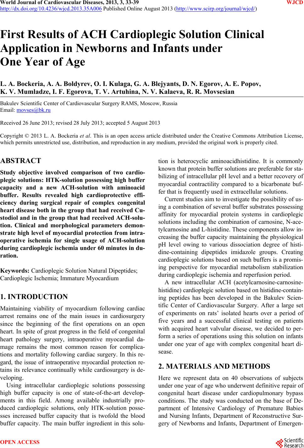 World Journal of Cardiovascular Diseases, 2013, 3, 33-39 WJCD http://dx.doi.org/10.4236/wjcd.2013.35A006 Published Online August 2013 (http://www.scirp.org/journal/wjcd/) First Results of ACH Cardioplegic Solution Clinical Application in Newborns and Infants under One Year of Age L. A. Bockeria, A. A. Boldyrev, O. I. Kulaga, G. A. Blejyants, D. N. Egorov, A. E. Popov, K. V. Mumladze, I. F. Egorova, T. V. Artuhina, N. V. Kalaeva, R. R. Movsesian Bakulev Scientific Center of Cardiovascular Surgery RAMS, Moscow, Russia Email: movses@bk.ru Received 26 June 2013; revised 28 July 2013; accepted 5 August 2013 Copyright © 2013 L. A. Bockeria et al. This is an open access article distributed under the Creative Commons Attribution License, which permits unrestricted use, distribution, and reproduction in any medium, provided the original work is properly cited. ABSTRACT Study objective involved comparison of two cardio- plegic solutions: HTK-solution possessing high buffer capacity and a new ACH-solution with aminoacid buffer. Results revealed high cardioprotective effi- ciency during surgical repair of complex congenital heart disease both in the group that had received Cu- stodiol and in the group that had received ACH-solu- tion. Clinical and morphological parameters demon- strate high level of myocardial protection from intra- operative ischemia for single usage of ACH-solution during cardioplegic ischemia under 60 minutes in du- ration. Keywords: Cardioplegic Solution Natural Dipeptides; Cardioplegic Ischemia; Immature Myocardium 1. INTRODUCTION Maintaining viability of myocardium following cardiac arrest remains one of the main issues in cardiosurgery since the beginning of the first operations on an open heart. In spite of great progress in the field of congenital heart pathology surgery, intraoperative myocardial da- mage remains the most common reason for complica- tions and mortality following cardiac surgery. In this re- gard, the issue of intraoperative myocardial protection re- tains its relevance continually while cardiosurgery is de- veloping. Using intracellular cardioplegic solutions possessing high buffer capacity is one of state-of-the-art develop- ments in this field. Among available industrially pro- duced cardioplegic solutions, only HTK-solution posse- sses increased buffer capacity that is twofold the blood buffer capacity. The main buffer ingredient in this solu- tion is heterocyclic aminoacidhistidine. It is commonly known that protein buffer solutions are preferable for sta- bilizing of intracellular pH level and a better recovery of myocardial contractility compared to a bicarbonate buf- fer that is frequently used in extracellular solutions. Current studies aim to investigate the possibility of us- ing a combination of several buffer substrates possessing affinity for myocardial protein systems in cardioplegic solutions including the combination of carnosine, N-ace- tylcarnosine and L-histidine. These components allow in- creasing the buffer capacity maintaining the physiological pH level owing to various dissociation degree of histi- dine-containing dipeptides imidazole groups. Creating cardioplegic solutions based on such buffers is a promis- ing perspective for myocardial metabolism stabilization during cardioplegic ischemia and reperfusion period. A new intracellular ACH (acetylcarnosine-carnosine- histidine) cardioplegic solution based on histidine-contain- ing peptides has been developed in the Bakulev Scien- tific Center of Cardiovascular Surgery. After a large set of experiments on rats’ isolated hearts over a period of five years and a successful clinical testing on patients with acquired heart valvular disease, we decided to per- form a series of operations using this solution on infants under one year of age with complex congenital heart di- sease. 2. MATERIALS AND METHODS Here we represent data on 40 observations of subjects under one year of age who underwent definitive repair of congenital heart disease under cardiopulmonary bypass conditions. The study was conducted on the base of De- partment of Intensive Cardiology of Premature Babies and Nursing Infants, Department of Reconstructive Sur- gery of Newborns and Infants, Department of Emergen- OPEN ACCESS 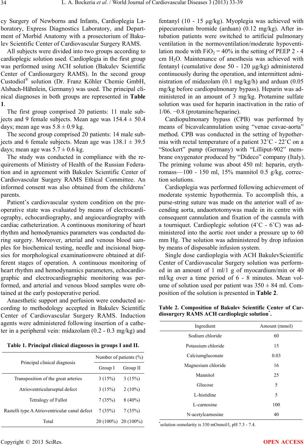 L. A. Bockeria et al. / World Journal of Cardiovascular Diseases 3 (2013) 33-39 34 cy Surgery of Newborns and Infants, Cardioplegia La- boratory, Express Diagnostics Laboratory, and Depart- ment of Morbid Anatomy with a prosectorium of Baku- lev Scientific Center of Cardiovascular Surgery RAMS. All subjects were divided into two groups according to cardioplegic solution used. Cardioplegia in the first group was performed using ACH solution (Bakulev Scientific Center of Cardiosurgery RAMS). In the second group Custodiol® solution (Dr. Franz Köhler Chemie GmbH, Alsbach-Hähnlein, Germany) was used. The principal cli- nical diagnoses in both groups are represented in Table 1. The first group comprised 20 patients: 11 male sub- jects and 9 female subjects. Mean age was 154.4 ± 50.4 days; mean age was 5.8 ± 0.9 kg. The second group comprised 20 patients: 14 male sub- jects and 6 female subjects. Mean age was 138.1 ± 39.5 days; mean age was 5.7 ± 0.6 kg. The study was conducted in compliance with the re- quirements of Ministry of Health of the Russian Federa- tion and in agreement with Bakulev Scientific Center of Cardiovascular Surgery RAMS Ethical Committee. An informed consent was also obtained from the childrens’ parents. Patient’s cardiovascular system condition on the pre- operative state was evaluated by means of electrocardi- ography, echocardiography, and angiocardiography with cardiac catheterization. A continuous monitoring of heart rhythm and hemodynamics parameters was conducted du- ring surgery. Moreover, arterial and venous blood sam- ples for biochemical testing, needle and incisional biop- sies for morphological examinationwere obtained at dif- ferent stages of operation. A continuous monitoring of heart rhythm and hemodynamics parameters, echocardio- graphic and electrocardiographic monitoring was per- formed, and arterial and venous blood samples were ob- tained at the early postoperative period. Anaesthetic support and perfusion were conducted ac- cording to methodology accepted in Bakulev Scientific Center of Cardiovascular Surgery RAMS. Induction agents were administered following insertion of a cathe- ter in a peripheral vein: midazolam (0.2 - 0.3 mg/kg) and Table 1. Principal clinical diagnoses in groups I and II. Number of patients (%) Principal clinical diagnosis Group I Group II Transposition of the great arteries 3 (15%) 3 (15%) Atrioventricularseptal defect 3 (15%) 2 (10%) Tetralogy of Fallot 7 (35%) 8 (40%) Rastelli type A Atrioventricular canal defect 7 (35%) 7 (35%) Total 20 (100%) 20 (100%) fentanyl (10 - 15 μg/kg). Myoplegia was achieved with pipecuronium bromide (arduan) (0.12 mg/kg). After in- tubation patients were switched to artificial pulmonary ventilation in the normoventilation/moderate hypoventi- lation mode with FiO2 = 40% in the setting of PEEP 2 - 4 cm H2O. Maintenance of anesthesia was achieved with fentanyl (cumulative dose 50 - 120 μg/kg) administered continuously during the operation, and intermittent admi- nistration of midazolam (0.1 mg/kg/h) and arduan (0.05 mg/kg before cardiopulmonary bypass). Heparin was ad- ministered in an amount of 3 mg/kg. Protamine sulfate solution was used for heparin inactivation in the ratio of 1/06. −0.8 (protamine/heparine). Cardiopulmonary bypass (CPB) was performed by means of bicavalcannulation using “venae cavae-aorta” method. CPB was conducted in the setting of hypother- mia with rectal temperature of a patient 32˚C - 22˚C on a “Stockert” pump (Germany) with “Lilliput-902” mem- brane oxygenator produced by “Dideco” company (Italy). The priming volume was about 450 ml: heparin, eryth- romass—100 - 150 ml, 15% mannitol 0.5 g/kg, correc- tion solutions. Cardioplegia was performed following achievement of moderate systemic hypothermia. To accomplish this, a purse-string suture was made on the anterior wall of as- cending aorta, andaortotomywas made in its centre with consequent cannulation and fixation of the cannula with a tourniquet. Cardioplegic solution (4˚C - 6˚C) was ad- ministered into the aortic root under a pressure up to 60 mm Hg. The solution was administered by drop infusion by means of disposable infusion system. Single dose cardioplegia with ACH BakulevScientific Center of Cardiovascular Surgery solution was perform- ed in an amount of 1 ml/1 g of myocardium/min or 40 ml/kg over a time period of 6 - 8 minutes. Mean vol- ume of solution used per patient was 350 ± 84 ml. Com- position of the solution is presented in Table 2. Table 2. Composition of Bakulev Scientific Center of Car- diosurgery RAMS ACH cardioplegic solution*. Ingredient Amount (mmol) Sodium chloride 60 Potassium chloride 15 Calciumgluconate 0.03 Magnesium chloride 16 Mannitol 25 Glucose 5 L-histidine 5 L-carnosine 100 N-acetylcarnosine 40 *solution osmolarity is 330 mOsmol/l, рН 7.3 - 7.4. Copyright © 2013 SciRes. OPEN ACCESS 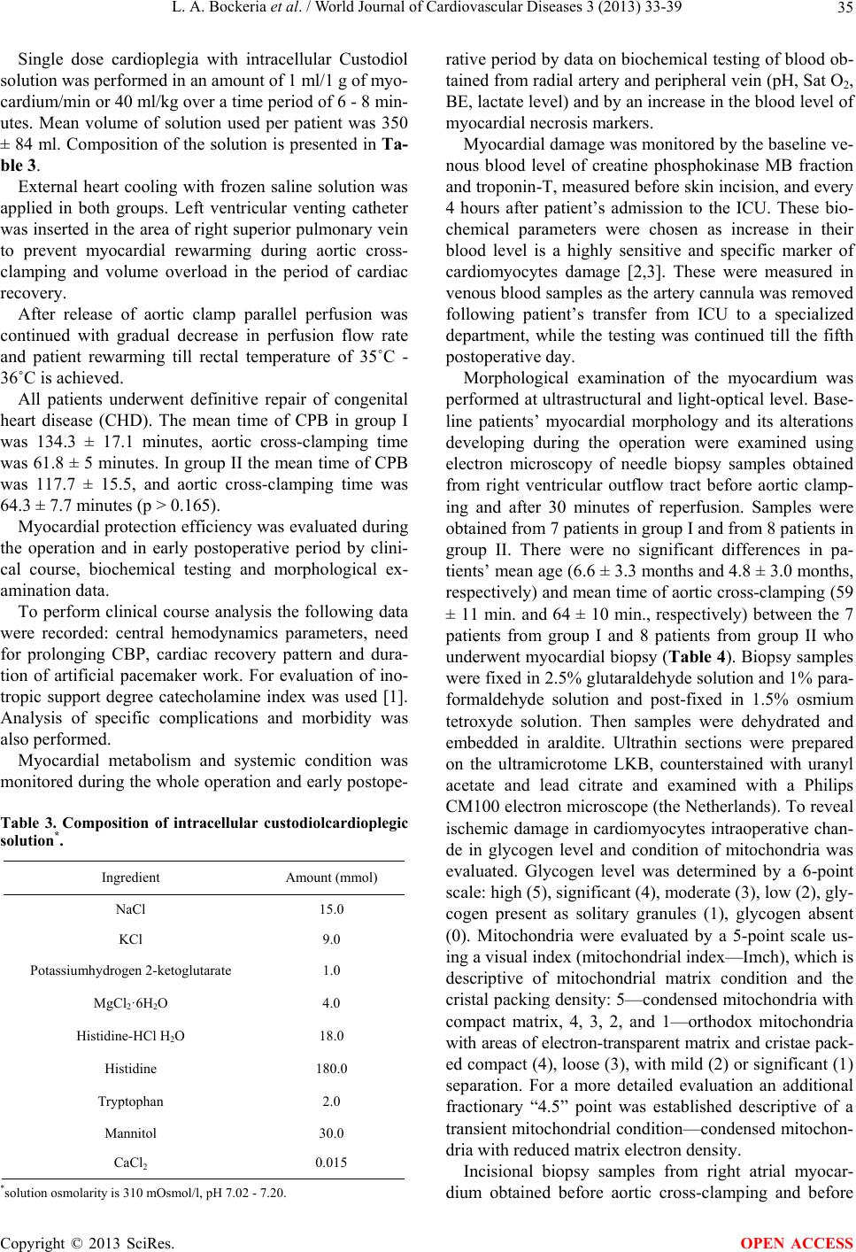 L. A. Bockeria et al. / World Journal of Cardiovascular Diseases 3 (2013) 33-39 35 Single dose cardioplegia with intracellular Custodiol solution was performed in an amount of 1 ml/1 g of myo- cardium/min or 40 ml/kg over a time period of 6 - 8 min- utes. Mean volume of solution used per patient was 350 ± 84 ml. Composition of the solution is presented in Ta- ble 3. External heart cooling with frozen saline solution was applied in both groups. Left ventricular venting catheter was inserted in the area of right superior pulmonary vein to prevent myocardial rewarming during aortic cross- clamping and volume overload in the period of cardiac recovery. After release of aortic clamp parallel perfusion was continued with gradual decrease in perfusion flow rate and patient rewarming till rectal temperature of 35˚C - 36˚C is achieved. All patients underwent definitive repair of congenital heart disease (CHD). The mean time of CPB in group I was 134.3 ± 17.1 minutes, aortic cross-clamping time was 61.8 ± 5 minutes. In group II the mean time of CPB was 117.7 ± 15.5, and aortic cross-clamping time was 64.3 ± 7.7 minutes (p > 0.165). Myocardial protection efficiency was evaluated during the operation and in early postoperative period by clini- cal course, biochemical testing and morphological ex- amination data. To perform clinical course analysis the following data were recorded: central hemodynamics parameters, need for prolonging CBP, cardiac recovery pattern and dura- tion of artificial pacemaker work. For evaluation of ino- tropic support degree catecholamine index was used [1]. Analysis of specific complications and morbidity was also performed. Myocardial metabolism and systemic condition was monitored during the whole operation and early postope- Table 3. Composition of intracellular custodiolcardioplegic solution*. Ingredient Amount (mmol) NaCl 15.0 KCl 9.0 Potassiumhydrogen 2-ketoglutarate 1.0 MgCl2·6Н2О 4.0 Histidine-НСl Н2О 18.0 Histidine 180.0 Tryptophan 2.0 Mannitol 30.0 CaCl2 0.015 *solution osmolarity is 310 mOsmol/l, рН 7.02 - 7.20. rative period by data on biochemical testing of blood ob- tained from radial artery and peripheral vein (рН, Sat O2, ВЕ, lactate level) and by an increase in the blood level of myocardial necrosis markers. Myocardial damage was monitored by the baseline ve- nous blood level of creatine phosphokinase MB fraction and troponin-T, measured before skin incision, and every 4 hours after patient’s admission to the ICU. These bio- chemical parameters were chosen as increase in their blood level is a highly sensitive and specific marker of cardiomyocytes damage [2,3]. These were measured in venous blood samples as the artery cannula was removed following patient’s transfer from ICU to a specialized department, while the testing was continued till the fifth postoperative day. Morphological examination of the myocardium was performed at ultrastructural and light-optical level. Base- line patients’ myocardial morphology and its alterations developing during the operation were examined using electron microscopy of needle biopsy samples obtained from right ventricular outflow tract before aortic clamp- ing and after 30 minutes of reperfusion. Samples were obtained from 7 patients in group I and from 8 patients in group II. There were no significant differences in pa- tients’ mean age (6.6 ± 3.3 months and 4.8 ± 3.0 months, respectively) and mean time of aortic cross-clamping (59 ± 11 min. and 64 ± 10 min., respectively) between the 7 patients from group I and 8 patients from group II who underwent myocardial biopsy (Table 4). Biopsy samples were fixed in 2.5% glutaraldehyde solution and 1% para- formaldehyde solution and post-fixed in 1.5% osmium tetroxyde solution. Then samples were dehydrated and embedded in araldite. Ultrathin sections were prepared on the ultramicrotome LKB, counterstained with uranyl acetate and lead citrate and examined with a Philips CM100 electron microscope (the Netherlands). To reveal ischemic damage in cardiomyocytes intraoperative chan- de in glycogen level and condition of mitochondria was evaluated. Glycogen level was determined by a 6-point scale: high (5), significant (4), moderate (3), low (2), gly- cogen present as solitary granules (1), glycogen absent (0). Mitochondria were evaluated by a 5-point scale us- ing a visual index (mitochondrial index—Imch), which is descriptive of mitochondrial matrix condition and the cristal packing density: 5—condensed mitochondria with compact matrix, 4, 3, 2, and 1—orthodox mitochondria with areas of electron-transparent matrix and cristae pack- ed compact (4), loose (3), with mild (2) or significant (1) separation. For a more detailed evaluation an additional fractionary “4.5” point was established descriptive of a transient mitochondrial condition—condensed mitochon- dria with reduced matrix electron density. Incisional biopsy samples from right atrial myocar- dium obtained before aortic cross-clamping and before Copyright © 2013 SciRes. OPEN ACCESS 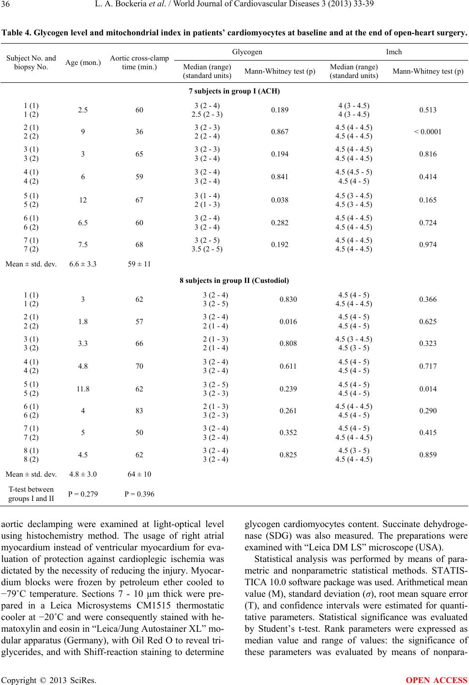 L. A. Bockeria et al. / World Journal of Cardiovascular Diseases 3 (2013) 33-39 Copyright © 2013 SciRes. 36 OPEN ACCESS Table 4. Glycogen level and mitochondrial index in patients’ cardiomyocytes at baseline and at the end of open-heart surgery. Glycogen Imch Subject No. and biopsy No. Age (mon.) Aortic cross-clamp time (min.) Median (range) (standard units) Mann-Whitney test (р)Median (range) (standard units) Mann-Whitney test (р) 7 subjects in group I (АСН) 1 (1) 1 (2) 2.5 60 3 (2 - 4) 2.5 (2 - 3) 0.189 4 (3 - 4.5) 4 (3 - 4.5) 0.513 2 (1) 2 (2) 9 36 3 (2 - 3) 2 (2 - 4) 0.867 4.5 (4 - 4.5) 4.5 (4 - 4.5) < 0.0001 3 (1) 3 (2) 3 65 3 (2 - 3) 3 (2 - 4) 0.194 4.5 (4 - 4.5) 4.5 (4 - 4.5) 0.816 4 (1) 4 (2) 6 59 3 (2 - 4) 3 (2 - 4) 0.841 4.5 (4.5 - 5) 4.5 (4 - 5) 0.414 5 (1) 5 (2) 12 67 3 (1 - 4) 2 (1 - 3) 0.038 4.5 (3 - 4.5) 4.5 (3 - 4.5) 0.165 6 (1) 6 (2) 6.5 60 3 (2 - 4) 3 (2 - 4) 0.282 4.5 (4 - 4.5) 4.5 (4 - 4.5) 0.724 7 (1) 7 (2) 7.5 68 3 (2 - 5) 3.5 (2 - 5) 0.192 4.5 (4 - 4.5) 4.5 (4 - 4.5) 0.974 Mean ± std. dev. 6.6 ± 3.3 59 ± 11 8 subjects in group II (Custodiol) 1 (1) 1 (2) 3 62 3 (2 - 4) 3 (2 - 5) 0.830 4.5 (4 - 5) 4.5 (4 - 4.5) 0.366 2 (1) 2 (2) 1.8 57 3 (2 - 4) 2 (1 - 4) 0.016 4.5 (4 - 5) 4.5 (4 - 5) 0.625 3 (1) 3 (2) 3.3 66 2 (1 - 3) 2 (1 - 4) 0.808 4.5 (3 - 4.5) 4.5 (3 - 5) 0.323 4 (1) 4 (2) 4.8 70 3 (2 - 4) 3 (2 - 4) 0.611 4.5 (4 - 5) 4.5 (4 - 5) 0.717 5 (1) 5 (2) 11.8 62 3 (2 - 5) 3 (2 - 3) 0.239 4.5 (4 - 5) 4.5 (4 - 5) 0.014 6 (1) 6 (2) 4 83 2 (1 - 3) 3 (2 - 3) 0.261 4.5 (4 - 4.5) 4.5 (4 - 5) 0.290 7 (1) 7 (2) 5 50 3 (2 - 4) 3 (2 - 4) 0.352 4.5 (4 - 5) 4.5 (4 - 4.5) 0.415 8 (1) 8 (2) 4.5 62 3 (2 - 4) 3 (2 - 4) 0.825 4.5 (3 - 5) 4.5 (4 - 4.5) 0.859 Mean ± std. dev. 4.8 ± 3.0 64 ± 10 T-test between groups I and II Р = 0.279 Р = 0.396 aortic declamping were examined at light-optical level using histochemistry method. The usage of right atrial myocardium instead of ventricular myocardium for eva- luation of protection against cardioplegic ischemia was dictated by the necessity of reducing the injury. Myocar- dium blocks were frozen by petroleum ether cooled to −79˚C temperature. Sections 7 - 10 μm thick were pre- pared in a Leica Microsystems CM1515 thermostatic cooler at −20˚C and were consequently stained with he- matoxylin and eosin in “Leica/Jung Autostainer XL” mo- dular apparatus (Germany), with Oil Red O to reveal tri- glycerides, and with Shiff-reaction staining to determine glycogen cardiomyocytes content. Succinate dehydroge- nase (SDG) was also measured. The preparations were examined with “Leica DM LS” microscope (USA). Statistical analysis was performed by means of para- metric and nonparametric statistical methods. STATIS- TICA 10.0 software package was used. Arithmetical mean value (M), standard deviation (σ), root mean square error (T), and confidence intervals were estimated for quanti- tative parameters. Statistical significance was evaluated by Student’s t-test. Rank parameters were expressed as median value and range of values: the significance of these parameters was evaluated by means of nonpara-  L. A. Bockeria et al. / World Journal of Cardiovascular Diseases 3 (2013) 33-39 37 metric Mann-Whitney test for pair-wise group compari- son. In our study we established the significance level of p < 0.05. 3. RESULTS There was no significant difference between two study groups in mean values of age, weight, left ventricular ejection fraction (LV EF), systolic and diastolic arterial pressure, level of creatine phosphokinase MB fraction (CPK-MB) and troponin-T (TN-T) (Table 5). During the operation electromechanical cardiac arrest in the ACH-solution group occurred at second 15.3 ± 2.7, while in HTK-solution group cardiac arrest occurred at second 27.4 ± 4 after the beginning of cardioplegia. This parameter was significantly different between study groups (p < 0.0001). We reported spontaneous cardiac recovery following aortic declamping in both groups. In the first group car- diac recovery occurred in 57 ± 22 seconds, and in the se- cond group—in 54.1 ± 11.7 seconds (р > 0.8). Based on echocardiographic data, LV EF on the first day following surgery in groups I and II was 54.0 ± 4.1% and 55.2 ± 2.65, respectfully (р > 0.654), while on the second day following surgery—58.1 ± 2.6 and 56.4 ± 3.3. The difference between groups was not significant (p > 0.41). According to monitoring data the values of systolic and diastolic pressure were not significantly different be- tween study groups throughout the observation (p > 0.05) (Figures 1(a) and (b)). Catecholamine index following discontinuation of CPB in the first and second groups was 13.5 ± 7.2 μg/kg/min and 11 ± 4.6 μg/kg/min, respectively (p > 0.654). There was no difference between groups (p > 0.05) (Figure 2). According to biochemical data analysis, no significant differences were reported beyween groups in the level of CPK-MB and TN-T (р > 0.05) (Figures 3(A) and (B)). Table 5. Mean values of age, weight, left ventricular ejec- tion fraction (LV EF), systolic and diastolic arterial pres- sure, level of creatine phosphokinase MB fraction (CPK- MB) and troponin-T (TN-T). АСН Custodiol Student’s test Age (days) 154.4 ± 50.4 138.1 ± 39.5 p > 0.62 Weight (kg) 5.8 ± 0.9 5.7 ± 0.6 p > 0.809 Baseline EF (%) 67.9 ± 0.4 67.6 ± 1.2 p > 0.631 Baseline SAP (mmHg) 77.8 ± 6.7 84.9 ± 4.7 p > 0.098 Baseline DAP (mmHg) 42.9 ± 3.5 49.5 ± 2.8 p < 0.006 Baseline CPK-MB (ng/ml) 8.1 ± 5.1 4.9 ± 1 p > 0.337 Baseline Tn-Т (ng/ml) 0.32 ± 0.54 0.07 ± 0.06 p > 0.437 (A) (B) Figure 1. (A) Systolic AP in dynamics; (B) Diastolic AP in dynamics. Figure 2. Inotropic support degree in dynamics. Duration of artificial pulmonary ventilation and dura- tion of stay in ICU in the first group were 110 ± 32.68 hours and 131.8 ± 36.36 hours, respectively, while in the second group—91.28 ± 40.9 and 132.9 ± 60.56 hours, respectively. There also was not significant difference in these parameters between groups (p > 0.488). 64 biopsies were investigated with light-optical mi- croscope in children from both groups of which 18 sam- ples were obtained from children with Atrioventricular canal defect diagnosis, 22—from Tetralogy of Fallot pa- tients, 6—from atrioventricularseptal defect patients, 12—from children with transposition of great arteries. Copyright © 2013 SciRes. OPEN ACCESS  L. A. Bockeria et al. / World Journal of Cardiovascular Diseases 3 (2013) 33-39 38 Notably, myocardial lipidosis was not reported in any of the biopsies regardless of the cardioplegic solution used during surgery. Generally, glycogen content in the first biopsy samples obtained from patients in both groups was high, while in decreased in the second sample. How- ever glycogen content was typically focal, that is myo- cardial areas with low glycogen content were found along with areas rich in glycogen. SDG activity was reported to be high in the first samples in both groups, and did not significantly reduce in the second samples obtained from all subjects (Figures 4(A) and (B)). Electron microscopy of patients’ RV outflow tract my- ocardium revealed cariomyocytes different in size and the degree of ultra structural maturity. Most of cardio- myocytes appeared to be small cells with severely curved (A) (B) Figure 3. (A) Troponin-T in dynamics; (B) CPK-MB in dy- namics. (A) (B) Figure 4. (A) High glycogen content in the first biopsy of patient K. Shiff-reaction staining. Magnification 10 oc. × 20 obj; (B) Focal glycogen content decrease in the second bi- opsy of patient K. Shiff-reaction staining. Magnification 10 oc. × 20 obj. nuclear membrane and a layer of sarcoplasm, in which a structured contractile apparatus is located with focuses of continuing myofibrils assembly. In some cells Golgi complex and, at a lesser extent, rough endoplasmic reti- culum tubules were well developed. Specific atrial gran- ules were often present in Golgi complex, while centri- oles were extremely rare. We reported signs of dystrophy in many cells: deposition of lipofuscin granules, glyco- somes different in size, lipid droplets, myelin-like bodies. Glycogen content was variable in cardiomyocytes. Gen- erally, it was moderate, and less frequentlyit was low. Mitochondria varied in conformation: in most cases they were condensed with low matrix electron density (Imch = 4, 5); much less frequently mitochondria were in or- thodox condition (Imch = 4 or Imch = 3) (Table 4). Comparison of glycogen level and mitochondrial in- dex in patients’ cardiomyocytes before aortic cross-clam- ping and after 30 minutes of reperfusion allowed us to identify significant decrease in glycogen level during the operation from moderate to low in 1 patient out of 7 pa- tients from group I (age—1 year, aortic cross-clamping time—67 minutes) and in 1 patient out of 8 patients in group II (age—1.8 months, aortic cross-clamping time— 57 minutes). We reported significant decrease in mito- chondrial index during surgery in 1 patient’s cardiomyo- cytes out of 7 patients from group I (age—9 months, aor- tic cross-clamping time—36 minutes) and in 1 patient’s cardiomyocytes out of 8 patients in group II (age—11.8 months, aortic cross-clamping time—62 minutes). The reported alterations in glycogen level or mitochondrial condition were mild and found only in solitary patients in both groups, which confirms good protective properties of cardioplegic solutions used (Figure 5). 4. DISCUSSION The results of our study demonstrate high efficiency of myocardial protection from one-hour intraoperative is- chemia in children less than one year of age with conge- nital heart disease when using the new cardioplegic ACH- solution. The results are based on hemodynamics para- meters analysis after recovery of cardiac function such as systolic and diastolic arterial pressure, catecholamine in- dex, spontaneous cardiac recovery. Comparative analysis of clinical, biochemical, and morphological data did not reveal any significant difference in parameters studied between patients who received ACH cardioplegic solu- tion and patients who received Custodiol. The new cardioplegic three natural dipeptides-based so- lution showed high efficiency comparable to Custodiol- cardioplegic solution, the latter having long ago gained a reputation of a highly effective medicinal product in many clinics worldwide that is used not only as a cardioplegic solution but also a solution for organ transplantation. Hopefully, further clinical studies of this solution will Copyright © 2013 SciRes. OPEN ACCESS  L. A. Bockeria et al. / World Journal of Cardiovascular Diseases 3 (2013) 33-39 Copyright © 2013 SciRes. 39 reveal new opportunities with the solution developed in our center. REFERENCES [1] Cruz, D.N., Antonelli, M., Fumagalli, R., et al. (2009) Early use of polymyxin B hemoperfusion in abdominal septic shock. JAMA, 301, 2445-2452. (A) (B) [2] Reithmann, C., Reber, D., Kozlik-Feldmann, R., et al. (1997) A post-receptor defect of adenylyl cyclase in se- verely failing myocardium from children with congenital heart disease. European Journal of Pharmacology, 330, 79-86. [3] Ross, R.D. (2001) Grading the graders of congestive heart failure in children. Journal of Pediatrics, 138, 618-620. (C) (D) Figure 5. (A) Longitudinal section of a cardiomyocyte: nu- cleus with dispersed chromatin and severelycurved nuclear membrane, solitary structured myofibrils, mitochondrial clumps and chains, glycogen granules. Patient M., 9 months. Magnification × 4800. (B) Mitochondrial ultrastructure evaluation according to scale using mitochondrial index: а—Imch = 5; б—Imch = 4, 5; в—Imch = 4; г—Imch = 3. (C) Nuclear fragment, cross and diagonally cut myofibrils, con- densed mitochondria with low matrix electron density (Imch = 4, 5), solitary condensed mitochondria with high matrix electron density (Imch = 5), moderate glycogen con- tent (Gl = 3), vacuoles. Condition before aortic cross-clam- ping. Magnification × 15,000. (D) Cross and diagonally cut myofibrils, clumps of condensed mitochondria with low ma- trix electron density (Imch = 4, 5), moderate glycogen con- tent (Gl = 3), vacuoles. Condition after 30 minutes of re- perfusion, in 59 minutes after aortic cross-clamping. Patient K., 6.5 months. Magnification × 15,000. OPEN ACCESS |