Paper Menu >>
Journal Menu >>
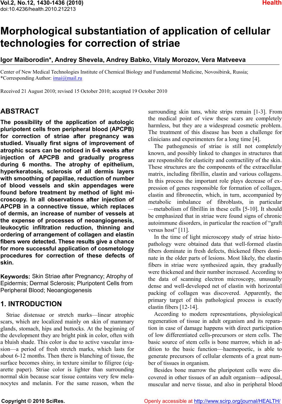 Vol.2, No.12, 1430-1436 (2010) Health doi:10.4236/health.2010.212213 Copyright © 2010 SciRes. Openly accessible at http://www.scirp.org/journal/HEALTH/ Morphological substantiation of application of cellular technologies for correction of striae Igor Maiborodin*, Andrey Shevela, Andrey Babko, Vitaly Morozov, Vera Matveeva Center of New Medical Technologies Institute of Chemical Biology and Fundamental Medicine, Novosibirsk, Russia; *Corresponding Author: imai@mail.ru Received 21 August 2010; revised 15 October 2010; accepted 19 October 2010 ABSTRACT The possibility of the application of autologic pluripotent cells from peripheral blood (APCPB) for correction of striae after pregnancy was studied. Visually first signs of improvement of atroph ic scars can be noticed in 6-8 w eeks a fter injection of APCPB and gradually progress during 6 months. The atrophy of epithelium, hyperkeratosis, sclerosis of all dermis layers with smoothing of papillae, reduction of number of blood vessels and skin appendages were found before treatment by method of light mi- croscopy. In all observations after injection of APCPB in a connective tissue, which replaces of dermis, an increase of number of vessels at the expense of processes of neoangiogenesis, leukocytic infiltration reduction, thinning and ordering of arrangement of collagen and elastin fibers were detected. These results give a ch a nc e for more successful application of cosmetology procedures for correction of these defects of skin. Keywords: Skin Striae after Pregnancy; Atrophy of Epidermis; Dermal Sclerosis; Pluripotent Cells from Peripheral Blood; Ne oangiogenesis 1. INTRODUCTION Striae distensae or stretch marks—linear atrophic scars, which are localized mainly on skin of mammary glands, stomach, hips and buttocks. At the beginning of the development they are bright pink in color, often with a bluish shade. This color is due to active vascular inva- sion—a period of fresh stretch marks, which lasts for about 6-12 months. Then there is blanch ing of tissue, the surface becomes shin y, in texture similar to filigree (cig- arette paper). Striae color is lighter than surrounding normal skin because scar tissue contains very few mela- nocytes and melanin. For the same reason, when the surrounding skin tans, white strips remain [1-3]. From the medical point of view these scars are completely harmless, but they are a widespread cosmetic problem. The treatment of this disease has been a challenge for clinicians and experimenters for a long time [4]. The pathogenesis of striae is still not completely known, and possibly linked to changes in structures that are responsible for elasticity a nd con tractility of the sk in. These structures are the components of the extracellular matrix, including fibrillin, elastin and various collagens. In this process the important role plays decrease of ex- pression of genes responsible for formation of collagen, elastin and fibronectin, which, in turn, accompanied by metabolic imbalance of fibroblasts, in particular —metabolism of fibrillin in these cells [5-10]. It should be emphasized that in striae were found signs of chronic autoimmune disorders, in particular the reaction of “graft versus host” [11]. In the time of light microscopy study of striae histo- pathology were obtained data that well-formed elastin fibers dominate in fresh defects, thickened fibers domi- nate in the older parts of lesions. Most likely, the elastin fibers in striae were synthesized again, they gradually were thickened and their number increased. According to the data of scanning electron microscopy, unusually dense and well-developed net of elastin with horizontal packing of collagen was discovered. Apparently, the primary target of this pathological process is exactly elastin fibers [12-14]. According to modern representations, physiological regeneration of tissue in adult organism and its repara- tion in case of damage happens with direct participation of low differentiated cells-precursors or stem cells. The basic source of stem cells is bone marrow, which in ad- dition to the basic function—haemopoetic, is able to generate precursors of cellular elements of a great num- ber of tissues in organism. Besides bone marrow the pluripotent cells were dis- covered in other tissues of an adult organism—adiposal, muscular and nerve tissue, and also in peripheral blood 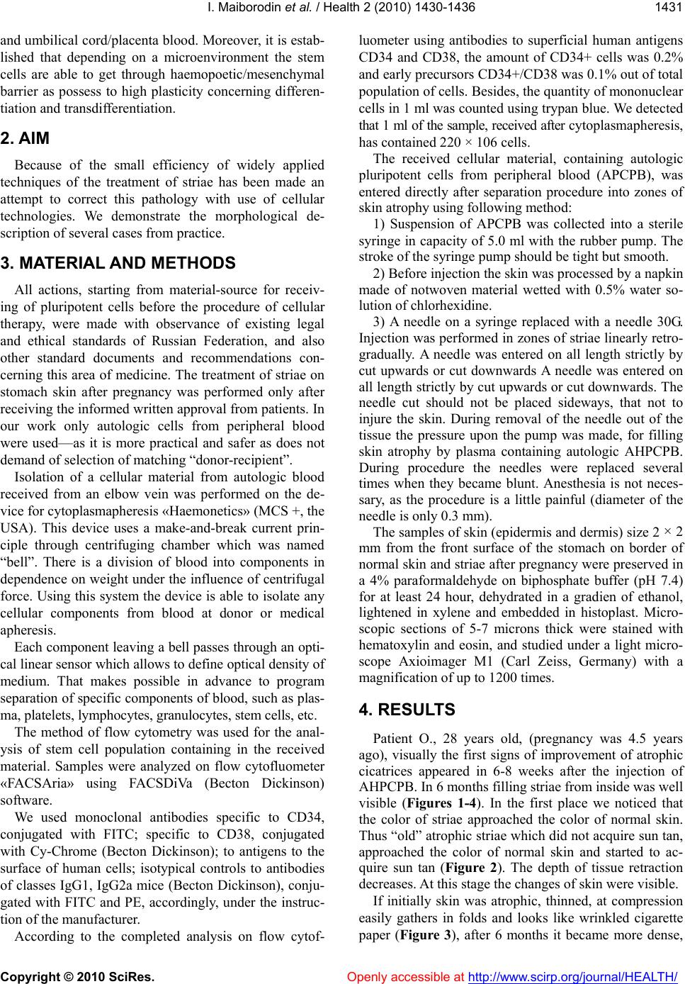 I. Maiborodin et al. / Health 2 (2010) 1430-1436 Copyright © 2010 SciRes. Openly accessible at http://www.scirp.org/journal/HEALTH/ 1431 and umbilical cord/placen ta blood. Moreover, it is estab- lished that depending on a microenvironment the stem cells are able to get through haemopoetic/mesenchymal barrier as possess to high plasticity concerning differen- tiation and transdifferentiation. 2. AIM Because of the small efficiency of widely applied techniques of the treatment of striae has been made an attempt to correct this pathology with use of cellular technologies. We demonstrate the morphological de- scription of several cases from practice. 3. MATERIAL AND METHODS All actions, starting from material-source for receiv- ing of pluripotent cells before the procedure of cellular therapy, were made with observance of existing legal and ethical standards of Russian Federation, and also other standard documents and recommendations con- cerning this area of medicine. The treatment of striae on stomach skin after pregnancy was performed only after receiving the informed written approval from patients. In our work only autologic cells from peripheral blood were used—as it is more practical and safer as does not demand of selection of matching “donor-recipient”. Isolation of a cellular material from autologic blood received from an elbow vein was performed on the de- vice for cytoplasmapheresis «Haemonetics» (MCS +, the USA). This device uses a make-and-break current prin- ciple through centrifuging chamber which was named “bell”. There is a division of blood into components in dependence on weight under the influence of centrifugal force. Using this system the device is able to isolate any cellular components from blood at donor or medical apheresis. Each component leaving a bell passes through an opti- cal linear sensor which allows to define optical density of medium. That makes possible in advance to program separation of specific components of blood, such as plas- ma, platelet s, lym phocy t es, granul ocy t es, st em cell s , et c. The method of flow cytometry was used for the anal- ysis of stem cell population containing in the received material. Samples were analyzed on flow cytofluometer «FACSAria» using FACSDiVa (Becton Dickinson) software. We used monoclonal antibodies specific to CD34, conjugated with FITC; specific to CD38, conjugated with Cy-Chrome (Becton Dickinson); to antigens to the surface of human cells; isotypical controls to antibodies of classes IgG1, IgG2a mice (Becton Dickinson), conju- gated with FITC and PE, accordingly, under the instruc- tion of the manufacturer. According to the completed analysis on flow cytof- luometer using antibodies to superficial human antigens CD34 and CD38, the amount of CD34+ cells was 0.2% and early precurso rs CD34+/CD38 was 0.1% out of total population of cells. Besides, the quantity of mononuclear cells in 1 ml was counted using trypan blue. We detected that 1 ml of the sample, received after c yt op l as ma p h er e si s, has contained 220 × 106 cells. The received cellular material, containing autologic pluripotent cells from peripheral blood (APCPB), was entered directly after separation procedure into zones of skin atrophy using following method: 1) Suspension of APCPB was collected into a sterile syringe in capacity of 5.0 ml with the rubber pump. The stroke of the syringe pump should be tight but smooth. 2) Before injection the skin was processed by a napkin made of notwoven material wetted with 0.5% water so- lution of chlorhexidine. 3) A needle on a syringe replaced with a needle 30G. Injection was performed in zones of striae linearly retro- gradually. A needle was entered on all length strictly by cut upwards or cut downwards A needle was entered on all length strictly by cut upward s or cut downwards. The needle cut should not be placed sideways, that not to injure the skin. During removal of the needle out of the tissue the pressure upon the pump was made, for filling skin atrophy by plasma containing autologic AHPCPB. During procedure the needles were replaced several times when they became blunt. Anesthesia is not neces- sary, as the procedure is a little painful (diameter of the needle is only 0.3 mm). The samples of skin (epidermis and dermis) size 2 × 2 mm from the front surface of the stomach on border of normal skin and striae after pregnancy were preserved in a 4% paraformaldehyde on biphosphate buffer (pH 7.4) for at least 24 hour, dehydrated in a gradien of ethanol, lightened in xylene and embedded in histoplast. Micro- scopic sections of 5-7 microns thick were stained with hematoxylin and eosin, and studied under a light micro- scope Axioimager M1 (Carl Zeiss, Germany) with a magnification of up to 1200 times. 4. RESULTS Patient O., 28 years old, (pregnancy was 4.5 years ago), visually the first signs of improvement of atrophic cicatrices appeared in 6-8 weeks after the injection of AHPCPB. In 6 months filling striae from inside was well visible (Figures 1-4). In the first place we noticed that the color of striae approached the color of normal skin. Thus “old” atrophic striae which did not acquire sun tan, approached the color of normal skin and started to ac- quire sun tan (Figure 2). The depth of tissue retraction decreases. At this stage the changes of ski n were visible. If initially skin was atrophic, thinned, at compression easily gathers in folds and looks like wrinkled cigarette paper (Figure 3), after 6 months it became more dense, 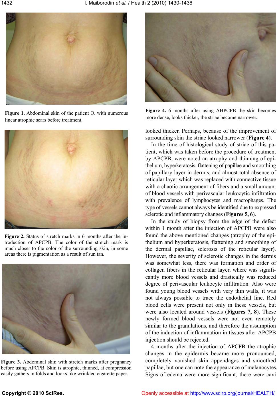 I. Maiborodin et al. / Health 2 (2010) 1430-1436 Copyright © 2010 SciRes. Openly accessible at http://www.scirp.org/journal/HEALTH/ 1432 Figure 1. Abdominal skin of the patient O. with numerous linear atrophic scars before treatment. Figure 2. Status of stretch marks in 6 months after the in- troduction of APCPB. The color of the stretch mark is much closer to the color of the surrounding skin, in some areas there is pigmentation as a result of sun tan. Figure 3. Abdominal skin with stretch marks after pregnancy before using APCPB. Skin is atrophic, thinned, at compression easily gathers in folds and looks like wrinkled cigarette paper. Figure 4. 6 months after using AHPCPB the skin becomes more dense, looks thicker, the striae become narrower. looked thicker. Perhaps, because of the improvement of surrounding skin the striae looked narrower (Figure 4). In the time of histological study of striae of this pa- tient, which was taken before the procedure of treatment by APCPB, were noted an atrophy and thinning of epi- thelium, hy perkerat osis, fl atteni ng of papil lae and smoothing of papillary layer in dermis, and almost total absence of reticular layer which was replaced with connective tissue with a chaotic arrangement of fibers and a small amount of blood vessels with perivascular leukocytic infiltration with prevalence of lymphocytes and macrophages. The type of vessels cannot always be identified due to expressed sclerotic and inflammatory changes (Figures 5, 6). In the study of biopsy from the edge of the defect within 1 month after the injection of APCPB were also found the above mentioned changes (atrophy of the epi- thelium and hyperkeratosis, flattening and smoothing of the dermal papillae, sclerosis of the reticular layer). However, the severity of sclerotic changes in the dermis was somewhat less, there was formation and order of collagen fibers in the reticular layer, where was signifi- cantly more blood vessels and drastically was reduced degree of perivascular leukocyte infiltration. Also were found young blood vessels with very thin walls, it was not always possible to trace the endothelial line. Red blood cells were present not only in these vessels, but were also located around vessels (Figures 7, 8). These newly formed blood vessels were not even remotely similar to the granulations, an d therefore the assumption of the induction of inflammation in tissues after APCPB injection sh ould be rejected. 4 months after the injection of APCPB the atrophic changes in the epidermis became more pronounced, completely vanished skin appendages and smoothed papillae, but one can note the appearance of melanocytes. Signs of edema were more significant, there were cavi 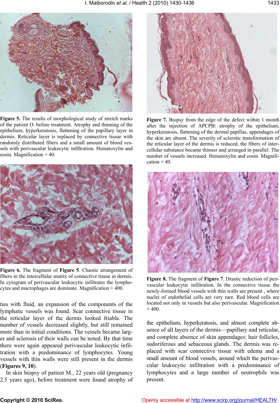 I. Maiborodin et al. / Health 2 (2010) 1430-1436 Copyright © 2010 SciRes. Openly accessible at http://www.scirp.org/journal/HEALTH/ 1433 Figure 5. The results of morphological study of stretch marks of the patient O. before treatment. Atrophy and thinning of the epithelium, hyperkeratosis, flattening of the papillary layer in dermis. Reticular layer is replaced by connective tissue with randomly distributed fibers and a small amount of blood ves- sels with perivascular leukocytic infiltration. Hematoxylin and eosin. Magnification × 40. Figure 6. The fragment of Figure 5. Chaotic arrangement of fibers in the intercellular matrix of connective tissue in dermis. In cytogram of perivascular leukocytic infiltrates the lympho- cytes and macrophages are dominate. Magnification × 400. ties with fluid, an expansion of the components of the lymphatic vessels was found. Scar connective tissue in the reticular layer of the dermis looked friable. The number of vessels decreased slightly, but still remained more than in initial conditions. The vessels became larg- er and sclerosis of their walls can be noted. By that time there were again appeared perivascular leukocytic infil- tration with a predominance of lymphocytes. Young vessels with thin walls were still present in the dermis (Figur es 9, 10). In skin biopsy of patient M., 22 years old (pregnancy 2.5 years ago), before treatment were found atrophy of Figure 7. Biopsy from the edge of the defect within 1 month after the injection of APCPB: atrophy of the epithelium, hyperkeratosis, flattening of the dermal papillae, appendages of the skin are absent. The severity of sclerotic transformation of the reticular layer of the dermis is reduced, the fibers of inter- cellular substance became thinner and arranged in parallel. The number of vessels increased. Hematoxylin and eosin. Magnifi- cation × 40. Figure 8. The fragment of Figure 7. Drastic reduction of peri- vascular leukocytic infiltration. In the connective tissue the newly-formed blood vessels with thin walls are present , where nuclei of endothelial cells are very rare. Red blood cells are located not only in vessels but also perivascular. Magnification × 400. the epithelium, hyperkeratosis, and almost complete ab- sence of all layers of the dermis—papillary and reticular, and complete absence of skin appendages: hair follicles, sudoriferous and sebaceous glands. The dermis was re- placed with scar connective tissue with edema and a small amount of blood vessels, around which the perivas- cular leukocytic infiltration with a predominance of lymphocytes and a large number of neutrophils was present.  I. Maiborodin et al. / Health 2 (2010) 1430-1436 Copyright © 2010 SciRes. Openly accessible at http://www.scirp.org/journal/HEALTH/ 1434 In the study of biopsy specimen from the edge of the Figure 9. Striae area in 4 months after injection APCPB. Atrophic changes and hyperkeratosis of the epidermis are more pronounced. Skin appendages vanished completely and papil- lae are smoothed. Instead of scar connective tissue in the reti- cular layer of the dermis there is friable connective tissue where significant signs of edema and expansion of the compo- nents of the lymphatic vessels are noted. The number of ves- sels is decreased, but the vessels became larger and have scle- rosed walls with perivascular leukocytic infiltration. Hematox- ylin and eosin. Magnification × 40. Figure 10. The fragment of Figure 9. The newly-formed blood vessels with thin walls and red cells located in the connective tissue on the place of reticular layer of dermis. In these vessels the endothelial lining can be seen along the full length, the remaining layers of the wall (muscle, adventitia) are absent. Magnification × 400. defect 6 weeks after the introduction of APCPB were also found atrophy of the epidermis and hyperkeratosis, but the severity of atrophy was significantly less. Also skin appendages and all layers of the dermis were absent. However, there was a formatting and some ordering of collagen fibers in the connective tissue on the place of dermis, there was significantly more vessels, reduced the severity of edema and the degree of perivascular leuko- cyte infiltration. In cytogram of such small infiltrates the lymphocytes and macrophages dominated. In 5 months after treatment, sclerosis of the dermis was still decreased. The number of vessels decreased, but the remaining vessels were large. Leukocytic infil- trates around the vessels were present again and ap- peared signs of edema. Patient S., 30 years old (pregnancy 9 years ago): in skin before the injection of APCPB were found hyper- keratosis, atrophy of the papillary and reticular layers of the dermis and smoothing, to almost complete disap- pearance, of dermal papillae. Also hair follicles, sudori- ferous and sebaceous glands were absent completely. The dermis was replaced by scar connective tissue with a chaotic arrangement of thick fibers and large cavities with a transparent content and a small amount of blood vessels, around which were noted perivascular edema and leukocytic infiltration with a predominance of macro- phages. In the study of biopsy specimen from the edge of the defect in 6 weeks after the injections of APCPB there were found almost normal skin structure—epidermis, papillary and reticular layers of the dermis. In this case, only hyperkeratosis, small lymphocytic-macrophage infil- tration of the vascular walls, a moderate perivascular and tissue edema attracted attention. Also the number of small vessels increased. 5. DISCUSSIONS Thus, all patients had similar initial state o f skin striae after pregnancy, (of course, the severity of the skin changes was different) and was consistent with the lite- rature data devoted to the description of “old” striae, which are characterized by the thinning of the connec- tive tissue stroma, which leads to the formation of lines of atrophied skin, and the presence of a large number of thickened elastin fibers [5,6,8,13]. Due to thinning and sclerosis of the dermis, which remains over a long period of time, its blood supply is disturbed. As a result of vascular disturbance there is no proper nutrition and oxygenation of cells. There are formed leukocytic infiltrates for lysis of nonviable tissue. Scar changes gradually pr ogress, and all stru ctures of the dermis, as the result of fibroblasts functional d isturbance [5-10,13], with time are replaced by randomly arranged thick fibers of collagen and elastin. After the introduction of APCPB the edema of the dermis and atrophy of the epithelium may become stronger or weaker. However, after the procedure, in all observa- tions in the connective tissue on the place of dermis there is an increase of number of vessels, thinning and ordering of distribution of collagen and elastin fibers.  I. Maiborodin et al. / Health 2 (2010) 1430-1436 Copyright © 2010 SciRes. Openly accessible at http://www.scirp.org/journal/HEALTH/ 1435 Apparently, injected cells somehow stimulate the growth of blood vessels, perhaps participate in this process because of the presence of pluripotent cells which were already stimulated to differentiate into en- dothelial or pericytal directions. After increasing the number of vessels, especially the “young” with thin walls, metabolic processes in the diseased tissues are improve. As a result of optimizing cond itions of life and functioning of the fibroblasts and tissue leukocytes the metabolism of extracellular matrix components of the connective tissue of the dermis intensifies, there is a re- juvenation of collagen and elastin fibers, which is mani- fested in the appearance of finer structures and ordering their location [13]. Also , wh en metabolism impro v es and therefore the volume of nonviable tissue reduces, perivas- cular infiltration and its activity are decreased. It should be noted that in the remote period of up to 5 months after the introduction of APCPB the number of vessels somewhat is decreased, but their size still re- mains bigger than original. The processes of neoangi- ogenesis at this time does not stop and is quite active, evidence of that can be the presence of wide blood ves- sels with thin walls even at this period. As a re sult of these therapeu tic procedur es some po si- tive aesthetic results were obtained due to the fact that stretch marks appear not as “sunken, hollow”, become narrower. In addition, the state of the surrounding skin improves, it becomes denser. Making this procedure, we did not attempt to achiev e a significant therapeutic effect, which manifests itself in full or partial disappearance of skin defects. Use of cellular technology improved the microcirculation in “old” stretch marks. The scars, ex- isting for many years, according to the morphological pattern following the introduction of APCPB, in particu- lar, in number and size of vessels, in appearance fibers of extracellular matrix of the dermis, are similar to “young” scar. After rejuvenation of the stretch marks it is possible to perform some cosmetic procedures (mechanical, chemi- cal or laser microdermabrasion (grinding) of the skin), aimed at making these scars to less visible or even com- pletely to eliminate them, as only fresh stretch marks, which appeared less than a year ago, can be corrected effectively [15,16]. Despite the fact that there are possibilities to struggle against stretch marks such as laser grinding, and peeling with the use of corrosive acids and microdermabrasion, as well as their combinations, the prevention of stretch marks is difficult process, but this is more effective than treatment of already existing stretch marks [17-19]. 6. CONCLUSIONS Thereby, in abdominal skin striae after pregnancy, completed several years ago, were found atrophy of the epithelium, hyperkeratosis and sclerosis of all layers of the dermis with smoothing of papillae, the decrease of number of blood vessels and skin appendages. After the introduction of APCPB the first visual signs of im- provement of appearance of atrophic scars were detected in 6-8 weeks. In 6 months there is well visible th e filling of scars from the inside, the color of stretch marks be- come closer to the color of normal skin, the “old” atrophic scars begin to get sun tan. The depth of the sunken tissue is reduced, scar skin becomes more dense, less thinned on appearance. According to morphological data after the application of APCPB the edema of the dermis and atrophy of the epithelium may become stronger or weaker. But in all cases in the connective tissue on the place of dermis there is increasing of num- ber of blood vessels due to the process of neoangiogene- sis, reducing the degree of leukocytic infiltration, thin- ning and ordering distribution of collagen and elastin fibers, which enables more successful application of cosmetic procedures for the correction of skin defects. REFERENCES [1] Goldberg, D.J., Marmur, E.S., Schmults, C., Hussain, M. and Phelps, R. (2005) Histologic and ultrastructural analysis of ultraviolet b laser and light source treatment of leukoderma in striae distensae. Dermatologic Surgery, 31, 385-387. [2] Kim, B.J., Lee, D.H., Kim, M.N., Song, K.Y., Cho, W.I., Lee, C.K., Kim, J.Y. and Kwon, O.S. (2008) Fractional photothermolysis for the treatment of striae distensae in asian skin. American Journal of Clinical Dermatology, 9, 33-37. [3] Y.K., Tay, Kwok, C. and Tan, E. (2006) Non-ablative 1,450-nm diode laser treatment of striae distensae. Lasers in Surgery and Medicine, 38, 196-199. [4] Nouri, K., Romagosa, R., Chartier, T., Bowes, L. and Spencer, J.M. (1999) Comparison of the 585 nm pulse dye laser and the short pulsed Co2 laser in the treatment of striae distensae in skin types IV and VI. Dermatologic Surgery, 25, 368-370. [5] Aoyama, T., Francke, U., Gasner, C. and Furthmayr, H. (1995) Fibrillin abnormalities and prognosis in marfan syndrome and related disorders. American Journal of Medical Genetics, 58, 169-176. [6] Lee, K.S., Rho, Y.J., Jang, S.I., Suh, M.H. and Song, J.Y. (1994) decreased expression of collagen and fibronectin genes in striae distensae tissue. Clinical and Experimen- tal Dermatology, 19, 285-288. [7] Shuster, S. (1979) The cause of striae distensae. Acta Dermato-Venereologica. Supplementum (Stockh), 59, 161 -169. [8] Viennet, C., Bride, J., Cohen-Letessier, A. and Humbert, P. (2001) Comportement mecanique de fibroblastes de vergetures inclus dans des lattices de collagene. Journal  I. Maiborodin et al. / Health 2 (2010) 1430-1436 Copyright © 2010 SciRes. Openly accessible at http://www.scirp.org/journal/HEALTH/ 1436 de la Societe de Biologie, 195, 427-430. [9] Viennet, C., Bride, J., Armbruster, V., Aubin, F., Gabiot, A.C., Gharbi, T. and Humbert, P. (2005) Contractile forc es gen era ted by str iae di stensae fibroblasts embedded in collagen lattices. Archives of Dermatological Research, 297, 10-17. [10] Watson, R.E., Parry, E.J., Humphries, J.D., Jones, C.J., Polson, D.W., Kielty, C.M. and Griffiths, C.E. (1998) Fi- brillin microfibrils are reduced in skin exhibiting striae distensae. The British Journal of Dermatology, 138, 931 -937. [11] Girault, P., Waton, J., Barbaud, A., Bordigoni, P. and Schmutz, J. (2009) Isomorphic disposition of chronic graft-versus-host disease in striae distensae. Journal of theEuropean Academy of Dermatology and Venereology, 23, 574-575. [12] Sheu, H.M., Yu, H.S. and Chang, C.H. (1991) Mast cell degranulation and elastolysis in the early stage of striae distensae. Journal of Cutaneous Pathology, 18, 410-416. [13] Tsuji, T. and Sawabe, M. (1988) Elastic fibers in striae distensae,” journal of cutaneous pathology, 15, 215-222. [14] Zheng, P., Lavker, R.M. and Kligman, A.M. (1985) Anatomy of striae. The British Journal of Dermatology, 112, 185-193. [15] Singh, G. and Kumar, L.P. (2005) Striae distensae. Indian Journal of Dermatology, Venere ology and Leprology, 71, 370-372. [16] Tehrani, R. (2006) Microdermabrasion for striae disten- sae. Indian Journal of Dermatology, Venereology and Leprology, 72, 59. [17] De Buman, M., Walther, M. and De Weck, R. (1987) Wirksamkeit der alphastria-creme bei der vorbeugung von schwangerschaftsstreifen. Gynakologische Rund- schau, 27, 79-84. [18] Walther, M. (1981) Il Trattamento preventivo delle “striae distensae” durante la gravidanza. Minerva Gine- cologica, 33, 497-499. [19] Wierrani, F., Kozak, W., Schramm, W. and Grünberger, W. (1992) Versuch einer vorbeugenden behandlung der striae gravidarum mittels prophylaktischer massagesal- benapplikation. Wiener Klinische Wochenschrift, 104, 42- 44. |