Vol.2, No.5, 285-290 (2013) Case Reports in Clinical Medicine
http://dx.doi.org/10.4236/crcm.2013.25077
The use of fiberoptic bronchoscope to remove
aspirated tracheobronchial foreign bodies:
Our experience
Abdulsalam Y. Taha
Department of Cardiothoracic and Vascular Surgery, School of Medicine, University of Sulaimania, Sulaimania, Iraq;
salamyt_1963@hotmail.com
Received 22 May 2013; revised 23 June 2013; accepted 15 July 2013
Copyright © 2013 Abdulsalam Y. Taha. This is an open access article distributed under the Creative Commons Attribution License,
which permits unrestricted use, distribution, and reproduction in any medium, provided the original work is properly cited.
ABSTRACT
Background: Foreign body (FB) aspiration is a
common emergency in our practice. The routine
method of removal is via rigid bronchoscopy
(RB) under general anesthesia. This is the pre-
ferred procedure particularly in children who
form the major affected population. Fiberoptic
bronchoscopy (FOB) has also been used for FB
removal in many countries, though in Iraq, the
standard mean remains rigid bronchoscopy. Ob-
jective: Herein, we present 5 cases of FB inhala-
tion in adults in whom FOB was used for re-
moval. The aim is to test its feasibility with lit-
erature review. Setting: the department of tho-
racic surgery/Sulaimania Teaching Hospital/Su-
laimania/Iraq. Study Design: a pros pective study
of 5 p atien ts. Patients an d methods : 5 patients (3
females and 2 males) with different bronchial or
laryngeal FBs in whom FOB was used as a me-
thod for removal are presented. The age ranged
from 16 to 71 years. The clinical and radiogra-
phic features are recorded. In all these patients,
initial FOB examination under local anesthesia
transorally or via tracheotomy stoma was done.
When removed by this method failed, RB under
GA was used and when this failed, thoracotomy
was the last resort. Results and Conclusions:
FBs encountered in this paper consisted of pins
(n = 2), sewing needle (n = 1), speec h valve (n = 1)
and a medical leach (n = 1). Three FBs (medical
leach, speech valve and one pin) were success-
fully removed by FOB. A pin in RMB was visual-
ized but failed to be removed by FOB and there-
fore, RB was required for its retrieval, while a
needle in left lower lobe was invisible by both
FOB and RB and thus surgery was necessary to
remove it. We conclude that in adolescent or
adult patients with bronchial FBs, FOB should
be tried first for re moval. If this fails th en R B ca n
be used. To increase it s succes s, FOB should be
combined with certain accessories like special
FB forceps and fluoroscopy.
Keywords: Aspirated Foreign Bodies; Fiberoptic
Bronchoscopy
1. INTRODUCTION
Gustav Killian, a laryngolog ist in G ermany, removed a
FB from an airway in 1897. The procedure was done in
an awake patient using a RB and topical cocaine as local
anesthetic. This marked the beginning of history of RB.
In 1966, Shigeto Ikeda from Japan introduced the first
flexible fiberoptic bronchoscope at the International Con-
gress on Diseases of the chest held in Copenhagen [1].
Each instrument has its own advantages and limitations
[1]. The standard technique to remove bronchial foreign
bodies in children used to be RB. This applies to our
practice in Iraq and worldwide [2-4]. The situatio n seems
to be different in adult patients whom infrequently pre-
sent with FB aspiration. Review of relevant studies from
different parts of the world reveals that FOB is a safe and
an effective method in the management of this problem
[5-7]. In Iraq, plenty of studies were done on bronchial
FBs managed by RB in all except one study [8] which
involved 12 patients with bronchial FBs removed by
FOB; though details were lacking. Herein, we present 5
adult Iraqi patients with different bronchial and laryn-
geal FBs successfully removed by FOB. The aim of this
paper is to present our small experience with a review of
the recent literature worldwide.
Copyright © 2013 SciRes. OPEN ACCESS
A. Y. Taha / Case Reports in Clinical Medicine 2 (2013) 285-290
286
2. PATIENTS
5 patients (3 females and 2 males) aged 16 - 71 years
admitted to the unit of Thoracic Surgery/Sulaimania
Teaching Hospital with bronchial or laryngeal FBs to
whom FOB was used for FB removal are studied. See
Figures 1-5.
3. METHODS
History and physical examination were performed to
all patients. The presenting signs and symptoms were
recorded. Plain chest radiograph (PA and lateral views)
were done to all to locate the radio-opaque FBs. FOB
was used as the initial method of FB removal. It was
done using Olympus video FOB except one patient who
had been examined by the standard FOB (SFOB) when
no videoscope was available in th e unit. Local anesthesia
was used. The patients were examined in sitting position.
Figure 1. A leach attached to vocal cords.
Figure 2. (a) A piece of speech valve in a patient with permanent tracheotomy for laryngeal cancer; (b) Visible on
CXR in LMB; (c) Removed by FOB.
Figure 3. (a) A teenager girl; (b) Aspirated a pin into LMB visible on CXR; (c)
and (d) Retrieved via FOB under local anesthesia.
Copyright © 2013 SciRes. OPEN ACCESS
A. Y. Taha / Case Reports in Clinical Medicine 2 (2013) 285-290 287
Figure 4. (a) and (b) A pin in RMB visualized but failed to be removed by FOB; (c) Thus RB under general anesthetic was
necessary for its removal.
Figure 5. (a) A sewing needle in LLL; neither FOB nor RB visualized it; (b) and (c) Thus thoracotomy was
needed for its removal.
The FOB was introduced through a mouth gag except
one patient with previous laryngectomy and permanent
tracheotomy for cancer in whom the FOB was intro-
duced through the tracheotomy stoma. The FBs were
removed using the flexible biopsy forceps introduced via
the instrumentation channel as we had not the proper FB
forceps. RB o r RB and surgery were us ed in case of fail-
ure of FOB.
4. RESULTS
All patients were teenagers except one old man. The
presenting symptoms were SOB, cough and hoarseness
of just few hours. All FBs were visible on CXR apart
from one. The FBs were pins (n = 2), sewing needle (n =
1), speech valve (n = 1) and medical leach (n = 1). FOB
visualized 4 FBs (80%) and was successful in removing
3 (60%: a pin, speech valve and the medical leach). It
visualized but failed to remove a pin (20%); this required
RB for removal. In one patient (with a needle in LLL),
both FOB and RB failed to visualize the FB and thus left
thoracotomy was needed to incise the lung parenchyma
and remove the FB.
The details of patients, management are shown in Ta-
ble 1.
5. DISCUSSION
Regarding the Iraqi studies relevant to the subject of
this paper, Muhammad TD et al. reported on 12 patients
with bronchial FBs removed by FOB [8], but there were
no relevant details. Therefore, the present study might be
the first detailed report on this subject. The number of
patients with adult FBs managed b y FOB in this study is
small if compared with studies from different parts of the
world. All patients were initially examined by video
FOB except one (in which the standard FOB was used).
Three FBs were located in left bronchial tree, one in
RMB and one in the larynx. All were visible to FOB
except the sewing needle which was deeply seated in
LLL. RB was necessary to remove 1 FB and surgery was
necessary in another one. No complications were re-
corded.
The types of FBs encountered in this paper were of 3
types: leech, pins and a needle and a tracheotomy speech
valve. Each represents a unique category with specific
predisposing factors and management issues.
The leech is a haemophagic parasite, living on occa-
sional meals of blood obtained by attaching to fish, am-
phibians and mammals. Live leeches when ingested not
only act as FBs in aerodigestive tract but also harm by
sucking blood causing severe anaemia [9]. The leech as a
FB and a parasite in the human respiratory tract occurs
principally in the Mediterranean countries, in Africa and
Asia. It reaches the respiratory tract when water is drunk
directly from rivers, lakes, etc. Treatment consists of en-
doscopic removal of the parasite, which may be tech-
Copyright © 2013 SciRes. OPEN ACCESS
A. Y. Taha / Case Reports in Clinical Medicine 2 (2013) 285-290
288
nically difficult, especially when the leech is in the re-
gion of the larynx. Our patient was a teenager with a
leech attached to vocal cords removed by FOB under LA.
Sunarays Akhtar and Inam ul Hak reported 4 children
aged 5 to 9 years from Baluchistan with laryngeal leeches
removed by direct laryngoscopy under GA [9].
Table 2 displays the results of using FOB for removal
of bronchial FBs from USA [5,10], Mexico [11], Spain
[7], Croatia [12], Turkey [13-15], Jordan [16,17], Sul-
tanate of Oman [18], Taiwan [6], Vietnam [19] and Honk
Kong [1].
Studies from countries close to Iraq showed that a
headscarf pin was a common subject retrieved by FOB
[13,14,16-18 ]. This is similar to our study in which 3 out
of 5 FBs were pins. The headscarf is a kind of head cover,
worn for religious intentions. In Islamic countries, girls
start to wear a headscarf with the onset of puberty [15]
and sometimes even earlier. The head scarf pins are used
for attaching the layers of the headscarf to each other in
order to keep it in a steady position around the head [15].
The aspiration takes place when the straight pin is held in
the mouth [18]. This recently recognized aspiration haz-
ard can be minimized using adhesive bands or snap fas-
teners, instead of pins when wearin g a headscarf [14, 1 5] .
Regarding the method of removal, FOB was very suc-
cessful and safe in most of these studies. Al-Ali M.A. et al.
from Jordan reported results close to ours; 75% success,
19% RB required and one patient managed surgically
Table 1. Details of management.
Case No. Gender Age (yr) S & S Type of FB Site Method of removal OutcomeFigures
Case 1 M* 17 Hoarseness Medical leach Attached to vocal cordsFOB Good 1
Case 2 M 71 Cough, SOB Piece of speech valveLMB^ FOB Good
2(a)-(c)
Case 3 F** 17 Cough, SOB Pin LMB FOB Good
3(a)-(d)
Case 4 F 18 Cough, SOB Pin RMB^^ RB (FOB failed) Good 4(a)-(c)
Case 5 F 16 Cough, SOB Sewing needle LLL^^^ Surgery (invisible to FOB
& RB) Good 5(a)-(c)
*M: male, **F: female; ^LMB: le ft main bronchus; ^^RMB: right main bronchus; ^^^LLL: left lower lobe.
Table 2. Results of relevant studies.
Author(s) Country Year No. of pts.FOB (n & %) Type of FB RB (n & %) Surgery
1 Muhammad T.D. et al. [8] Iraq 2010 100 12 out of 15(80%)? 87 out of 88
(98.9%) 1(1%)
2 Abdul-Ameer M.H. [4] Iraq 2010 248 0 - 248 (100%) 0
3 El-Kushman H.M. et al .
[16] Jordan 2007 60 32 out of 32
(100%) Pins 28 out of 28
(100%) 0
4 Al-Ali M.A. et al. [17] Jordan 2007 16 12 (75%) Pins 3 (19%) 1 (6%)
5 Murthy P.S. et al. [18] Sultanate of
Oman 2001 6 No details Pins No details No details
6 Gencer M. et al. [13] Turkey 2007 23 23 (100%) Pins 0 0
7 Hasdiraz L. et al. [14] Turkey 2006 98 4 out of 16 (25%)Pins 93 out of 94 (99%)1 (1%)
8 Kaptanoglu M. et al. [15] T urkey 1999 63 2 (3.2%) Pins 57 (90%) 1 (1.6%)
9 Donado Una J.R. et al. [7] Spain 1998 56 53 (95%) Food items in 71% 2 (3.6%) 1 (1.8%)
10 Mise K. et al. [12] Croatia 2009 86 90.7% (SFOB)
8.1% (FOB via
ETT)
Mainly animal &
fish bones 0 1 (1.2%)
11 Lan R.S. et al. [6] Taiwan 1989 33 33 (100%) Solid matter 0 0
12 Chin-Wing Y.U. [1] Hong Kong 2012 The author states that most FBs in adults can be removed with FOB,
RB is occasionally needed.
13 Lan Huu Nguyen et al . [19] Vietnam 2010 100 9 8 (98%) Sapote fruit
14 Ramirez-Figueroa J.L.
et al. [11] Mexico This is a very interesting study. 59 children aged 9 m - 16 yr with different FBs all received FOB.
It was successful in 91.3% of patients.
15 Boyd M. et al. [10] USA 2009 The author c o n c l u d e s t h a t FOB is effective both i n the diagnosis & treatment of F B s .
16 Swanson K.L. et al. [5] USA 2001 The aut h or believes that FBs often can be removed with a FOB under LA.
Copyright © 2013 SciRes. OPEN ACCESS
A. Y. Taha / Case Reports in Clinical Medicine 2 (2013) 285-290 289
[17]. El-Kushmal et al. achieved 100% success rate with
FOB, moreover, rigid videobronchoscopy was used by
the same authors from Jordan in a group of 28 patients
and could achieve a 100% success as well [16].
Looking at the Turkish experience with headscarf pin
aspiration, we find that a 100% su ccess was achieved by
Gencer M. et al. [13] while it was much lower in the
other 2 studies (25% [14] and 3.2% [15]). The RB, on
the other hand was more frequently used and more suc-
cessful in the latter 2 studies (99% [14] and 90% [15]).
These differences may be related to different training an d
skills of the authors. Surgery was the last resort and very
occasionally needed in the reviewed studies, a finding
similar to ours [14,15,17].
The experience in the west highly supports the utility
and safety of FOB for removal of bronchial FBs [5,10,
11]. Both Swanson K.L. et al. and Boyd M. et al.; Ame-
rican authors, conclude that FOB is effective both in the
diagnosis & treatment of FBs [5,10]. The study from
Mexico is even more interesting. Fifty n ine ch ild ren ag ed
9 months to 16 years with different bronchial FBs were
all bronchoscoped using FOB to remove FBs. It was suc-
cessful in 91.3% of patients. The authors thus conclude
that FOB must be taken into account as an initial thera-
peutic method for FB removal in infants and chil dren [11].
The other reviewed studies from Asia and Europe re-
port excellent results with FOB and bronch ial FBs [1,6 ,7 ,
12,19]. Chin-Wing Y.U. from Honk Kong states that
most FBs in adults can be removed with FOB; RB is
occasionally needed [1].
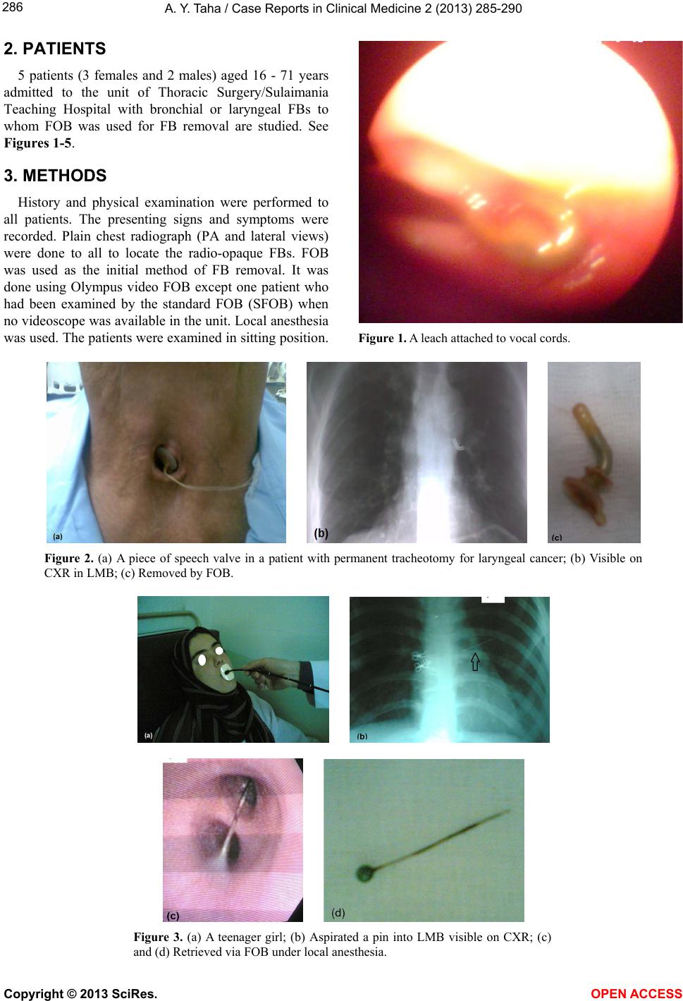
The second patient in this study was a man of 71 with
total laryngectomy and permanent tracheotomy done for
cancer of the larynx 10 years earlier. He had presented
with an aspirated piece of speech valve. This FB was re-
moved successfully by FOB through the tracheotomy
stoma. The literature confirms the difficulties encoun-
tered in the fixation of prostheses for voice rehabilitation
after laryngectomy [20].
6. CONCLUSION
Though RB is a time tested safe and effective proce-
dure for FB removal which is in use all over the world;
FOB can also be used safely provided the operator has
adequate skill in RB, the latter should be readily avail-
able in case a difficulty is encountered with FOB. It is
especially helpful when an adult patient has a doubtful
diagnosis of FB aspiration and the risk of GA necessary
for RB is to be avoided. To increase its success rate,
FOB use should be coupled with the necessary equip-
ments like special FB forceps and fluoroscopy.
REFERENCES
[1] Chin-Wing, Y.U. (2012) Rigid bronchoscopy—A physic-
cian’s perspective. Hong Kong Respiratory Medicine. The
official website of HKTS, ACCP HK & Macau, HKLF.
[2] Elhassani, N.B. (1978) Aspirated tracheobronchial for-
eign bodies in infants. Annals of the Royal College of
Surgeons of England, 23, 310-314.
[3] Hussein. W.M. (1984) Tracheobronchial foreign bodies in
pediatric age group. Journal of the Faculty of Medicine,
Baghdad, 26, 63-72.
[4] Abdulameer, M.H. (2010) Foreign bodies inhalation. Jour-
nal of the Faculty of Medicine, Baghdad, 52, 255-258.
[5] Swanson, K.L. and Edell, E.S. (2001) Tracheobronchial
foreign bodies. Chest Surgery Clinics of North America,
11, 861-872.
[6] Lan, R.S., Lee, Ch., Chiang, Y.C. and Wang, W.J. (1989)
Use of fiberoptic bronchoscopy to retrieve bronchial for-
eign bodies in adults. American Review of Respiratory
Disease, 140, 1734-1737. doi:10.1164/ajrccm/140.6.1734
[7] Donado Una, J.R., de Miguel Poch, E., Casado Lopez,
M.E. and Alfaro Abreu, J.J. (1998) Fiberoptic broncho-
scopy in extraction of tracheobronchial foreign bodies in
adults. Arch Bronchopneumo l, 34, 76-81.
[8] Muhammad, T.D., Saad, M.M., Bahaa, M.A. and Omer,
R.H. (2010) Tracheo-bronchial foreign bodies inhalation
in Al-Anbar Governorate. Kufa Medical Journal, 13, 248-
262.
[9] Akhtar, S. and Ul Hak, I. (2006) An experience with
leeches in the aero-digestive tract.
http://www.pafmj.org/showdetails.php?id=60&t=c
[10] Boyd, M., Chatterjee, A., Chiles, C. and Chin Jr., R.
(2009) Tracheobronchial foreign body aspiration in adults.
Southern Medical Journal, 102, 171-174.
doi:10.1097/SMJ.0b013e318193c9c8
[11] Ramirez-Figueroa, J.L., Gochicoa-Rangel, L.G, Ramirez-
San Juan, D.H. and Vargas, M.H. (2005) Foreign body
removal by flexible fiberoptic bronchoscopy in infants
and children. Pediatric Pulmonology, 40, 392-397.
doi:10.1002/ppul.20242
[12] Mise, K., Jurcev Savicevic, A., Pavlov, N. and Jankovic,
N. (2009) Removal of tracheobronchial foreign bodies in
adults using flexible bronchoscopy: Experience 1995-
2006. Surgical Endoscopy, 23, 1360-1364.
doi:10.1007/s00464-008-0181-9
[13] Gencer, M., Ceylan, E. and koksal, N. (2007) Extraction
of pins from the airway with flexible bronchoscopy. Res-
piration, 74, 674-679.
[14] Hasdiraz, L., Bicer, C., Bilgin, M. and Oguzkaya, F. (2006)
Turban pin aspiration: Non-asphyxiating tracheobronchial
foreign body in young Islamic women. Thoracic and
Cardiovascular Surgeon, 54, 273-275.
doi:10.1055/s-2006-923954
[15] Kaptanoglu, M., Dogan, K., Onen, A. and Kunt, N. (1999)
Turban pin aspiration; a potential risk for young Islamic
girls. International Journal of Pediatric Otorhinolaryn-
gology, 48, 131-135.
[16] El-Kushman, H.M., Sharara, A.M., Sa Ada, M.A. and
Hijazi, Ma. (2007) Tracheobronchial straight metallic pin
aspiration in young Jordanian females. Saudi Medical
Journal, 28, 913-916.
Copyright © 2013 SciRes. OPEN ACCESS
A. Y. Taha / Case Reports in Clinical Medicine 2 (2013) 285-290
290
[17] Al-Ali, M.A., Khassawneh, B. and Alzoubi, F. (2007)
Utility of fiberoptic bronchoscopy for retrieval of aspi-
rated headscarf pins. Respiration, 74, 309-313.
doi:10.1159/000098477
[18] Murthy, P.S., Ingle, V.S., George, E., Ramakrishna, S. and
Shah, F.A. (2001) Sharp foreign bodies in the tracheo-
bronchial tree. American Journal of Otolaryngology, 22,
154-156. doi:10.1053/ajot.2001.22585
[19] Nguyen, L.H., Huy, D., Nogoc, T., Nguyen, P.T., Thi,
Q.H., Aelony, Y. and Homasson, J.P.D. (2010) Endobron-
chial foreign bodies in Vietnamese adults are related to
eating habits. Respirology, 15, 491-494.
[20] Ten Hallers, E.J., Marres, H.A., Rakhorst, G., Hagen, R.,
Staffieri, A., Van Der Laan, B.F., Van Der Houwen, E.B.
and Verkerke, G.J. (2005) Difficulties in the fixation of
prostheses for voice rehabilitation after laryngectomy.
Acta Oto-Laryngologica, 125, 804-813.
doi:10.1080/00016480510031506
Copyright © 2013 SciRes. OPEN ACCESS