Open Journal of Gastroenterology, 2013, 3, 227-230 OJGas
doi:10.4236/ojgas.2013.34038 Published Online August 2013 (http://www.scirp.org/journal/ojgas/)
Rapidly enlarged inflammatory hepatocellular adenoma:
A case report*
Kenji Koneri1#, Hidetaka Kurebayashi1, Katsuji Sawai1, Mitsuhiro Morikawa1, Makoto Murakami1,
Yasuo Hirono1, Takanori Goi1, Atsushi Iida1, Kanji Katayama1, Hiroshi Itoh2, Motoko Sasaki3,
Yasuni Nakanuma3, Akio Yamaguchi1
1Department of Gastroenterological Surgery, University of Fukui, Fukui, Japan
2Department of Tumor Pathology, University of Fukui, Fukui, Japan
3Department of Human Pathology, Kanazawa University Graduate School of Medicine, Kanazawa, Japan
Email: #koneri@hotmail.co.jp
Received 9 June 2013; revised 9 July 2013; accepted 24 July 2013
Copyright © 2013 Kenji Koneri et al. This is an open access article distributed under the Creative Commons Attribution License,
which permits unrestricted use, distribution, and reproduction in any medium, provided the original work is properly cited.
ABSTRACT
We present a rare case of rapidly enlarging inflame-
matory hepatocellular adenoma (IHCA) in a 60-year-
old Japanese man. Screening abdominal computed to-
mography (CT) for the fatty liver patient revealed a
1.7-cm liver mass in the anterior segment of the liver.
After 19 months, the lesion had rapidly enlarged to 6
cm in diameter and the patient was referred to our
hospital. On perflubutane microbubble contrast-enhan-
ced ultrasonography, the tumor showed a characteris-
tic centripetal filling pattern in the vascular phase. We
performed hepatic anterior segment resection because
we could not rule out ma lignant tumor. Histopat holo-
gical examination sho wed hyp erplasia of mildly at ypi-
cal hepatocytes and sinusoidal dilatation with marked
inflamm ator y cell infil tr ati on . I mmun ohi sto logic al s tain -
ing revealed positive staining for serum amyloid A
and C-reactive protein; therefore, we diagnosed this
tumor as IHCA. The patient remains alive 42 months
after operation without evidence of recurrence.
Keywords: Inflammatory Hepatocellular Adenoma;
Perflubutane Microbubble Contrast-Enhanced
Ultrasonography; Surgery; Rapid Enlargement
1. INTRODUCTION
A new classification for hepatocellular adenoma (HCA)
was described in 2006 in the World Health Organization
(WHO) classification of tumors of the digestive system.
According to this classification, HCA is grouped into
three subtypes based on genotype and phenotype as fol-
lows: hepatocyte nuclear factor (HNF) 1α-inactivated
HCA, β-catenin-activated HCA and inflammatory HCA
(IHCA) [1-3]. Among these subtypes, IHCA results from
a molecular gain-of-function mutation of the interleukin
(IL)-6 pathway, and shows characteristic pathological
features such as inflammatory cell infiltration, sinusoidal
dilatation and increased expression of inflammatory pro-
teins such as serum amyloid A (SAA) and C-reactive
protein (CRP) [4]. A non-negligible number of IHCA pa-
tients have recently been reported in Western countries
[2], but very few in Asian countries. We now report a
case of an IHCA Asian male patient, who showed rapid
enlargement of tumor and underwent surgical resection
after preoperative perflubutane microbubble (Sonazoid,
Daiichi-Sankyo, Tokyo, Japan) contrast-enhanced ultra-
sonography (Sonazoid-CEUS) and gadolinium ethoxy-
benzyl diethylenetriaminepentaacetic acid (Primovist,
Bayer, Leverkusen, Germany) contrast-enhanced magne-
tic resonance imaging (Primovist-MRI).
2. CASE REPORT
The patient was followed by a local doctor for fatty liver
and mild hypertension. In August 2007, a 1.7 cm tumor
was detected in the anterior segment of the liver by ab-
dominal computed tomography (CT) for screening of
fatty liver. After diagnosis as nodular hyperplasia (FNH),
follow-up observations were planned. In May 2009, fol-
low-up CT revealed rapid enlargement of the tumor up to
6 cm in diameter, so the patient was referred to our hos-
pital. The patient was a Japanese male who was 60 years
*Author contributions: K. Koneri, H. Kurebayashi and A. Yamaguchi
treated the patient and wrote the manuscript; T. Sawai, M. Morikawa,
M. Murakami, Y. Hirono, T. Goi, A. Iida and K. Katayama treated the
p
atient; H. Itoh, M. Sasaki and Y. Nakamura contributed to the patholo-
gical examination.
N
o support has been received from any company.
#Corresponding author.
Published Online August 2013 in SciRes. http://www.scirp.org/journal/ojgas
K. Koneri et al. / Open Journal of Gastroenterology 3 (2013) 227-230
228
old upon admission, and had no history of diabetes,
blood transfusion, steroid use or heavy drinking. His
height, weight and body mass index were 165 cm, 72 kg
and 26.11 kg/m2, respectively. Laboratory studies showed
mildly elevated levels of aspartate aminotransferase (40
IU/L), alanine aminotransferase (49 IU/L) and alkaline
phosphatase (629 IU/L), but white blood cell count, se-
rum C-reactive protein and feasting blood glucose were
normal. Carcinoembryonic antigen, feto protein and des-
gamma-carboxy prothrombin levels were also normal,
and negative results were obtained for HBs antigen and
HCV antibody.
Enhanced computed tomography (CT) in August 2007
showed an ill-defined 1.7 cm tumor in the anterior seg-
ment of the liver. In May 2009, plain CT revealed slight-
ly low-density tumor and enhanced CT revealed diffuse
enhancement at the arterial phase and homogeneous en-
hancement at the delayed phase. The tumor had rapidly
enlarged over1 year and 7 months (Figure 1).
For qualitative diagnosis, we performed US study.
Brightness-mode (B-mode) US showed a well-demar-
cated hypoechoic tumor. We performed CEUS using 0.5
ml of Sonazoid followed by 10 ml saline flush through a
peripheral vein. At 18 seconds (sec) after injection, rapid
peripheral enhancement was shown. Subsequently, cen-
tripetal filling was shown at 20 sec and complete filling
at 28 sec. After 12 minutes (Kupffer phase), delayed wash
out and peripheral rim of sustained enhancement were
observed. Re-enhancement was observed after re-inject-
ing 0.5 ml of Sonazoid (Figure 2).
Figure 1. Enhanced computed tomography findings. (A) An
ill-defined 1.7 cm tumor (arrow) was first detected in the ante-
rior segment of the liver; (B) The tumor had rapidly enlarged to
6 cm after 1 year and 7 months. The lesion was slightly hy-
pointense; (C) The entire tumor showed diffuse enhancement at
the arterial-dominant phase; (D) The tumor showed uniform
enhancement at the equilibrium phase.
Plain MRI showed slightly hyperintense tumor on T1-
and T2-weighted imaging, and hyperintensity on diffu-
sion-weighted imaging. After enhancement using Pri-
movist, diffuse enhancement was seen in the arterial
phase. The enhancement effect continued in the portal
phase. At 15 min after injection, in the hepatocyte phase,
the tumor was hypointense compared with the back-
ground liver (Figure 3).
On the basis of these findings, hepatocellular adenoma
was suspected, but we could not rule out the risk of rup-
ture and malignant transformation. A biopsy was not
considered because of a high risk of bleeding and dis-
semination of the tumor cells. Therefore we selected sur-
gical resection. Intraoperative findings showed no ascites
and no liver cirrhosis. The tumor was growing with he-
Figure 2. B-mode US and Sonazoid CEUS findings. (A) Well-
demarcated hypoechoic mass was observed at B-mode; (B) The
tumor showed initial peripheral enhancement at 18 sec after So-
nazoid injection; (C) The tumor showed centripetal filling at 20
sec; (D) The entire tumor showed complete filling at 28 sec; (E)
Delayed washout and peripheral rim of sustained enhancement
were observed in the Kupffer phase; (F) Strong re-enhancement
was observed after re-injection of Sonazoid.
Figure 3. EOB-MRI findings. (A) The tumor was detected as
slight hyperintensity on T1-weighted imaging; (B) The tumor
showed slight hyperintensity on T2-weighted imaging; (C)
Strong hyperintensity was evident on diffusion-weighted im-
aging; (D) After Primovist enhancement, diffuse strong en-
hancement was seen within the lesion in the arterial phase; (E)
The entire tumor was enhanced in the portal phase; (F) The
lesion was hypointense compared with the background in the
hepatocyte phase.
Copyright © 2013 SciRes. OJGas
K. Koneri et al. / Open Journal of Gastroenterology 3 (2013) 227-230 229
mispheric expansion from the liver surface and anterior
segmentectomy was performed.
Macroscopically, the tumor was soft, the cut surface
was brown and it was 6 × 5 cm in size with a thin cap-
sule. A dark purple congested area was evident at the
center of the tumor. Histologically, hematoxylin and eo-
sin (H&E) staining revealed trabecular hyperplasia of
mildly atypical hepatocytes and increased cell density,
and there were marked sinusoidal dilatation and hemor-
rhage, thick-walled arteries and infiltration of inflamma-
tory cells, which were predominantly lymphocytes (Fig-
ure 4). Immunohistological staining showed positive re-
sults for SAA and CRP (Figure 5). On the basis of these
findings, we diagnosed the tumor as IHCA. The postop-
erative course was satisfactory, and the patient was dis-
charged on day 16 after hepatectomy. The patient re-
mains alive 42 months after operation, without evidence
of recurrence.
3. DISCUSSION
Currently, HCA is classified into 3 subtypes based on ge-
notype and phenotype: HNF 1α-inactivated HCA, β-cate-
nin-activated HCA and IHCA [1-3]. In Western countries,
the incidence of HCA is 3 - 4 per 1 million population
and the “inflammatory” subtype accounts for approxi-
mately half of HCAs [3]. But the incidence of this sub-
type is exceptionally low in Asian countries, only 7 IHCA
cases were reported by Sasaki from Japan [5].
Use of oral contraceptives, glycogen storage disease
and anabolic hormone treatment such as steroid and an-
Figure 4. Macroscopic and microscopic findings. (A) The tu-
mor was soft, the cut surface was brown and it was 6 × 5 cm in
size with a thin capsule. A dark purple congested area was evi-
dent at the center of the tumor; (B) It showed trabecular hyper-
plasia of mildly atypical hepatocytes and increased cell density
(H&E, ×40); (C) Sinusoidal dilatation (black arrow) and hem-
orrhage (white arrow) were evident (H&E, ×100); (D) Abnor-
mally thick artery (black arrow) and filtration of inflammatory
cells (white arrow) were evident (H&E, ×100).
Figure 5. Immunohistological findings. (A) It showed positiv-
ity for SAA (SAA, ×40); (B) High magnification of SAA stain-
ing revealed positivity for SAA in atypical hepatocytes (SAA,
×100); (C) It showed strong positivity for CRP (CRP, ×40).
drogens are well known as the major risk factors for
HCAs [3,5]. Classically, about 90% of HCA cases occur
in young women, with low incidences in children, males
and the elderly [5]. However, recent studies showed that
IHCA have more strong correlation with heavy alcohol
intake or obesity [5,6] than classically-known patho-
genesis.
On diagnostic imaging, differentiating HNF 1α-inac-
tivated HCA from other type of HCA is clinically impor-
tant. The reason is HNF 1α-inactivated HCA seldom pro-
gresses to hepatocellular carcinoma, whereas malignant
transformation will occur in 5% - 10% of cases among
β-catenin-activated HCA and IHCA [7]. Laumonier et al.
evaluated the possibility of distinguishing subtypes of
HCA using sulfur hexafluoride microbubbles (SonoVue,
Bracco, Milan, Italy) CEUS. They retrospectively evalu-
ated 17 cases of IHCA versus 16 cases of HNF1α-HCA,
and concluded that the most valuable criteria for the di-
agnosis of IHCA were the higher rates of hypervascular-
ity (IHCA 100% vs. HNF1α-HCA 62.5%, p < 0.01), cen-
tripetal filling (94.1% vs. 25%, p < 0.01), delayed wash-
out (64.7% vs. 12.5%, p < 0.01) and peripheral rim of
sustained enhancement (64.7% vs. 0%, p < 0.01) [8].
There are no previous reports for availability of Sonazoid
to perform real-time assessment of vascularity in IHCA.
In our case, it seems that the CEUS findings using Sona-
zoid is quite similar to the previously-reported findings
using SonoVue agent. These findings indicate the po-
tential of Sonazoid for use in the differential diagnosis of
HCA subtypes.
It was reported that IHCA shows distinctive patho-
logical features. Histologically, it exhibits focal or dif-
fuse inflammation, sinusoidal dilatation, congestion and
an area of peliosis, along with numerous thick-walled
arteries with ductular reaction [3]. Immunohistologically,
Copyright © 2013 SciRes. OJGas
K. Koneri et al. / Open Journal of Gastroenterology 3 (2013) 227-230
Copyright © 2013 SciRes.
230
[3] Bioulac-Sage, P., Balabaud, C. and Wanless, I. (2000)
Focal nodular hyperplasia and hepatocellular adenoma. In:
Bosman, F., Ed., WHO classification of tumours of the
digestive system. 4th Edition, IARC, Lyon, 198-204.
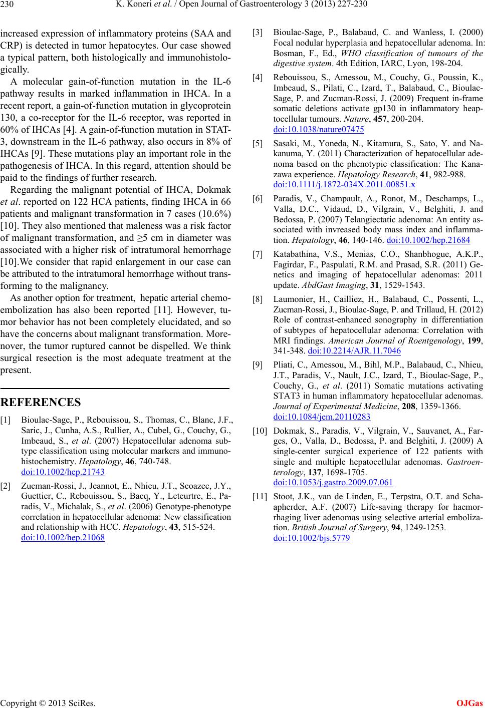
increased expression of inflammatory proteins (SAA and
CRP) is detected in tumor hepatocytes. Our case showed
a typical pattern, both histologically and immunohistolo-
gically. [4] Rebouissou, S., Amessou, M., Couchy, G., Poussin, K.,
Imbeaud, S., Pilati, C., Izard, T., Balabaud, C., Bioulac-
Sage, P. and Zucman-Rossi, J. (2009) Frequent in-frame
somatic deletions activate gp130 in inflammatory heap-
tocellular tumours. Nature, 457, 200-204.
doi:10.1038/nature07475
A molecular gain-of-function mutation in the IL-6
pathway results in marked inflammation in IHCA. In a
recent report, a gain-of-function mutation in glycoprotein
130, a co-receptor for the IL-6 receptor, was reported in
60% of IHCAs [4]. A gain-of-function mutation in STAT-
3, downstream in the IL-6 pathway, also occurs in 8% of
IHCAs [9]. These mutations play an important role in the
pathogenesis of IHCA. In this regard, attention should be
paid to the findings of further research.
[5] Sasaki, M., Yoneda, N., Kitamura, S., Sato, Y. and Na-
kanuma, Y. (2011) Characterization of hepatocellular ade-
noma based on the phenotypic classification: The Kana-
zawa experience. Hepatology Research, 41, 982-988.
doi:10.1111/j.1872-034X.2011.00851.x
Regarding the malignant potential of IHCA, Dokmak
et al. reported on 122 HCA patients, finding IHCA in 66
patients and malignant transformation in 7 cases (10.6%)
[10]. They also mentioned that maleness was a risk factor
of malignant transformation, and 5 cm in diameter was
associated with a higher risk of intratumoral hemorrhage
[10].We consider that rapid enlargement in our case can
be attributed to the intratumoral hemorrhage without trans-
forming to the malignancy.
[6] Paradis, V., Champault, A., Ronot, M., Deschamps, L.,
Valla, D.C., Vidaud, D., Vilgrain, V., Belghiti, J. and
Bedossa, P. (2007) Telangiectatic adenoma: An entity as-
sociated with invreased body mass index and inflamma-
tion. Hepatology, 46, 140-146. doi:10.1002/hep.21684
[7] Katabathina, V.S., Menias, C.O., Shanbhogue, A.K.P.,
Fagirdar, F., Paspulati, R.M. and Prasad, S.R. (2011) Ge-
netics and imaging of hepatocellular adenomas: 2011
update. AbdGast Imaging, 31, 1529-1543.
As another option for treatment, hepatic arterial chemo-
embolization has also been reported [11]. However, tu-
mor behavior has not been completely elucidated, and so
have the concerns about malignant transformation. More-
nover, the tumor ruptured cannot be dispelled. We think
surgical resection is the most adequate treatment at the
present.
[8] Laumonier, H., Cailliez, H., Balabaud, C., Possenti, L.,
Zucman-Rossi, J., Bioulac-Sage, P. and Trillaud, H. (2012)
Role of contrast-enhanced sonography in differentiation
of subtypes of hepatocellular adenoma: Correlation with
MRI findings. American Journal of Roentgenology, 199,
341-348. doi:10.2214/AJR.11.7046
[9] Pliati, C., Amessou, M., Bihl, M.P., Balabaud, C., Nhieu,
J.T., Paradis, V., Nault, J.C., Izard, T., Bioulac-Sage, P.,
Couchy, G., et al. (2011) Somatic mutations activating
STAT3 in human inflammatory hepatocellular adenomas.
Journal of Experimental Medicine, 208, 1359-1366.
doi:10.1084/jem.20110283
REFERENCES
[1] Bioulac-Sage, P., Rebouissou, S., Thomas, C., Blanc, J.F.,
Saric, J., Cunha, A.S., Rullier, A., Cubel, G., Couchy, G.,
Imbeaud, S., et al. (2007) Hepatocellular adenoma sub-
type classification using molecular markers and immuno-
histochemistry. Hepatology, 46, 740-748.
doi:10.1002/hep.21743
[10] Dokmak, S., Paradis, V., Vilgrain, V., Sauvanet, A., Far-
ges, O., Valla, D., Bedossa, P. and Belghiti, J. (2009) A
single-center surgical experience of 122 patients with
single and multiple hepatocellular adenomas. Gastroen-
terology, 137, 1698-1705.
doi:10.1053/j.gastro.2009.07.061
[2] Zucman-Rossi, J., Jeannot, E., Nhieu, J.T., Scoazec, J.Y.,
Guettier, C., Rebouissou, S., Bacq, Y., Leteurtre, E., Pa-
radis, V., Michalak, S., et al. (2006) Genotype-phenotype
correlation in hepatocellular adenoma: New classification
and relationship with HCC. Hepatology, 43, 515-524.
doi:10.1002/hep.21068
[11] Stoot, J.K., van de Linden, E., Terpstra, O.T. and Scha-
apherder, A.F. (2007) Life-saving therapy for haemor-
rhaging liver adenomas using selective arterial emboliza-
tion. British Journal of Surgery, 94, 1249-1253.
doi:10.1002/bjs.5779
OJGas