International Journal of Clean Coal and Energy, 2013, 2, 24-26
doi:10.4236/ijcce.2013.22B006 Published Online May 2013 (http://www.scirp.org/journal/ijcce)
The Effects of N-acetyl-seryl-aspartyl-lysyl-proline on
Expression of NF-κb and MCP-1 in Rats with Silicosis
Qian Li, Fang Yang*, Lijuan Zhang, Hong Xu, Wenli Zhang
Medical research center Hebei United University, Thangshan, China
Email: liqian324@yahoo.com.cn
Received 2013
ABSTRACT
In the present study, we developed silicosis of rat model by bronchial perfusion SiO2 dust, and intervenes with AcSDKP,
immunohisto chemistry was used to detect NF-κb and MCP-1 expression in lung tissue, and positive cells were counted.
We found that compared with silicotic model group, the positive cells of NF-κb and MCP-1 were decreased signifi-
cantly in anti-fibrosis treatment of AcSDKP group. The findings suggest that AcSDKP could inhibit the expression of
NF-κb and MCP-1 in lung tissue of silicosos, this may be related to AcSDKP inhibit of macrophage infiltration in lung
tissue and reduced the degree of dust alveolitis.
Keywords: N-acetyl-seryl-aspartyl-lysyl-proline; Silicosis; Monocyte Chemotactic Protein-1; Nuclear Factor-κb
1. Introduction
The main pathological change of silicosis is the fibro-
blast proliferation and collagen deposition in lung tissue.
Recent studies have shown that cytokines play an impor-
tant role in the development of silicosis, they constitute a
complex signal transduction molecules network system
and the chain reaction system, leading to infiltration of
inflammatory cells, fibroblasts and mesenchymal con-
nective tissue deposition is characterized by a chronic
immune-mediated inflammation[1]. Rat silicosis model
adopted by the bronchial perfusion SiO2 dust, and inter-
venes with AcSDKP, study the therapeutic effects of
AcSDKP on silicotic fibrosis, provides new method for
silicosis preventing and controlling.
2. Materials and Methods
2.1. Ethics Statement
Male Wistar rats, weighting 180 ± 10 g, were purchased
from Vital River Laboratory Animal Technology Co. Ltd.
(Beijing, China). All animal experiments were reviewed
and approved by the Institutional Animal Care and Use
Committee at the Hebei United University. Animals were
given free access to food and water and were cared for
according to guidelines set by the National Institute of
Health (NIH)
2.2. Induction of Silicosis and AcSDKP
Treatment
Rats were anesthetized with Isoflurane and then received
either silica solution (50 mg/rat, 1ml) or 0.9% saline (as a
vehicle control) by trachea. Prior to instillation the 5 μm
silica particles (Sigma , St. Louis, MO, USA) were baked
at 180℃ for 6 hours. AcSDKP [800 μg/(kg d), Bachem
AG company, USA] or control (0.9% saline) was given
via a miniosmotic pump (Alzer 2 ml4, DURECT Co. Ltd,
USA) planted into the abdominal cavity. Rats were di-
vided into 3 groups (n = 10), 1) control group (instilled
with 0.9% saline, and then treated with 0.9% saline for 4
w) ; 2) silicotic model group (instilled of SiO2,, and then
treated with 0.9% saline for 4 w); 3) anti-fibrosis treat-
ment of AcSDKP group (instilled with SiO2, and treated
with AcSDKP for 4 w) [2].
2.3. Immunohistochemistry for NF-κb and
MCP-1
Take the lungs to organize 4% paraformaldehyde to be
fixed, conventional paraffin wax embedding, 6 μm serial
section, for immunohistochemical method dyeing. After
incubation with 5% horse serum, sections were incubated
with primary antibodies against NF-κb and MCP-1 (Santa
cruz Biotechnology, USA) followed by the biotinylated
secondary antibody and finally the ABC reagent (Wuhan
Boshide Biological Engineering Co. Ltd, China). Immu-
noreactivity was visualized with DAB. A brown color
staining was considered a positive result. Counts the
*Corresponding author.
Copyright © 2013 SciRes. IJCCE
Q. LI ET AL. 25
positive cells of NF-κb and MCP-1.
3. Results
3.1. Morphological Observation
Surfaces of lung are smooth and no nodule formation in
control group. Canous nodules distribute sporadically on
the surface and cutting surface in silicotic model group.
Compared with silicotic mode group, the numbers of
canous nodules decreased on the surface and cutting sur-
face in the groups with AcSDKP treatment.
3.2. Expression of NF-κb and MCP-1 in Lung of
Rat with Silicosis
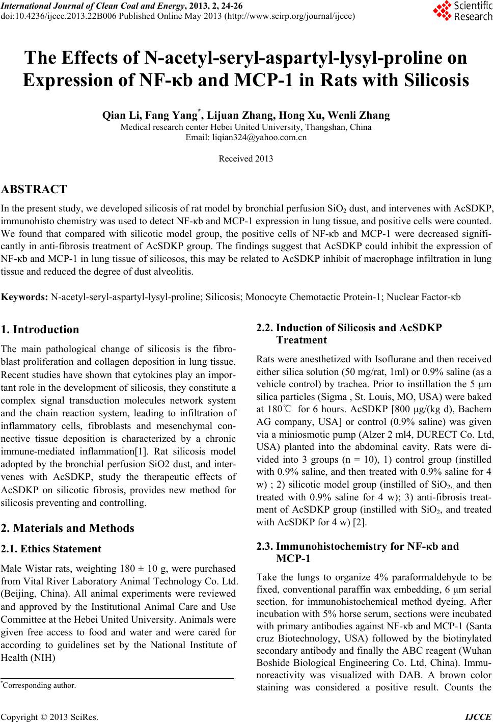
Immunohistochemistry staining shows that NF-κb and
MCP-1 only express in a small number of alveolar
macrophages and fibroblasts in control group (Figure 1).
NF-κb and MCP-1 expressions increase in silicotic nod-
ules and interstitial fibrotic area. The expressions of
NF-κb and MCP-1 in AcSDKP treatment are less than
that in silicotic model. Positive cells counting shows that
NF-κb and MCP-1 expressions in silicotic model group
are increases by 2.76 fold and 5.40 fold compared with
control group. AcSDKP treatment decreases the expres-
sions of NF-κb and MCP-1. The positive cells counting
of NF-κb and MCP-1 in anti-fibrotic treatment group is
70.12% and 65.37% of silicotic model group respectively.
Variance analysis reveals that differences are significant
(p < 0. 05).
a b c
d e f
*
*
#
#
A
45
40
35
30
25
20
15
10
5
0
control silicotic
model
AcSDKP
anti-fibrotic
p
ositive cells
NF-κb
MCP-1
(a) control group for NF-κb; (b) silicotic model group for NF-κb; (c)
AcSDKP anti-fibrotic group for NF-κb; (d) control group for MCP-1; (e)
silicotic model group for MCP-1; (f) AcSDKP anti-fibrotic group for MCP-1.
(A) The positive cells expression of NF-κb and MCP-1 in the lung of rat. *p
0.05 vs contro; #p 0.05 vs silicotic model.
Figure 1. Effect of Ac-SDKP on NF-κb and MCP-1 analyzed
by Immunohistochemistry in rats with silicosis. (× 400).
4. Discussion
N-acetyl-seryl-aspartyl-lysyl-proline(AcSDKP) is a the
physiological hematopoietic growth inhibition factor,
studies have shown that AcSDKP inhibit proliferation
and collagen synthesis of cardiac fibroblasts [3,4]. In
recent years NF-κB prepared in the silicosis fibrosis
pathogenesis research is paid attention, NF-κb is a tran-
scription activation function of proteins, with the wide
range of promoter or Enhancer combining parts of κb site
specificity and promote their transcription, NF-κB in-
volved in many of the former transcriptional regulation
of inflammatory mediators molecules promoting cyto-
kines, adhesion molecules, chemokines, inflammatory
factors, oxidative stress-related enzymes released in large
quantities, so as to promote organ inflammation and fi-
brosis[5]. MCP-1 is a chemokine CC subfamily, its main
function is the monocyte-macrophage cell chemotaxis, in
addition, MCP-1 except took one kind of front inflam-
mation cell factor participates in the adjustment white
blood cell outside the chemotaxy, it also has stimulates
the ciliary cell to multiply, the collagen synthesizes as
well as induces certain presses the function which the
fibrosis cell factor or the medium produce[6]. Our studies
found that positive cells of NF-κb and MCP-1 expres-
sions increase in silicotic model group compared with
control group, it is shown that in the stimulation of SiO2
dust, the production and expression of NF-κb and MCP-1
increased in silicosis lungs of rats. More important dis-
coveries found that in silicosis lungs of rats, the positive
cells of NF-κb and MCP-1 were significantly decreased
after given AcSDKP intervention. This indicated that,
AcSDKP can suppress the production and expression of
NF-κb and MCP-1 in the lung with the SiO2 dust stimu-
lation, reduced the inflammatory response in the lungs of
silicotic rats.
5. Conclusions
Our data indicate that AcSDKP possibly through inhibi-
tion of NF-κb and MCP-1 production and expression,
reduce the synthesis and release of pro-fibrotic cytokines,
thereby inhibiting or reducing extracellular matrix syn-
thesis and deposition of the lungs, reducing the extent of
silicosis fibrosis and inhibiting the progression of fibro-
sis.
6. Acknowledgements
This work was funded by National Natural Science
Foundation of China (No. 81072254) and Hebei Province
Natural Science Foundation of China (No. C 20114010
24).
REFERENCES
[1] G. Semenzalo, F. Adami, N. Maschio, et al., “Immune
Copyright © 2013 SciRes. IJCCE
Q. LI ET AL.
Copyright © 2013 SciRes. IJCCE
26
Mechanisms in Interstitial Lung Disease,” Allergy, Vol.
55, 2000, pp. 1103-1120.
doi:10.1034/j.1398-9995.2000.00127.x
[2] S. Ying, Y. Fang, Y. Jingbo, et al., ”New Anti-fibrotic
Mechanisms of N-acetyl-seryl-aspartyl-lysyl-proline in
Silicon Dioxide-induced Silicosis,” Life Sciences, Vol. 87,
No. 3, 2010, pp. 232-239.
[3] M. D. Gunn, N. A. Nelken, X. Liao, et al., “Monocyte
Chemo Attractant Protein-1 Is Sufficient for the Chemo-
taxis of Monocyte and Lymphocytes in Transgenic Mice
but Requires an Additional Stimulus for Inflammatory
Activation, Journal of Immunol, Vol. 158, No. 1, 1997,
pp. 376-383.
[4] I. Inoshima, Kuwano K, Hamada N, et al.,
Anti-monocyte Chemo Attractant Protein-1 Gene Ther-
apy Attenuates Pulmonary Fibrosis in Mice Am,” Journal
of Physiol Lung Cell Mol Physiol, Vol. 286, No. 5, 2004,
pp. L1038 - L1044.
[5] P. A. Henriksen, M. Hitt, Z. Xing, et al., “Adenoviral
Gene Delivery of Elafin and Secretory Leukocyte Prote-
ase Inhibitor Attenuates NF-kappa B-dependent Inflam-
matory Responses of Human Endothelial Cells and
Macrophages to Atherogenic Stimuli,” Journal of Immu-
nol, Vol. 172, No. 7, 2004, pp. 4535-4544.
[6] Z. Y. Tao, A. Maria Cavasin, F. Yang, Y. H. Liu and X. P.
Yang, “Temporal Changes in Matrix Metalloproteinase
Expression and Inflammatory Response Associated with
Cardiac Rupture after Myocardial Infarction in Mice,”
Life Science, Vol. 74, No. 12, 2004, pp. 1561-1572.
doi:10.1016/j.lfs.2003.09.042