Surgical Science, 2013, 4, 350-353
http://dx.doi.org/10.4236/ss.2013.48069 Published Online August 2013 (http://www.scirp.org/journal/ss)
Colonoscopically Assisted Laparoscopic Polypectomy—An
Alternative to Right Hemi colectomy for Large Right-Sided
Benign Polyps*
A. Z. Kaleem#, C. Strachan, L. Whittaker, S. M. Ahmad
Department of Colorectal Surgery, Northern Lincolnshire and Goole Hospital NHS Foundation Trust,
Scunthorpe General Hospital, Scunthorpe, UK
Email: #ahmed.kaleem@nhs.net
Received April 28, 2013; revised May 30, 2013; accepted June 8, 2013
Copyright © 2013 A. Z. Kaleem et al. This is an open access article distributed under the Creative Commons Attribution License,
which permits unrestricted use, distribution, and reproduction in any medium, provided the original work is properly cited.
ABSTRACT
Introduction: Laparoscopic assisted colonoscopic polypectomies have been well described in the literature and are
well established in surgical practice, for removal of large, inaccessible, or flat based polyps. Laparoscope allows the
endoscopist a serosal viewpoint and thus clear indication of perforation, in addition to enhancing endoscopic position-
ing through colonic mobilisation, facilitating polypectomy. We describe a previously rarely published technique, in
which the colonoscope directs the surgeon to polyps and laparoscopy enables wedge resection of benign polyps using
Endo GIA staplers. Using this method, the colonoscope provides an intra-luminal view ensuring adequate excision with
margins whilst the laparoscope provides intra-peritoneal access for the wedge resection. Methods: This is a case series
of 12 patients with large tubulovillous adenomas, found and biopsied at colonoscopy. Under a general anaesthetic, an
on table colonoscopy was performed to identify and reassess the polyp, whilst a laparoscopy was performed to excise
the polyp via wedge resection, using the endoscopic view as guidance. Results: The polyp was identified and com-
pletely resected in our 12 patients. All patients were discharged on the first post-operative day. Of the polyps excised, a
focus of adenocarcinoma was detected in one and an adjacent endocrine tumour was found in another patient in histol-
ogy along with tubulovillous adenoma. Rest were all tubulovillous adenomas only. Conclusion: We propose that this
technique should be regarded as an alternative to Right hemicolectomies and difficult endoscopic mucosal resections for
large adenomas, and be regarded as a definitive and safe procedure in its own right.
Keywords: Colonoscopically Assisted Laparoscopic Polypectomy
1. Introduction
Colonoscopic polypectomies are the treatment of choice
for most benign colonic polyps, and are the most com-
monly performed therapeutic intervention in colonoscopy.
Polypectomy techniques and established practice has
evolved considerably in the last 10 years; providing
many options for the varying morphology of colonic
polyps.
Snare polypectomies (cold/hot) are performed com-
monly and routinely for most accessible, pedunculated
and small polyps. If a broad based polyp is found, more
advanced procedures such as endoscopic mucosal resec-
tions (EMR), endoscopic sub mucosal dissections or
formal operative colonic resections are performed to re-
move them. EMR techniques are growing in popularity
given the improved accuracy and safety profiles associ-
ated with raising a lesion via injection to facilitate ease of
removal and thus identification of an invading lesion.
EMR is now recommended by the British Society of
Gastroenterology guidelines for flat and depressed le-
sions. The risk of perforation of the colon during colono-
scopic polypectomies varies between 0.1% and 3%, in
some studies [1] and between 0% - 0.19% for therapeutic
interventions, according to the British society of gastro-
enterology guidelines [2]. A recent Japanese study has
reported figures of 0.91% perforation risk associated
with EMR, compared with 0.17% with simple Polypec-
tomy and 3.3% with Endoscopic Mucosal resection [3].
Such figures rise with the size and location of the polyp.
*Source of funding: N/A (Department of Colorectal Surgery, Scunthorpe).
Disclosure Statement: Mr Ahmed Zaman Kaleem, Miss Caroline Stra-
chan, Miss Laura Whittaker and Mr Syed Muzaffar Ahmad have no
conflicts of interest or financial ties to disclose.
#Corresponding author.
C
opyright © 2013 SciRes. SS
A. Z. KALEEM ET AL. 351
Perforation risk is greater with polyps > 3 cm and when
located in the thin walled caecum or ascending colon.
This is due to the high wall tension in the caecum. In
addition there is a risk of thermal transmural damage to
the colonic mucosal surface in coagulating the polyp
during snare removal.
The well documented and established method of com-
bining laparoscopy with colonoscopy to perform colono-
scopic polypectomies, allows the direct visualization of
the polyp and the extra-luminal surface with the laparo-
scope [4,5]. Thus if perforation were to occur, the laparo-
scope allows this to be viewed and immediately repaired,
or indeed proceed to laparoscopic resection.
We describe an alternative technique of polypectomy,
for benign colonic polyps, utilizing the principles of si-
multaneous laparoscopy and colonoscopy. We propose
the colonoscopically assisted laparoscopic polypectomy
as a scarcely described technique and improved method
of polypectomy using a laparoscopic wedge resection of
the segment of colon containing the benign polyp.
2. Materials and Methods
Thus far, we have successfully carried out this procedure
on 12 patients, 7 male and 5 female, with an age range
between 66 and 92. Pathological examination from initial
biopsies showed tubulovillous adenoma in all 12 patients.
7 of the 12 polyps were located in the caecum, and 5
polyps were located in the ascending colon and the pa-
tients were referred for this procedure, as the polyps were
deemed unsuitable for colonoscopic resection.
The resected polyps ranged in size from 15 mm in
maximum diameter to 50 mm from colonoscopic assisted
laparoscopic polypectomy. The pathology of the wedge
resections of the polyps showed tubulovillous adenoma
in 11 of the 12 patients and a focus of adenocarcinoma in
1 patient, where the tumour had been completely excised
at all margins. This case was taken to the multi-disciple-
nary team meeting (MDT) where the decision to perform
a laparoscopic Right hemicolectomy was taken. In one
patient, the appendix was removed in the wedge resec-
tion given the proximity of the appendiceal opening to
the polyp. The pathological analysis of the polyp was a
completely excised TVA; however, the pathological
analysis of the appendix was that of a well differentiated
endocrine tumour, confined to the sub mucosa, and a
pathological staging of pT1, completely excised. Thus,
histological analysis confirms complete excision of ade-
noma in all performed polypectomies.
3. Technique
The patient receives full bowel preparation for colono-
scopy. The patients are admitted the day before surgery
and are consented and counselled for colonoscopically
assisted laparoscopic polypectomy, +/ R hemicolec-
tomy (laparoscopic/ open), with all the potential risks
involved in such a procedure for example; leak, need for
further surgery if malignancy is suspected or confirmed
on histology, incomplete resection, further polyps, an-
aesthetic risks. It is explained to the patient that this is a
novel procedure in this country, and permission is taken
for publication and photography during laparoscopy/
colonoscopy.
The patient is positioned in the lithotomy position on
the operating table, after a general anaesthetic is admin-
istered, prepped and draped for a laparoscopic procedure.
A colonoscopy is performed and the polyp is identified
with the colonoscope. A tattoo may also be used to fa-
cilitate polyp identification at laparoscopy. At this point,
laparoscopy is performed, via a 10 mm umbilical port. A
further 2 ports are inserted under vision, one 12 mm port
in the hypo gastric and another 5 mm port in the paraum-
blical region as shown in Figure 1.
Tattoo on bowel or light of colonoscope is used to find
polyp from abdominal side (Figures 2(a) and 3). If visu-
alization from abdominal side via laparoscope is difficult
or polyp is located posteriorly then caecum and ascend-
ing colon are mobilized from lateral aspect. When polyp
is identified both endoscopically and laparoscopically
then by using an endoscopic GIA linear stapler, the seg-
ment of bowel containing the polyp is stapled off (Fig-
ures 2(b), 4 and 5). Before firing the stapler using
colonoscope, it is made sure that polyp is completely
excised. Once divided it is removed in a Burt bag
through the umbilical port (Figure 6). The role of the
colonoscopy here is to directly visualize the lumen to
identify that the polyp is being completely enveloped by
the stapler along with good margins. The stapler will not
be closed and fired before the surgeon is satisfied with
the colonoscopic image demonstrating the complete
Figure 1. Port positioning.
Copyright © 2013 SciRes. SS
A. Z. KALEEM ET AL.
352
(a) (b)
Figure 2. (a) Demonstration of the right sided polyp; (b)
Demonstration of the wedge resection.
Figure 3. Double view of linear stapler and colonoscopic
view.
Figure 4. Firing of the stapler and the colonoscopic view.
Figure 5. Post wedge stapling of the polyp.
Figure 6. Retrieval of the wedge of caecum in a Burt bag.
involvement of the polyp in the stapler. The right colon
has, in 11 out of our 12 cases not required mobilization
in order to carry out the segmental resection. In one case,
the polyp was located on the posterior wall and thus the
right colon had to be laparoscopically mobilized in order
to angle the laparoscopic stapler to achieve polypectomy.
The colonoscope is then withdrawn with intermittent
suction to deflate the colon, and the ports are removed
under direct vision and the wounds are closed in the
usual manner. Patients remain in hospital overnight for
monitoring and are discharged the following day if well.
All patients were discharged the following day.
Colonoscopically assisted laparoscopic polypectomy
was successfully achieved in 12 cases. All patients were
able to tolerate oral intake following the procedure and
there were no complications. The pathological analysis
has been detailed above. Of the excised polyps, 11 were
confirmed as benign adenomatous polyps and 1 polyp
showed a focus of moderately differentiated adenocarci-
noma arising from Tubulovillous adenomatous lesion.
This patient went on to have a laparoscopically assisted
R hemicolectomy.
4. Discussion
Adenomatous colonic polyps are amongst the most
Copyright © 2013 SciRes. SS
A. Z. KALEEM ET AL.
Copyright © 2013 SciRes. SS
353
common disorders of the colon and regarded as pre-ma-
lignant conditions. The majority of colonic polyps are
amenable to endoscopic snare excision. Large flat or in-
accessible right sided polyps pose a problem to safe en-
doscopic excision. The danger of incomplete excision or
perforation of the colon is significant, particularly in thin
walled caecum and ascending colon, and varies amongst
reports to as much as 3% [2]. Laparoscopic assisted colono-
scopic polypectomies are now well established for the
removal of such polyps and provide the extra security of
an intra-abdominal, serosal view of the colon whilst the
endoscopist is removing the polyp via snare [5]. Thus in
the case of perforation, it may be identified and treated
immediately. In addition, the use of the colonoscope al-
lows for accurate location of the polyp and thus mini-
mizing the need for colonic mobilization, with the excep-
tion of posterior/lateral wall located polyp. In this ap-
proach, the polyp is removed piecemeal and without sur-
rounding margins and thus histological comments on the
complete excision are difficult. There is also the consid-
eration of a late perforation due to diathermy injury
which may not be evident at the time of laparoscopy/
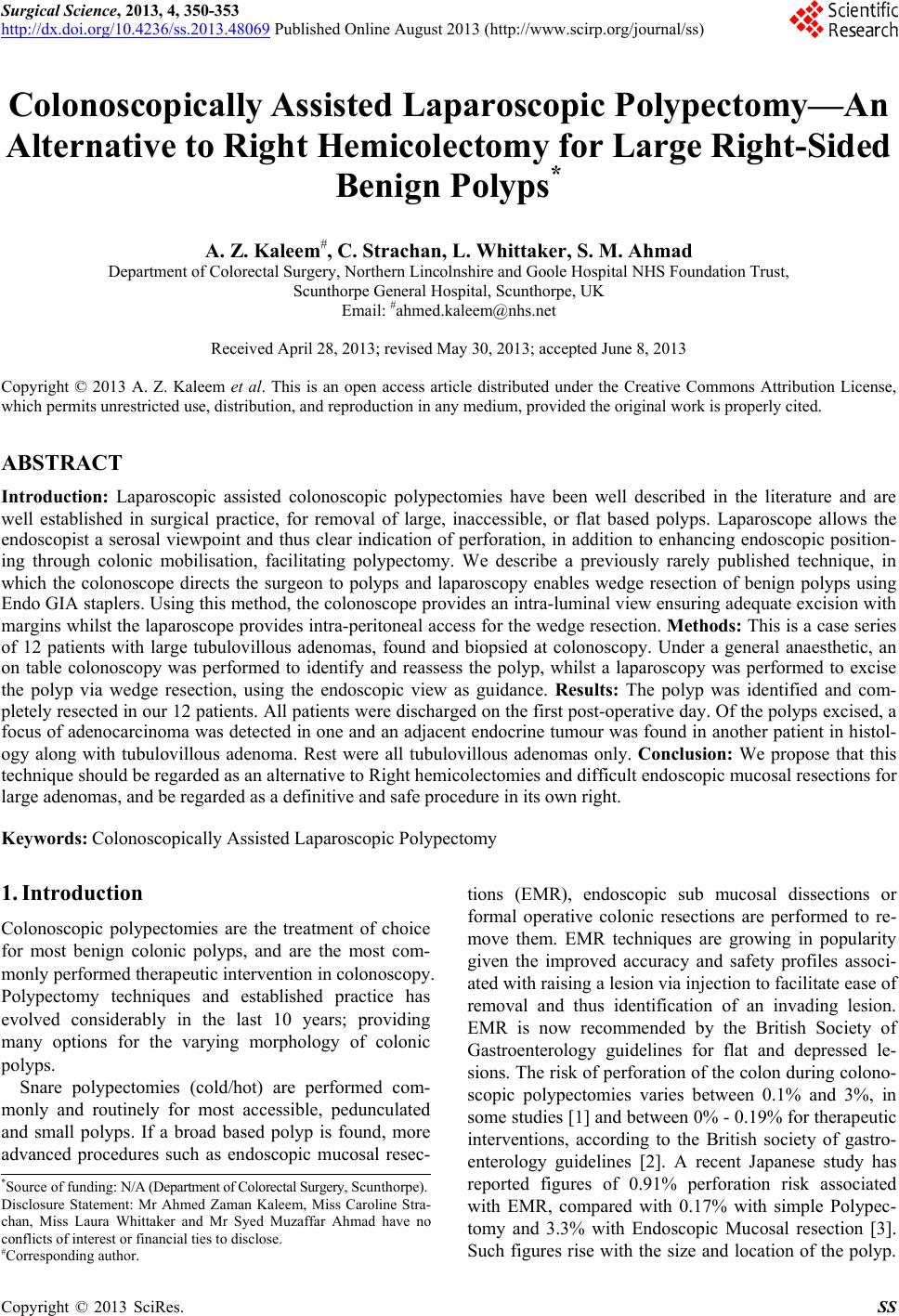
colonoscopy. We describe an alternative technique to
ensure complete polyp and margin removal, as a simple
wedge resection with the combination of laparoscopy and
colonoscopy. This technique has previously been de-
scribed, in association with the laparoscopic assisted
EMR, but not as a definitive procedure in its own right
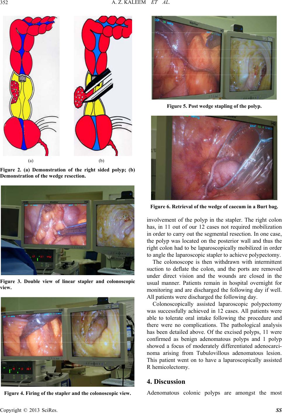
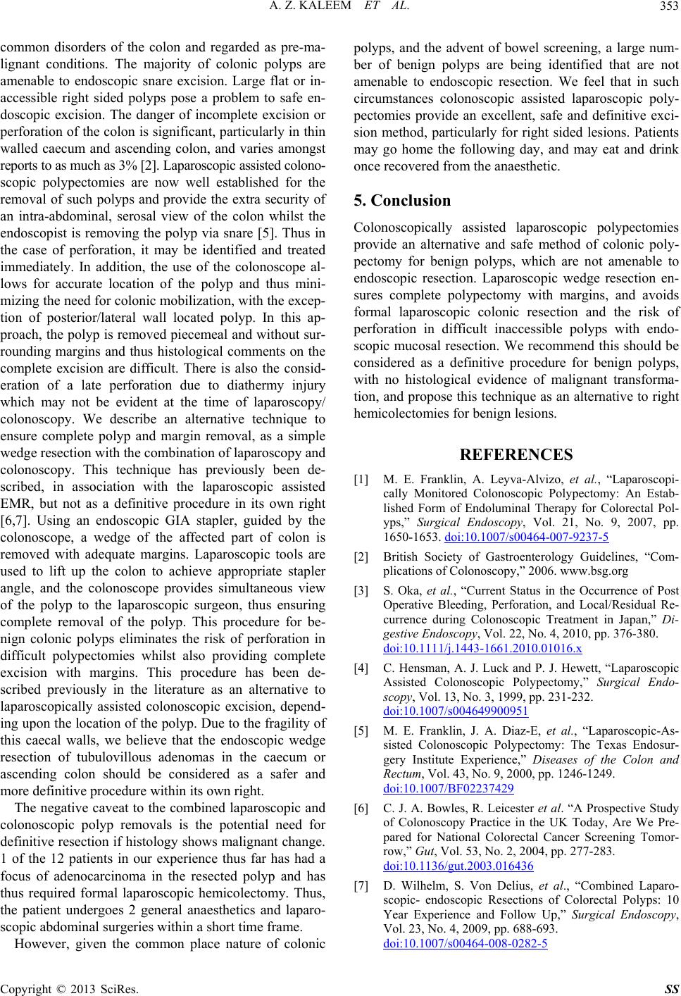
[6,7]. Using an endoscopic GIA stapler, guided by the
colonoscope, a wedge of the affected part of colon is
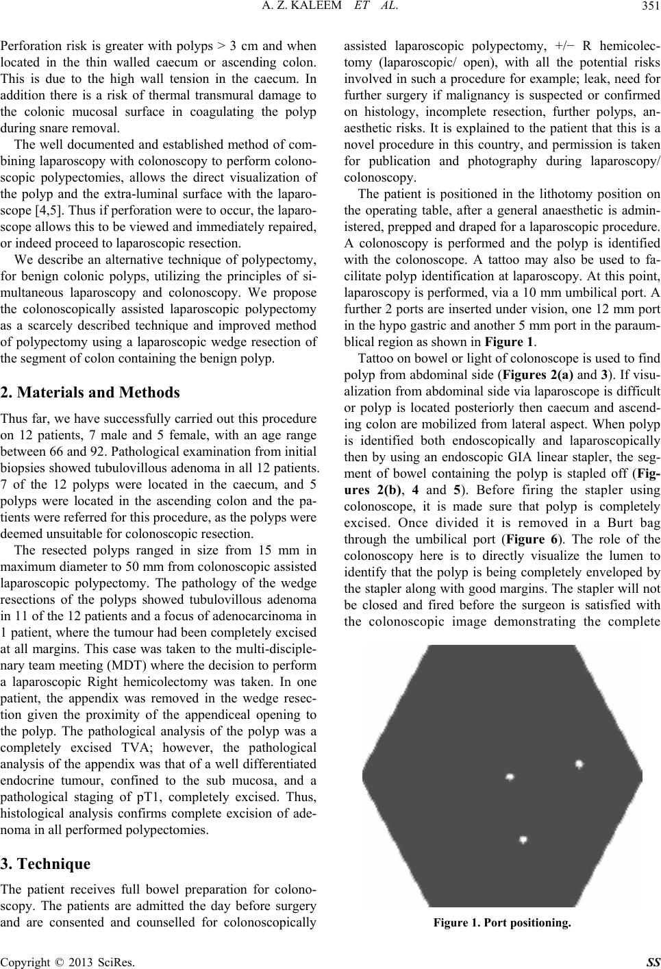
removed with adequate margins. Laparoscopic tools are
used to lift up the colon to achieve appropriate stapler
angle, and the colonoscope provides simultaneous view
of the polyp to the laparoscopic surgeon, thus ensuring
complete removal of the polyp. This procedure for be-
nign colonic polyps eliminates the risk of perforation in
difficult polypectomies whilst also providing complete
excision with margins. This procedure has been de-
scribed previously in the literature as an alternative to
laparoscopically assisted colonoscopic excision, depend-
ing upon the location of the polyp. Due to the fragility of
this caecal walls, we believe that the endoscopic wedge
resection of tubulovillous adenomas in the caecum or
ascending colon should be considered as a safer and
more definitive procedure within its own right.
The negative caveat to the combined laparoscopic and
colonoscopic polyp removals is the potential need for
definitive resection if histology shows malignant change.
1 of the 12 patients in our experience thus far has had a
focus of adenocarcinoma in the resected polyp and has
thus required formal laparoscopic hemicolectomy. Thus,
the patient undergoes 2 general anaesthetics and laparo-
scopic abdominal surgeries within a short time frame.
However, given the common place nature of colonic
polyps, and the advent of bowel screening, a large num-
ber of benign polyps are being identified that are not
amenable to endoscopic resection. We feel that in such
circumstances colonoscopic assisted laparoscopic poly-
pectomies provide an excellent, safe and definitive exci-
sion method, particularly for right sided lesions. Patients
may go home the following day, and may eat and drink
once recovered from the anaesthetic.
5. Conclusion
Colonoscopically assisted laparoscopic polypectomies
provide an alternative and safe method of colonic poly-
pectomy for benign polyps, which are not amenable to
endoscopic resection. Laparoscopic wedge resection en-
sures complete polypectomy with margins, and avoids
formal laparoscopic colonic resection and the risk of
perforation in difficult inaccessible polyps with endo-
scopic mucosal resection. We recommend this should be
considered as a definitive procedure for benign polyps,
with no histological evidence of malignant transforma-
tion, and propose this technique as an alternative to right
hemicolectomies for benign lesions.
REFERENCES
[1] M. E. Franklin, A. Leyva-Alvizo, et al., “Laparoscopi-
cally Monitored Colonoscopic Polypectomy: An Estab-
lished Form of Endoluminal Therapy for Colorectal Pol-
yps,” Surgical Endoscopy, Vol. 21, No. 9, 2007, pp.
1650-1653. doi:10.1007/s00464-007-9237-5
[2] British Society of Gastroenterology Guidelines, “Com-
plications of Colonoscopy,” 2006. www.bsg.org
[3] S. Oka, et al., “Current Status in the Occurrence of Post
Operative Bleeding, Perforation, and Local/Residual Re-
currence during Colonoscopic Treatment in Japan,” Di-
gestive Endoscopy, Vol. 22, No. 4, 2010, pp. 376-380.
doi:10.1111/j.1443-1661.2010.01016.x
[4] C. Hensman, A. J. Luck and P. J. Hewett, “Laparoscopic
Assisted Colonoscopic Polypectomy,” Surgical Endo-
scopy, Vol. 13, No. 3, 1999, pp. 231-232.
doi:10.1007/s004649900951
[5] M. E. Franklin, J. A. Diaz-E, et al., “Laparoscopic-As-
sisted Colonoscopic Polypectomy: The Texas Endosur-
gery Institute Experience,” Diseases of the Colon and
Rectum, Vol. 43, No. 9, 2000, pp. 1246-1249.
doi:10.1007/BF02237429
[6] C. J. A. Bowles, R. Leicester et al. “A Prospective Study
of Colonoscopy Practice in the UK Today, Are We Pre-
pared for National Colorectal Cancer Screening Tomor-
row,” Gut, Vol. 53, No. 2, 2004, pp. 277-283.
doi:10.1136/gut.2003.016436
[7] D. Wilhelm, S. Von Delius, et al., “Combined Laparo-
scopic- endoscopic Resections of Colorectal Polyps: 10
Year Experience and Follow Up,” Surgical Endoscopy,
Vol. 23, No. 4, 2009, pp. 688-693.
doi:10.1007/s00464-008-0282-5