Effects on Cell Viability and on Apoptosis in Tumoral (MCF-7) and in Normal (MCF10A) Epithelial Breast Cells
after Human Chorionic Gonadotropin and Derivated-Angiotensin Peptides Treatments
68
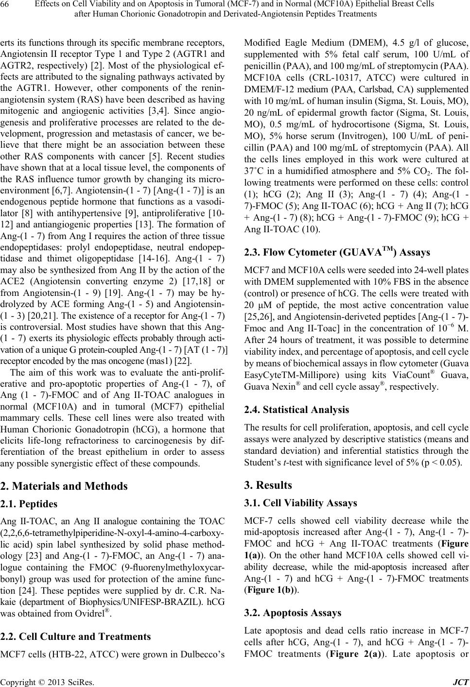
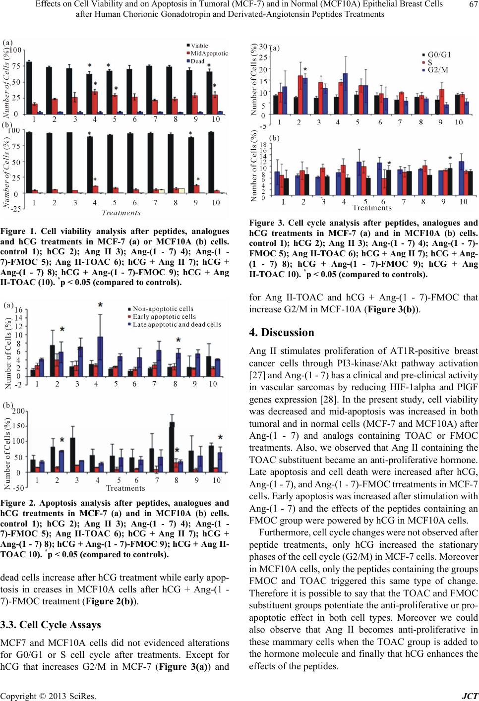
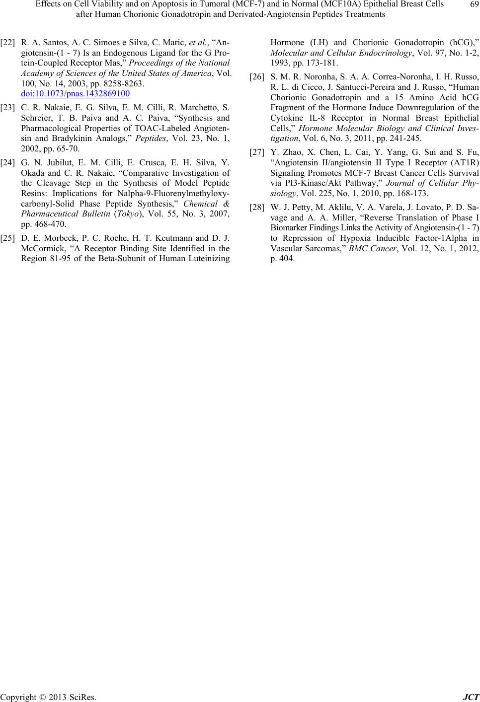
In summary, cell viability was decreased and apoptosis
(initial, mid and late) was increased after hCG and/or
Ang-(1 - 7) peptides treatments. These results point out
hCG and Ang-(1 - 7) as effective compounds to inhibit
cell proliferation, since they decrease cell viability and
increase apoptosis in both normal and tumoral breast cells,
being the effect more pronounced in the tumoral cell line.
Our results support the idea of investigating more closely
the putative use of these compounds as novel therapeutic
agents for breast cancer.
5. Acknowledgements
The work was supported by Grants number 2007 /56480-
0, 2008/54383-0 and 2011/10516-0 from the Sao Paulo
Research Foundation (FAPESP)-Brazil.
REFERENCES
[1] “What Are the Key Statistics about Breast Cancer?” 2013.
http://www.cancer.org/cancer/breastcancer/detailedguide/
breast-cancer-key-statistics
[2] P. M. Frossard, M. J. Malloy, G. G. Lestringant and J. P.
Kane, “Haplotypes of the Human Renin Gene Associated
with Essential Hypertension and Stroke,” Journal of Hu-
man Hypertension, Vol. 15, No. 1, 2001, pp. 49-55.
doi:10.1038/sj.jhh.1001107
[3] M. Fujita, I. Hayashi, S. Yamashina, M. Itoman and M.
Majima, “Blockade of Angiotensin AT1a Receptor Sig-
naling Reduces Tumor Growth, Angiogenesis, and Meta-
stasis,” Biochemical and Biophysical Research Commu-
nications, Vol. 294, No. 2, 2002, pp. 441-447.
doi:10.1016/S0006-291X(02)00496-5
[4] S. Greco, A. Muscella, M. G. Elia, et al., “Angiotensin II
Activates Extracellular Signal Regulated Kinases via Pro-
tein Kinase C and Epidermal Growth Factor Receptor in
Breast Cancer Cells,” Journal of Cellular Physiology,
Vol. 196, No. 2, 2003, pp. 370-377.
doi:10.1002/jcp.10313
[5] F. Deshayes and C. Nahmias, “Angiotensin Receptors: A
New Role in Cancer?” Trends in Endocrinology & Me-
tabolism, Vol. 16, No. 7, 2005, pp. 293-299.
doi:10.1016/j.tem.2005.07.009
[6] E. I. Ager, J. Neo and C. Christophi, “The Rennin-Angio-
Tensin System and Malignancy,” Carcinogenesis, Vol. 29,
No. 9, 2008, pp. 1675-1684. doi:10.1093/carcin/bgn171
[7] F. Deshayes and C. Nahmias, “Angiotensin Receptors: A
New Role in Cancer?” Trends in Endocrinology & Meta-
bolism, Vol. 16, No. 7, 2005, pp. 293-299.
doi:10.1016/j.tem.2005.07.009
[8] R. A. Santos, M. J. Campagnole-Santos and S. P. Andrade,
“Angiotensin-(1 - 7): An Update,” Regulatory Peptides,
Vol. 91, No. 1-3, 2000, pp. 45-62.
doi:10.1016/S0167-0115(00)00138-5
[9] I. F. Benter, D. I. Diz and C. M. Ferrari, “Pressor and
Reflex Sensitivity Is Altered in Spontaneously Hyperten-
sive Rats Treated with Angiotensin-(1 - 7),” Hypertension,
Vol. 26, No. 6, 1995, pp. 1138-1144.
doi:10.1161/01.HYP.26.6.1138
[10] W. B. Strawn, C. M. Ferrario and E. A. Tallant, “Angio-
tensin (1 - 7) Reduces Smooth Muscle Growth after Vas-
cular Injury,” Hypertension, Vol. 33, No. 1, 1999, pp.
207-211. doi:10.1161/01.HYP.33.1.207
[11] E. J. Freeman, G. M. Chisolm, C. M. Ferrario and E. A.
Tallant, “Angiotensin-(1 - 7) Inhibits Vascular Smooth Mus-
cle Cell Growth,” Hypertension, Vol. 28, No. 1, 1996, pp.
104-108. doi:10.1161/01.HYP.28.1.104
[12] E. A. Tallant, C. M. Ferrario and P. E. Gallagher, “An-
giotensin-(1 - 7) Inhibits Growth of Cardiac Myocytes
through Activation of the Mas Receptor,” American Jour-
nal of Physiology—Heart and Circulatory Physiology,
Vol. 289, No. 4, 2005, pp. H1560-H1566.
doi:10.1152/ajpheart.00941.2004
[13] R. D. Machado, R. A. Santos and S. P. Andrade, “Op-
posing Actions of Angiotensins on Angiogenesis,” Life
Sciences, Vol. 66, No. 1, 2000, pp. 67-76.
[14] R. A. Santos, K. B. Brosnihan, D. W. Jacobsen, P. E.
DiCorletto and C. M. Ferrario, “Production of Angio-
tensin-(1 - 7) by Human Vascular Endothelium,” Hyper-
tension, Vol. 19, No. 2, 1992, pp. 56-61.
[15] K. Yamamoto, M. C. Chappell, K. B. Broshinan and C.
M. Ferrario, “In Vivo Metabolism of Angiotensin I by
Neutral Endopeptidase (EC 3.4.24.11) in Spontaneously
Hypertensive Rats,” Hypertension, Vol. 19, No. 6, 1992,
pp. 692-696. doi:10.1161/01.HYP.19.6.692
[16] M. C. Chappell, E. A. Tallant, K. B. Brosnihan and C. M.
Ferrario, “Conversion of Angiotensin I to Angiotensin-(1 - 7)
by Thimet Oligopeptidase (E.C.3.4.24.15) in Vascular
Smooth Muscle Cells,” Journal of Vascular Medicine and
Biology, Vol. 5, No. 4, 1994, pp. 129-137.
[17] S. R. Tipnis, N. M. Hooper, R. Hyde, E. Karran, G. Christie
and A. J. Turner, “A Human Homolog of Angiotensin-
Converting Enzyme. Cloning and Functional Expression
as a Captopril-Insensitive Carboxypeptidase,” The Jour-
nal of Biological Chemistry, Vol. 275, No. 43, 2000, pp.
33238-33243. doi:10.1074/jbc.M002615200
[18] M. A. Crackower, R. Sarao, G. Y. Oudit, C. Yagil, I.
Kozieradzki, S. E. Scanga, et al., “Angiotensin-Convert-
ing Enzyme 2 Is an Essential Regulator of Heart Func-
tion,” Nature, Vol. 417, No. 6891, 2002, pp. 822-828.
doi:10.1038/nature00786
[19] M. Donoghue, F. Hsieh, E. Baronas, K. Godbout, M.
Gosselin, N. Stagliano, et al., “A Novel Angiotensin-Con-
verting Enzyme-Related Carboxypeptidase (ACE2) Con-
verts Angiotensin I to Angiotensin 1-9,” Circulation Re-
search, Vol. 87, No. 5, 2000, pp. E1-E9.
doi:10.1161/01.RES.87.5.e1
[20] M. C. Chappell, N. T. Pirro, A. Sykes and C. M. Ferrario,
“Metabolism of Angiotensin-(1 - 7) by Angiotensin-Con-
verting Enzyme,” Hypertension, Vol. 31, No. 1, 1998, pp.
362-367. doi:10.1161/01.HYP.31.1.362
[21] K. Yamada, S. N. Iyer, M. C. Chappell, D. Ganten and C.
M. Ferrario, “Converting Enzyme Determines the Plasma
Clearance of Angiotensin-(1 - 7),” Hypertension, Vol. 32,
No. 3, 1998, pp. 496-502. doi:10.1161/01.HYP.32.3.496
Copyright © 2013 SciRes. JCT