Journal of Cancer Therapy, 2013, 4, 1082-1084
http://dx.doi.org/10.4236/jct.2013.46123 Published Online August 2013 (http://www.scirp.org/journal/jct)
The Emergence of Rapid Counter Immunostaining in the
Controlled Narrow Excision of Malignant
Melanoma—How We Do It
Jordan Troxel, Grace Brummer, Kristin Cox, S. Ray Peterson
Department of Dermatology, Central Utah Clinic, Provo, USA.
Email: sraypeterson@mac.com
Received May 22nd, 2013; revised June 25th, 2013; accepted July 3rd, 2013
Copyright © 2013 Jordan Troxel et al. This is an open access article distributed under the Creative Commons Attribution License,
which permits unrestricted use, distribution, and reproduction in any medium, provided the original work is properly cited.
ABSTRACT
Mohs Micrographic Surgery (MMS) is widely employed in the treatment of non-melanoma skin cancer and is a pre-
ferred treatment for many cutaneous malignancies, particularly in high risk locations and tumors [1,2]. It has also been
used in the narrow excision of malignant melanoma with local control rates equivalent to standard margins [3]. It has
gained acceptance in the treatment of noninvasive melanoma where standard 0.5 cm margins may be inadequate for
local control [4]. The frozen section processing used in MMS has been assumed by some to be inadequate in assessing
melanocyte populations or residual melanoma within excision margins. This difficulty has likely led to a majority of
surgeons with fellowship training to process margins with slow, permanent hematoxylin and eosin sections (“slow-
mohs”) or to simply resort to standard 0.5, 1.0, or 2.0 cm margins with traditional excision and outside pathology con-
firmation of clear margins. A recent survey of practicing fellowship-trained Mohs surgeons revealed roughly one-third
(35.9%) of Mohs surgeons felt comfortable interpreting MART-1 immunostains, and far fewer were actually perform-
ing immunostains in their labs [5]. Some Mohs surgeons currently refer melanoma to a colleague experienced in proc-
essing and reading melanoma with available rapid immunostaining. The development of rapid immunohistochemistry,
which can be implemented into a traditional frozen section laboratory, has greatly improved the ease of interpreting
margins in the excision of melanoma. Although the process is considerably more complicated than staining with H&E
or Toluidine Blue (T-Blue), it easily falls within the skill-set and equipment of most busy frozen section laboratories.
The additional cost of biologic reagents may be fully recovered by proper billing of immunohistochemical laboratory
work and interpretation of slides.
Keywords: Moh’s Surgery; Malignant Melanoma; Rapid MART-1
1. Introduction
Multiple immunohistochemical staining protocols for the
melanoma antigen recognized by T-cells (MART-1) us-
ing frozen sections have been developed and presented
over the last decade. Historically, melanoma was proc-
essed with en-face frozen H&E staining, or with perma-
nent paraffin sections requiring overnight processing and
multiple days between each Mohs layer. In 2004, Bricca
[6] and associates presented a reproducible 1-hour MART-
1 protocol for melanoma frozen sections. Later, Asadi [7]
and colleagues reported a modified 20-minute protocol.
Currently, the American College of Mohs Surgery has a
suggested modified protocol available to its members
online [8]. The third protocol resembles a combination of
the Bricca and Asadi protocols.
While all three methods produce reliable staining, each
has its respective strengths and weaknesses. The 1-hour
protocol gives consistent dependable results, however the
lab processing time is dramatically longer than that of a
routine section stained with H&E. The 20-minute rapid
protocol requires considerably less time, but demands the
lab technician’s full attention, excluding other casework.
The clarity of the slides produced using this rapid proto-
col also may contain excessive chromogen “chatter” where
much of the melanocytic detail may be lost. The weak-
ness inherent in the Mohs College protocol is that it is
only available to its members, and remains unpublished
in the public domain.
We present an experimental 35-minute MART-1
staining protocol for melanoma frozen sections that
Copyright © 2013 SciRes. JCT
The Emergence of Rapid Counter Immunostaining in the Controlled Narrow
Excision of Malignant MelanomaHow We Do It
1083
combines the best of the previously presented protocols,
while eliminating many of the aforementioned obstacles
(Tables 1 and 2). In addition, we propose the use of
Table 1. Happy medium protocol.
1. Cut thin (2 - 4 µm) sections-minimum two
copies of each piece
2. Mount on a positively charged slide
facilitates better adhesion
3. Air dry-2 minutes—room temperature
4. Heat on a 60˚C hot plate (or equivalent)—3
minutes
5. Fix in Acetone(in a Coplin jar) —2 minutes
6. Air dry-2 minutes—room temperature
Mounting and
Preparation
7. Rehydrate in Tris-Buffered Saline (TBS)
(in a Coplin Jar)—2.5 minutes
1. Add protein blocking agent—3 minutes
2. Shake off, do not rinse
3. Apply “Ready to Use” MART-1 antibody
6 minutes
4. Rinse in Tris Buffered Saline (TBS)—2
minutes
5. Apply Polymer HRP (horse-radish
peroxidase)—6 minutes
6. Rinse in TBS–2 minutes
(during this processing-mix up DAB
Chromogen for step 7)
7. Apply pre-mixed
(1 drop substrate/1 drop solution/1 ml distilled
water) DAB Chromogen—2 minutes
Staining*
8. Rinse in distilled water for 1.5 - 2 minutes
(shorter time = darker chromogen)
1. Dip in T-Blue—10 seconds
2. Rinse with running water-30 - 45 seconds
3. Dip in 95% reagent alcohol-15 seconds
4. Dip in 3 changes of 100% reagent
alcohol—15 seconds each
Counterstain
(with Toulidine Blue)+
5. Dip in 3 changes containing a clearing
agent—15 seconds each
OR
1. Dip in Hematoxylin—2 seconds
2. Rinse with running water-30 - 45 secondsǂ
3. Dip in 95% reagent alcohol—20 seconds
4. Dip in 3 changes of 100% reagent
alcohol—20 seconds each
Counterstain
(with Hematoxylin)
5. Dip in 2 - 3 changes of a clearing agent—20
seconds each
*Steps in this section take place in a humidity chamber-to achieve humidifi-
cation by pouring 90˚C - 100˚C (boiling) water in the chamber beneath the
slides and shutting the lid. +We perform the counterstain in a linear auto-
mated stainercan be hand dipped using the same time increments. ǂIf you
require bluing in the H & E protocol, it will need to be added here.
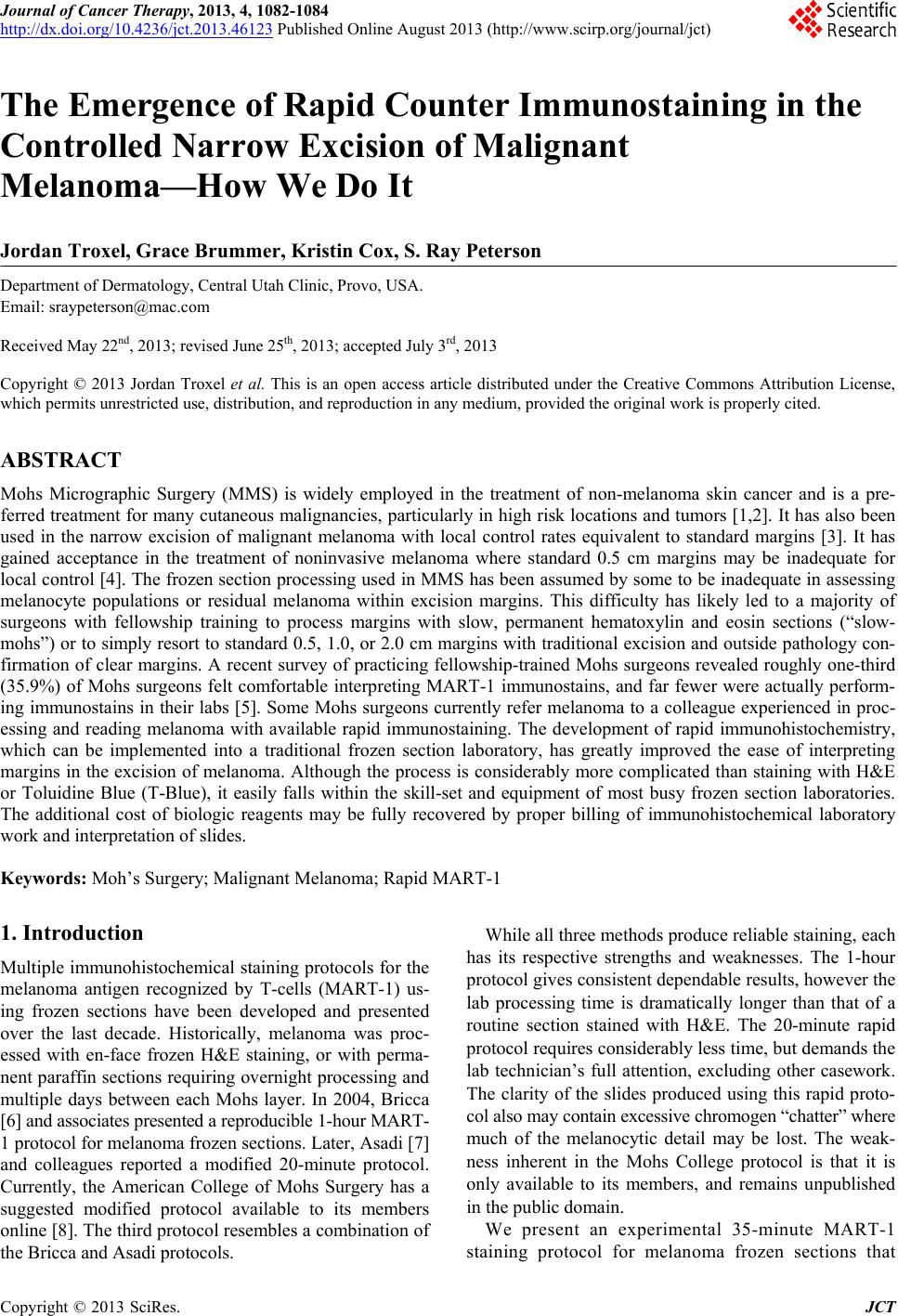
T-Blue as a reasonable alternative to the standard use of
Hematoxylin, as a counterstain, although Hematoxylin
may certainly be used. We have also substituted T-Blue
in all of the published protocols with reproducible stain-
ing compatible with the suggested reagents. Lastly, we
suggest the implementation of an automated stainer dur-
ing the counterstaining process for consistent, reproduci-
ble, easily readable slides. As an added benefit, the use of
the automated stainer unburdens the histo-technician
freeing him/her for additional lab work.
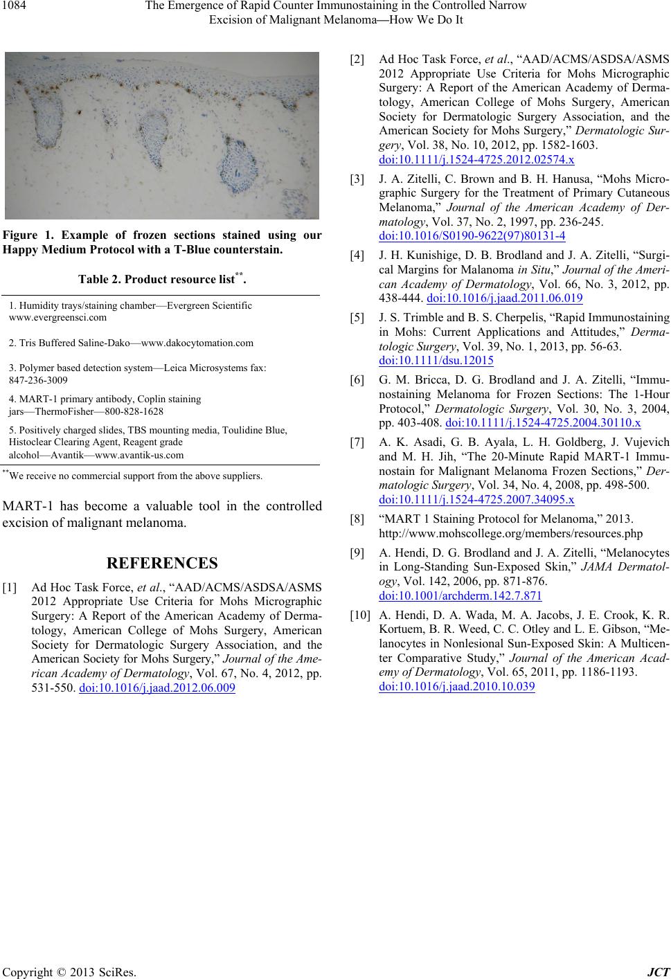
We have also found inexpensive ways to implement
this protocol. In addition to readily available supplies, we
were able to utilize commonly found items in lieu of
more costly lab equipment. Our 60˚C hot plate is a toast-
er oven in combination with a certified thermometer. A
small percolator of boiling water replaced the need for an
additional 100˚C hot plate that is used in the humidifica-
tion process. Likewise, we eliminated the problematic
issue of obtaining negative control tissue, that can be
costly and difficult to store, by utilizing non-melanoma
tissue from cases being treated at the same time as the
melanoma(s). Positive/negative controls are subjected to
the same protocol as the melanoma tissue, replacing the
MART-1 antibody with distilled water for the negative
control. By setting this as our standard operating proce-
dure, we were able to satisfy CLIA requirements for pos-
itive/negative controls.
Lastly, we introduced a linear automated stainer to
perform the counterstain. With our stainer set with T-
Blue, it delivers a pleasing alternative to the traditional
Hematoxylin counterstain. This simple step provides
more opportunity for the histotechnician to continue pro-
cessing other tissue while not sacrificing reproducible
slide quality.
2. Conclusion
Most practices currently performing standard Mohs
processing are well equipped to add rapid MART-1 im-
munostaining regardless of training or experience. Addi-
tional publications have elucidated simple interpretation
of normal melanocytes, atypical melanocytic prolifera-
tions, or malignant melanoma and other cells which will
stain positive with MART-1 [9,10]. Processing mela-
noma with MART-1 staining, due to the complexity and
intensive hands-on nature of the staining process, can
initially be a rate-limiting event, but may quickly become
routine, reproducible, and valuable in the treatment of
melanoma. As most practices have a single histotech,
multi-tasking is essential to the efficiency and flow of the
lab. Finding efficient, reproducible practices are central
to the continued adeptness of the Mohs laboratory. Our
experience with this protocol has produced well-defined,
consistent readable slides in which the melanocytic detail
is markedly distinct (Figure 1). Counterstaining with
Copyright © 2013 SciRes. JCT
The Emergence of Rapid Counter Immunostaining in the Controlled Narrow
Excision of Malignant MelanomaHow We Do It
Copyright © 2013 SciRes. JCT
1084
[2] Ad Hoc Task Force, et al., “AAD/ACMS/ASDSA/ASMS
2012 Appropriate Use Criteria for Mohs Micrographic
Surgery: A Report of the American Academy of Derma-
tology, American College of Mohs Surgery, American
Society for Dermatologic Surgery Association, and the
American Society for Mohs Surgery,” Dermatologic Sur-
gery, Vol. 38, No. 10, 2012, pp. 1582-1603.
doi:10.1111/j.1524-4725.2012.02574.x
[3] J. A. Zitelli, C. Brown and B. H. Hanusa, “Mohs Micro-
graphic Surgery for the Treatment of Primary Cutaneous
Melanoma,” Journal of the American Academy of Der-
matology, Vol. 37, No. 2, 1997, pp. 236-245.
doi:10.1016/S0190-9622(97)80131-4
Figure 1. Example of frozen sections stained using our
Happy Medium Protoc ol with a T-Blue counterstain. [4] J. H. Kunishige, D. B. Brodland and J. A. Zitelli, “Surgi-
cal Margins for Malanoma in Situ,” Journal of the Amer i-
can Academy of Dermatology, Vol. 66, No. 3, 2012, pp.
438-444. doi:10.1016/j.jaad.2011.06.019
Table 2. Product resource list**.
1. Humidity trays/staining chamberEvergreen Scientific
www.evergreensci.com
2. Tris Buffered Saline-Dakowww.dakocytomation.com
3. Polymer based detection systemLeica Microsystems fax:
847-236-3009
4. MART-1 primary antibody, Coplin staining
jarsThermoFisher800-828-1628
5. Positively charged slides, TBS mounting media, Toulidine Blue,
Histoclear Clearing Agent, Reagent grade
alcoholAvantikwww.avantik-us.com
[5] J. S. Trimble and B. S. Cherpelis, “Rapid Immunostaining
in Mohs: Current Applications and Attitudes,” Derma-
tologic Surgery, Vol. 39, No. 1, 2013, pp. 56-63.
doi:10.1111/dsu.12015
[6] G. M. Bricca, D. G. Brodland and J. A. Zitelli, “Immu-
nostaining Melanoma for Frozen Sections: The 1-Hour
Protocol,” Dermatologic Surgery, Vol. 30, No. 3, 2004,
pp. 403-408. doi:10.1111/j.1524-4725.2004.30110.x
[7] A. K. Asadi, G. B. Ayala, L. H. Goldberg, J. Vujevich
and M. H. Jih, “The 20-Minute Rapid MART-1 Immu-
nostain for Malignant Melanoma Frozen Sections,” Der-
matologic Surgery, Vol. 34, No. 4, 2008, pp. 498-500.
doi:10.1111/j.1524-4725.2007.34095.x
**We receive no commercial support from the above suppliers.
MART-1 has become a valuable tool in the controlled
excision of malignant melanoma. [8] “MART 1 Staining Protocol for Melanoma,” 2013.
http://www.mohscollege.org/members/resources.php
[9] A. Hendi, D. G. Brodland and J. A. Zitelli, “Melanocytes
in Long-Standing Sun-Exposed Skin,” JAMA Dermatol-
ogy, Vol. 142, 2006, pp. 871-876.
doi:10.1001/archderm.142.7.871
REFERENCES
[1] Ad Hoc Task Force, et al., “AAD/ACMS/ASDSA/ASMS
2012 Appropriate Use Criteria for Mohs Micrographic
Surgery: A Report of the American Academy of Derma-
tology, American College of Mohs Surgery, American
Society for Dermatologic Surgery Association, and the
American Society for Mohs Surgery,” Journal of the Ame-
rican Academy of Dermatology, Vol. 67, No. 4, 2012, pp.
531-550. doi:10.1016/j.jaad.2012.06.009
[10] A. Hendi, D. A. Wada, M. A. Jacobs, J. E. Crook, K. R.
Kortuem, B. R. Weed, C. C. Otley and L. E. Gibson, “Me-
lanocytes in Nonlesional Sun-Exposed Skin: A Multicen-
ter Comparative Study,” Journal of the American Acad-
emy of Dermatology, Vol. 65, 2011, pp. 1186-1193.
doi:10.1016/j.jaad.2010.10.039