Journal of Cancer Therapy, 2013, 4, 1074-1081
http://dx.doi.org/10.4236/jct.2013.46122 Published Online August 2013 (http://www.scirp.org/journal/jct)
Saporin Conjugated Monoclonal Antibody to the
Transcobalamin Receptor TCblR/CD320 Is Effective in
Targeting and Destroying Cancer Cells
Edward V. Quadros1,2, Yasumi Nakayama1, Jeffrey M. Sequeira1
1Department of Medicine, SUNY-Downstate Medical Center, Brooklyn, USA; 2Department Cell Biology, SUNY-Downstate Medical
Center, Brooklyn, USA.
Email: edward.quadros@downstate.edu
Received May 29th, 2013; revised June 30th, 2013; accepted July 8th, 2013
Copyright © 2013 Edward V. Quadros et al. This is an open access article distributed under the Creative Commons Attribution Li-
cense, which permits unrestricted use, distribution, and reproduction in any medium, provided the original work is properly cited.
ABSTRACT
Cobalamin uptake into cells is mediated by the CD320 receptor for transcobalamin-bound cobalamin. Optimum recep-
tor expression is associated with proliferating cells and therefore, in many cancers this receptor expression is up regu-
lated. Delivering drugs or toxins via this receptor provides increased targeting to cancer cells while minimizing toxicity
to the normal tissues. Saporin conjugated monoclonal antibodies to the extracellular domain of TCblR were effectively
internalized to deliver a toxic dose of Saporin to some cancer cell lines propagating in culture. Antibody concentration
of 2.5 nM was effective in producing optimum inhibition of cell proliferation. The cytotoxic effect of mAb-Saporin
appears to be dictated primarily by the level of receptor expression and therefore normal primary cells expressing low
levels of CD320 were spared while tumor cell lines with higher CD320 expression were destroyed. Targeting the path-
way for cellular uptake of vitamin B12 via the CD320 receptor with toxin-antibody conjugates appears to be a viable
treatment strategy for certain cancers that over expresses this receptor.
Keywords: Transcobalamin Receptor; CD320 Gene; Cobalamin; Cancer; Toxin
1. Introduction
The transcobalamin receptor TCblR/CD320 [1] is ubiq-
uitously expressed in most cell types and mediates up-
take of transcobalamin (TC), a plasma protein saturated
with cobalamin (Cbl, B12). Cbl in blood is bound to two
plasma proteins; haptocorrin (HC) which carries ~70% of
the vitamin and TC which is ~30% saturated with Cbl.
The latter, a nonglycosylated protein secreted by vascular
endothelial cells [2] has a relatively short half life and
plays the all important role of transporting the newly
absorbed Cbl in the distal ileum to all tissues where it
binds to the receptor on the cell surface with high affinity
[3]. TC-Cbl is internalized by receptor-mediated endo-
cytosis of the ligand [4,5]. It is generally accepted that
the TC is degraded in the lysosome and the Cbl is re-
leased [6]. Even though published data supports recy-
cling of the receptor [7,8], unequivocal evidence in sup-
port of this is lacking. TCblR expression appears to be
cell cycle associated with highest expression in actively
proliferating cells and is substantially down regulated in
quiescent cells [7-9]. This differential expression of the
receptor serves to provide optimum delivery of the vita-
min to cells during the early phase of DNA synthesis.
This process ensures adequate functioning of Cbl de-
pendent enzymes, especially the methionine synthase that
is essential for recycling of methyl folate to generate
folates needed for purine and pyrimidine biosynthesis.
The more proliferative a cell, higher the need for folates
and Cbl and this need for Cbl is met by the increased
expression of TCblR in cancer cells that may have in-
herently lost the ability to stop dividing and differentiate.
Selective targeting of cancer cells for destruction by de-
livering drugs and toxins preferentially to these cells has
been the ultimate objective of cancer therapy. The search
for tumor specific markers and the strategies to utilize
these in cancer therapy have been pursued for decades
with mixed results. Success in this approach has been
limiting and can be attributed to multiple factors that
include the lack of specificity of the target antigen, cel-
lular events that can alter this targeting and to the com-
plex and diverse nature of cancer itself. TCblR is struc-
turally related to the LDL receptor family with two
Copyright © 2013 SciRes. JCT
Saporin Conjugated Monoclonal Antibody to the Transcobalamin Receptor
TCblR/CD320 Is Effective in Targeting and Destroying Cancer Cells
1075
LDLR type A domains separated by a CUB like domain
and contains a single transmembrane region followed by
a short cytoplasmic tail [1]. We have generated mono-
clonal antibodies to the recombinant extracellular 200aa
domain of TCblR and have defined the epitope specific-
ity of these antibodies [10]. We have previously demon-
strated the utility of anti TCblR antibody to deliver sec-
ondary antibody conjugated to Saporin [11], an inhibitor
of ribosomal assembly [12] to cancer cells propagating in
culture [13,14]. The present study extends this prelimi-
nary observation by demonstrating enhanced targeting
and destruction of cancer cells by Saporin conjugated di-
rectly to monoclonal antibodies to the extracellular do-
main of TCblR/CD320.
2. Methods
All tumor cell lines obtained from ATCC were cultured
to passage 2 to 4 and frozen for use in this study. MCH
064, MCH 065 and RF peripheral skin fibroblast cultures
in passage 9 - 12 were from The Repository for Mutant
Human Cell Strains, Montreal Children’s Hospital, Can-
ada. Fresh human bone marrow mononuclear cells were
obtained from Lonza, Walkersville, MD. Human chori-
onic trophoblasts, ED cells were obtained as previously
described [15]. Cells were cultured in Dulbecco’s modi-
fied Eagle’s minimal essential medium (DMEM) with
10% fetal bovine serum (FBS) and antibiotics. Mono-
clonal antibodies generated to the extracellular domain of
TCblR [10] were purified by affinity chromatography on
a protein G agarose column and used for covalent conju-
gation of Saporin. The coupling of Saporin and purifica-
tion of monoconjugate of the antibody was done at Ad-
vanced Targeting Systems, San Diego, CA. The anti-
body-Saporin conjugates were stored in aliquots at 20˚C
and used in the present study. All data reported represent
mean results of duplicate analyses.
Determination of optimum concentration of mAb-
Saporin conjugate:
HEK293 cells (2 × 10³ in 100 ul complete DMEM)
were seeded in 96 well culture plates with 0.156 - 5 nM
mAb-Saporin for 72 hours and viable cells were quanti-
fied using the CellTiter96 Aqueous One solution cell
proliferation assay (Promega). Optical density at 495nm
was read in a microplate reader (Bio-Rad). Statistical
analysis was done using GraphPad software Prism 3.03.
Effect of Cell seeding density on efficacy of mAb-
Saporin:
Initial seeding density defines duration of the prolif-
erative phase in the culture and therefore cells seeded at
lower density would continue to divide for a longer pe-
riod compared to cells seeded at a higher density until the
cell population reaches confluency. Since TCblR expres-
sion is highest in actively dividing cells and down-regu-
lated in resting cells, we tested cell lines at seeding den-
sities ranging from 1000 - 10,000 cells/well with 2.5 nM
mAb-Saporin concentration.
Measuring functional receptor on cells:
Functional receptor activity in whole cells was deter-
mined by incubating cells with 57CoB12 labeled TC. For
suspension cultures, cells are collected by centrifugation,
resuspended in DMEM and 0.5 to 1 × 10⁶ cells are sus-
pended in 1 ml DMEM containing 10,000 CPM of radio-
labeled TC at 4˚C, or 37˚C. An identical tube containing
10 mM EGTA is included to show blocking of the uptake.
After 1 hour incubation, cells are pelleted by centrifuga-
tion at 2000 g for 5 min. washed once with buffer and
counted for radioactivity. Adherent cells are seeded in 6
well culture plates, washed with DMEM and incubated
with 57CoB12-TC in 1 ml DMEM for 1 hour. The cell
layer is washed with HBS, incubated with 0.5 ml trypsin/
EDTA for 5min at 37˚C, cells collected with solution and
counted for radioactivity.
Determination for mAb-Saporin concentration for in-
hibiting cell growth by 50% (IC50):
Since the toxic effect of mAb-Saporin was more pro-
nounced in cell cultures seeded at lower density, the
IC50 determinations were done with cells seeded at 2000
cells/well in 96 well plates with a mAb-Saporin concen-
tration of 0.156 to 5 nM for 96 hours. The number of
viable cells was determined by the MTS assay.
Specificity of TCblR pathway for delivering the mAb-
Saporin toxin:
The specificity of the TCblR-mediated pathway for
uptake and internalization of the mAb-Saporin complex
was determined by adding recombinant soluble extracel-
lular fragment of receptor to the culture medium. The
soluble receptor would compete with the cell surface
receptor for the antibody and this would reduce the Ab-
toxin available for cellular uptake resulting in a decrease
in percent inhibition. For these studies, K562 cells and
both normal and stably transfected HEK293 cells to over
express the receptor were used. Cells were seeded in 96
well plates at 2000 cells/well and the amount of mAb-
Saporin-Ab used was equivalent to the IC50 concentra-
tion.
Specificity of mAb-Saporin for the TCblR receptor A
100 fold excess primary mAb or normal mouse IgG was
added to the incubation medium containing a mAb-
Saporin concentration of 2.5 nM. A decrease in the mAb-
Saporin induced inhibition of cell growth should be ob-
served when excess primary Ab without Saporin is pre-
sent since the ratio of mAb-Saporin to unlabelled mAb
should be lower and this increases the probability of
unlabelled mAb binding to TCblR. The addition of nor-
mal mouse IgG should not result in a decrease in cell-kill
since this cannot bind to TCblR and therefore would not
Copyright © 2013 SciRes. JCT
Saporin Conjugated Monoclonal Antibody to the Transcobalamin Receptor
TCblR/CD320 Is Effective in Targeting and Destroying Cancer Cells
1076
compete with mAb-Saporin for binding to TCblR.
Effect of mAb-Saporin on normal and cancer cells in
culture:
In order to determine any differential effect of the
mAb-Saporin in targeting cancer cells in culture, various
normal and cancer cell lines seeded at 1000, 2000 and
4000 cells/well were exposed to mAb-Saporin at an an-
tibody concentration of 2.5 nM.
Immunohistochemical staining of TCblR on cells:
Cell lines cultured on gelatin-coated cover slips were
fixed in 70% ethanol for 2 hrs at 4˚C and washed in PBS.
Human placental tissue was fixed in paraformaldehyde,
embedded in Paraplast and 5 um sections were used for
immunostaining. Rabbit polyclonal Ab to human TCblR
(RPI) at 1:500 dilution was added for 1 hr, washed and
incubated with biotin conjugated secondary Ab for 1 hr
followed by ABC reagent (Vector Labs) for 30 min and
color development with DAB (Vector Labs).
3. Results
A number of adherent and suspension cell cultures were
propagated in medium containing 0.156 to 5 nM mAb-
Saporin. The effective concentration determined by per-
cent decrease in viable cell number showed maximum
effect at an antibody concentration of 2.5 to 5 nM. An
example of this is shown in Figure 1(a) whereby normal
HEK293 cells (293N) and those stably transfected to
over express TCblR (293OE) showed a dose dependent
effect with maximum inhibition of cell growth between
2.5 and 5 nM mAb-Saporin concentration. In addition, in
293N cells, inhibition of proliferation was influenced by
initial seeding density which influences the rate and du-
ration of proliferation and this effect was abrogated in
293OE cells which constitutively over express the re-
ceptor, independent of the cell cycle. In addition, 293OE
cells were more severely affected compared to 293N
cells due to sustained over expression of TCblR (Figure
1(b)). A mAb concentration of 2.5 nM was chosen for
subsequent studies to ensure adequate extra cellular con-
centration of mAb-Saporin for all cell lines tested. Un-
conjugated Saporin at 10 to 100 fold molar excess had no
effect on cell proliferation (data not shown).
Since initial seeding cell density is likely to affect the
rate of proliferation and time to confluence, we tested a
number of suspension and adherent cancer cell lines at
initial seeding densities of 2000 - 10,000 cells per well in
96 well plates. The effect of mAb-Saporin was least in-
fluenced by the initial seeding density in both suspension
and adherent tumor cells in culture, but the effect on cell
proliferation was more pronounced in suspension cul-
tures (Figure 2). A skin fibroblast cell line (RFP3) and a
trophoblast line (ED) derived from human placenta (15)
served as non-cancerous cell lines and were not affected
or minimally affected (Figure 2).
(a)
(b)
Figure 1. Effect of antibody concentration (a) and seeding
cell density (b) on cell proliferation. Normal HEK 293 cells
(N) and HEK 293 cells transfected to stably over express
TCblR (OE) were tested for inhibition of cell proliferation
during a 72hour exposure to anti TCblRmAb-Saporin con-
jugate in culture. Results shown are mean of duplicate
samples that varied <5%.
Since in normal cells, the receptor expression is cell
cycle assocated and lower seeding density and higher
receptor expression appear to have a greater inhibitory
effect on cell proliferation, the lack of this effect in can-
cer cells was puzzling and therefore, we evaluated the
expression of functional TCblR in a number of tumor and
normal cell lines. Since TCblR expression appears to be
cell cycle dependent, we determined cell surface receptor
expression by TC-Cbl binding at various time points
during a 96 hour culture period. As seen in Table 1,
TCblR expression varied considerably among the various
tumor lines. In some cell lines TCblR expression peaked
between 8 - 48 h and was followed by a substantial de-
crease in TC-Cbl binding by 96h, a profile observed for
Copyright © 2013 SciRes. JCT
Saporin Conjugated Monoclonal Antibody to the Transcobalamin Receptor
TCblR/CD320 Is Effective in Targeting and Destroying Cancer Cells
1077
Figure 2. Effect of initial seeding density on proliferation of
cells in the presence of antibody-Saporin conjugate. Normal
primary cell lines and neoplastic cell lines were seeded at a
density of 1000 - 10,000 cells per well in 96 well plates with
2.5 nM mAb-Saporin conjugate. Results shown are mean of
duplicate samples that varied <5%.
most normal cell lines. In some tumor lines, TC-Cbl was
substantially up regulated initially and did not decrease
to the level seen in normal cells. On the other hand, some
tumor lines expressed moderate level of TCblR through-
out the culture period without the peak and decrease seen
in normal cells. Immunohistochemical visualization of
the receptor showed specific membrane staining of
TCblR (Figure 3). While the level of TCblR expression
is too low to detect in most normal cell lines such as skin
fibroblasts, the expression is adequate to visualize in
human placenta and most tumor cell lines.
Because of the wide range of doubling time and the 8 -
48 h period for peak receptor expression in various tumor
lines, the cytotoxic effect of the mAb-saporin was deter-
mined at three different initial cell densities of 1000,
2000 and 4000 cells and we evaluated three monoclonals
with specificity for different regions of the protein (10).
As shown in Figure 4, typically, lower seeding density
showed higher inhibition of cell growth and the effect
varied with different cell lines. Table 1 provides the
IC50 inhibitory dose for the three mAb-Saporin conju-
Figure 3. Immunohistochemical identification of TCblR
expression in various human tissues. (A) KB epidermoid
carcinoma; (B) HL60 acute promyelocytic leukemia; (C)
U266 myeloma; (D) human placenta, E: U373 glioblas-
toma-as- trocytoma; (F) HEK293 embryonic kidney; (G)
PC3 prostate adenocarcinoma.
Figure 4. Inihibition of cell proliferation in the presence of
mAb-Saporin conjugates. Data shows various tumor cell
lines with differences in susceptibility to the toxin. Results
shown are mean of duplicate samples that varied <5%.
Copyright © 2013 SciRes. JCT
Saporin Conjugated Monoclonal Antibody to the Transcobalamin Receptor
TCblR/CD320 Is Effective in Targeting and Destroying Cancer Cells
Copyright © 2013 SciRes. JCT
1078
Table 1. TCblR expression profile and c oncentration of mAb-Saporin required for inhibiting cell proliferation.
Cbl-TC binding IC50/2000 cells (nM)
Cell Line Source Cell line discription Doublingpg/106 cells 1 - 10
saporin
1 - 19
saporin
1 - 25
saporin
Time hr8 - 18 hr24 hr48 hr72 hr 96 hr
K-562* ATCC CCL-243 chronic myelogenous
leukemia (CML) 24 - 4010 1910 7 3 <0.05 <0.05<0.05
HL-60 ATCC CCL-240 acute promyelocytic leukemia 24 4 6 5 4 3 11 10 10
Jurkat ATCC TIB-152 acute T-cell leukemia 25 - 3520 2115 6 4 2.4 2.5 2.08
U266B1
[U266]* ATCC TIB-196 myeloma plasmacytoma 55 10 107 7 8 0.8 <0.05<0.05
RPMI8226* ATCC CCL-155 plasmacytoma; myeloma,
B lymphocyte 60 - 706 1515 8 7 1.92 1.621.54
NCI-H929
[H929]* ATCC CRL-9068 Plasmacytoma, Myeloma 70 20 2424 3315 1.89 1.421.49
U4937
(U937)*
ATCC
CRL-1593.2 monocytic cell, histocytic lymphoma20 - 489 9 10 3 3 1.87 1.581.64
SW48
[SW-48]* ATCC CCL-231 colorectal adenocarcinoma, epithelial35 20 3718 107 <0.05 1.7 <0.05
SW480
[SW-480] ATCC CCL-228 colorectal adenocarcinoma, epithelial25 - 4819 1512 7 10 3.29 3.382.45
RKO* ATCC CRL-2577 carcinoma colon,epithelial 14 26 2711 8 8 1.71 1.811.52
LoVo ATCC CCL-229 colorectal adenocarcinoma, epithelial36 - 489 7 10 1214 7.81 41.76.94
Caco-2 ATCC HTB-37 colorectal adenocarcinoma 24 - 626 8 7 5 4 3.21 4.812.98
Hep G2 ATCC HB - 8065 hepatocellular carcinoma (epithelial)50 - 6017 149 3 3 10.4 10.44.81
Hep 3B ATCC HB - 8064 hepatocellular carcinoma (epithelial)40 - 5011 1110 9 6 5.95 6.254.03
KB ATCC CCL-17 HeLa Derivatives 30 - 4038 1615 115 4.03 4.812.84
MDA-MB-231 ATCC HTB-26 adenocarcinoma,
mammary gland; breast 50 - 6033 4510 6 8 11.3 17.911.3
MCF7 ATCC HTB-22 adenocarcinoma,
mammary gland; breast 50 12 2821 107 6.25 8.935
HeLa ATCC CCL-2 cervix,adenocarcinoma (epithelial)48 13 1213 9 9 2.11 2.551.84
A431NS ATCC CRL-1555 epidermal carcinoma: skin 80 - 1003 3 4 4 3 6.58 9.624.63
MIA PaCa-2 ATCC CRL-1420 Pancreatic carcinoma 40 22 1111 7 7 5 10.45.2
PC-3* ATCC CRL-1435 prostate adenocarcinoma 50 16 1618 1010 2.19 2.081.81
U-373 MG ATCC HTB-17 identities in question 24 - 4819 8 8 6 7 3.1 3.792.91
HEK-293
Normal ATCC CRL-1573 human embryonic kidney cells 24 - 309 9 9 5 6 4.3 4.1 3.7
EC304
(ECV 304) ATCC CRL-1998 T24 (human bladder cell) derivative48 13 166 5 5 2.91 3.292.5
ED Quadros lab embryonic cells from human placenta48 8 132 2 2 12 14 12
RFP3 Quadros lab human skin fibroblast 18 - 243 132 2 2 30 24 26
2M-125C Lonza 2M-125C human bone marrow MNC Not tested 5 4.8 4.8
Cells for TCblR expression were seeded in 6 well plates at a density of 0.2 × 10⁶ cells/well and Cbl-TC binding was determined by incubating with [57Co]
Cbl-TC (10,000 cpm) for 1hr at 37˚C in 1ml medium. The effect of mAb-Saporin on cells was determined at 72 hr following seeding of 2000 cells/well in a 96
well plate with 0.156 - 5 nM mAb-Saporin and IC50 (concentration for inhibition cell proliferation by 50%) was calculated from the slope of the growth curve.
Asterisk indicates cells most susceptible to mAb-Saporin.
*
Saporin Conjugated Monoclonal Antibody to the Transcobalamin Receptor
TCblR/CD320 Is Effective in Targeting and Destroying Cancer Cells
1079
gates on cell proliferation. All three antibodies were ef-
fective as carriers of Saporin and the average inhibition
observed for a specific cell line was similar for all three
antibodies. However, there were major differences in
susceptibility of different cell lines to the mAb-Saporin
conjugate. On average, most of the suspension cell lines
showed higher inhibition of cell proliferation with K562
and RPMI18266 showing the most inhibition. Among the
adherent cell lines also, the effect varied with RKO,
SW48, HeLa and PC3 showing the most inhibition and
LoVo. Hep G2, Hep 3B, MCF7, MDA-MB-231 and
MIA PaCa-2 cell lines, showing least inhibition. Inhibi-
tion of cell proliferation was more pronounced in sus-
pension cultures than in adherent cultures at all concen-
trations of the Saporin-antibody and cell density tested.
In cell lines with 50% or less inhibition of growth, in-
creasing the antibody concentration did not increase the
inhibition. Based on the above data, the IC 50 for most
tumor cell lines appears to be below 100 pM (Table 1).
The specificity of Saporin conjugated antibodies was
evident from a decrease in inhibition with the addition of
excess unconjugated specific antibody and lack of any
effect with excess normal mouse IgG (Figure 5). The
specificity of receptor targeting by the antibodies was
further confirmed by adding soluble recombinant TCblR
to the incubation, which reduced the effect of the Saporin
conjugated antibodies (Figure 6).
4. Discussion
Our previous study described the utility of monoclonal
antibodies to the extracellular domain of TCblR as effec-
tive carriers of Saporin conjugated secondary antibody
into cells [11]. Encouraged by this preliminary observa-
tion, we have extended the approach to evaluate the effi-
cacy of Saporin directly conjugated to primary antibodies.
Two of the three representative antibodies (TCblRKB1-
10 and TCblRKB 1-19) selected were binding antibodies,
in that binding of these antibodies to their epitopes on
TCblR did not prevent the binding and internalization of
the physiologic ligand, TC-Cbl. The third antibody
(TCblRKB1-25) was a blocking antibody since binding
of this antibody to TCblR, prevented the binding and
cellular uptake of TC-Cbl. The epitope specificity of
these antibodies and properties are described elsewhere
[10]. All three mAb-Saporin conjugates are fully func-
Figure 5. The specificity of mAb-Saporin conjugate for
TCblR. Figure shows excess unconjugated specific mAb
decreases the inhibitory effect of the mAb-Saporin conju-
gate while normal mouse IgG does not (A) & (C). This ef-
fect is less pronounced in suspension cultures (B) and in
cells over expressing TCblR (D). Results shown are mean of
duplicate samples that varied <5%.
Figure 6. The specificity of targeting TCblR by Mab-Saporin conjugate. The recombinant extracellular domain of TCblR
added to culture decreases the inhibitory effect of the mAb-Saporin conjugate in culture. Results shown are mean of dupli-
cate samples that varied <5%.
Copyright © 2013 SciRes. JCT
Saporin Conjugated Monoclonal Antibody to the Transcobalamin Receptor
TCblR/CD320 Is Effective in Targeting and Destroying Cancer Cells
1080
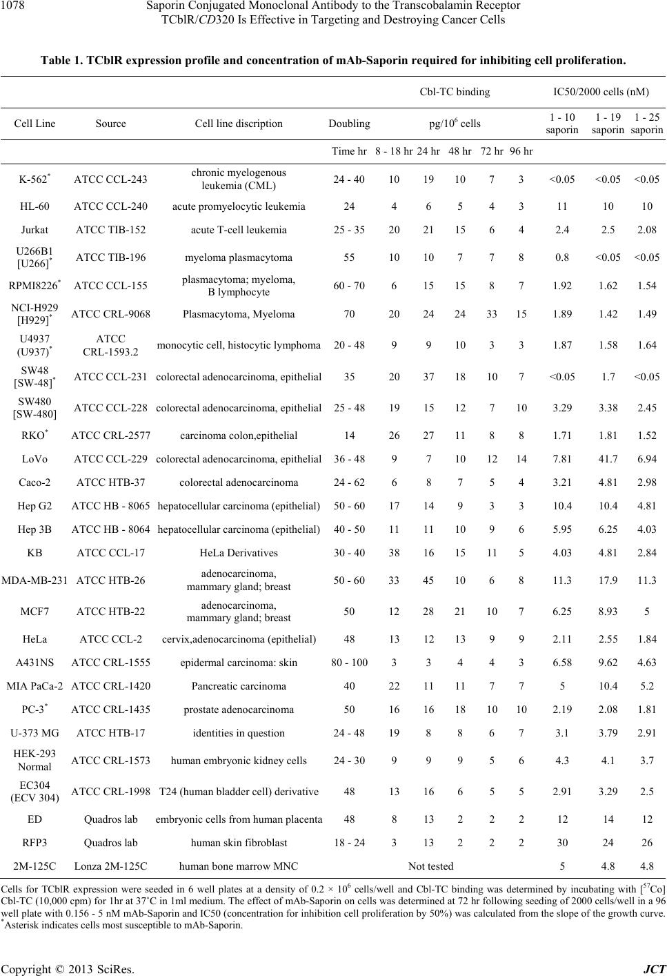
tional in recognizing the target antigen with high affinity,
an important requirement for mAbs to be used as carriers
of drugs and toxins. Even though a marginal increase in
internalization of mAb 1 - 10 and 1 - 19 is observed with
TC-Cbl in the culture medium, the binding of the native
ligand is not a pre requisite for antibody binding and in-
ternalization. Since in the absence of TC-Cbl, the apo re-
ceptor remains on the plasma membrane, TC-Cbl binding
appears to trigger the necessary response for internaliza-
tion. The effective binding and internalization of all an-
tibodies, including the ligand blocking antibody, mAb 1 -
25 is strong evidence that antibody binding triggers a
response similar to that of ligand TC-Cbl binding. Highly
potent toxins such as ricin, cholera toxin, gelonin and
saporin, drugs or radionuclides are very effective in de-
stroying cancer cells if a toxic dose can be delivered spe-
cifically to these cells [16,17]. This strategy requires a
tumor specific carrier to transport the toxin across the
plasma membrane into cells since these molecules cannot
cross the cell membrane by either specific or nonspecific
transport mechanisms. An ideal target protein would be a
receptor or cell surface protein that is expressed and in-
ternalized only in cancer cells. However, such proteins
are scarce and not easy to identify. Many proteins and
receptors are expressed in all cell types and some of
these are cell cycle associated or expressed only in ac-
tively dividing cells. Such proteins can be carriers for
drugs and toxins and may provide some degree of en-
hanced targeting to cancer cells. However selective tar-
geting to cancer cells and lack of toxicity to the normal
cell population will depend not only on the differential
expression but also on the density of the target protein in
the two cell types. For example, a protein with relatively
high expression such as the transferrin receptor [18],
even though differentially expressed in cancer and nor-
mal cells, would not be suitable for delivering a toxin
because the normal cells would internalize sufficient
toxin to kill the cell. The ideal target protein is one with
fairly low expression in normal cells and cannot inter-
nalize toxic amounts of drugs but is adequately over ex-
pressed in cancer cells to internalize cytotoxic amounts
of the drug. The TCblR is one such protein whose ex-
pression is sufficiently low to render any toxin internal-
ized in normal cells to be ineffective and is adequately
over expressed in some cancers to internalize sufficient
toxin to kill the cell. In addition, the cell cycle associated
expression of this protein makes highly proliferative
cancer cells with sustained expression, an ideal target for
this approach. Blocking Cbl uptake into cells with mono-
clonal antibody to TC can deplete cells of Cbl and ulti-
mately inhibit DNA synthesis leading to inhibition of cell
replication [19,20]. A specific antibody to TCblR that
blocks the binding of TC-Cbl could also have the same
effect [10]. This approach, even though less toxic, is
likely to be slow and many cancers require a faster effect
to destroy the malignant tissue before it metastasizes.
Inhibiting CD320 expression with siRNA also affects
proliferation by depleting intracellular Cbl [21]. The use
of potent drugs or toxins conjugated to antibody that can
deliver the payload to its target antigen is a highly effec-
tive strategy that can provide the specificity and speed of
action demanded in cancer therapy. The present data on
the use of mAb-Saporin conjugate to target TCblR ap-
pears to be specific for certain cancers and provides
proof of concept for utilizing this receptor for targeted
delivery of drugs and toxins and awaits confirmation of
in vivo targeting efficacy of this pathway.
5. Acknowledgements
This work was supported by NIH grant DK064732 to
EVQ and by KYTO Biopharma, Toronto, CA. EVQ and
JMS are inventors on patent applications US2011/
052154 and WO/2013/015821 by The Research Founda-
tion of SUNY. YN declared no potential conflicts.
REFERENCES
[1] E. V. Quadros, Y. Nakayama and J. M. Sequeira, “The
Protein and the Gene Encoding the Receptor for the Cel-
lular Uptake of Transcobalamin-Bound Cobalamin,” Blood,
Vol. 113, No. 1, 2009, pp. 186-192.
doi:10.1182/blood-2008-05-158949
[2] E. V. Quadros, S. P. Rothenberg and E. A. Jaffe, “Endo-
thelial Cells from Human Umbilical Vein Secrete Func-
tional Transcobalamin II,” American Journal of Physiol-
ogy, Vol. 256, No. 2, 1989, pp. C296-303.
[3] E. V. Quadros, Y. Nakayama and J. M. Sequeira, “The
Binding Properties of the Human Receptor for the Cellu-
lar Uptake of Vitamin B12,” Biochemical and Biophysi-
cal Research Communications, Vol. 327, No. 4, 2005, pp.
1006-1010. doi:10.1016/j.bbrc.2004.12.103
[4] K. Takahashi, M. Tavassoli and D. W. Jacobsen, “Rece-
ptor Binding and Internalization of Immobilized Transco-
balamin II by Mouse Leukaemia Cells,” Nature, Vol. 288,
No. 5792, 1980, pp. 713-715. doi:10.1038/288713a0
[5] T. Kishimoto, M. Tavassoli, R. Green and D. W. Jacob-
sen, “Receptors for Transferrin and Transcobalamin II
Display Segregated Distribution on Microvilli of Leuke-
mia L1210 Cells,” Biochemical and Biophysical Research
Communications, Vol. 146, No. 3, 1987, pp. 1102-1108.
doi:10.1016/0006-291X(87)90761-3
[6] E. V. Quadros, “Advances in the Understanding of Co-
balamin Assimilation and Metabolism,” British Journal of
Haematology, Vol. 148, No. 2, 2010, pp. 195-204.
doi:10.1111/j.1365-2141.2009.07937.x
[7] J. Lindemans, A. C. Kroes, J. van Geel, J. van Kapel, M.
Schoester, et al., “Uptake of Transcobalamin II-Bound
Cobalamin by HL-60 Cells: Effects of Differentiation In-
Copyright © 2013 SciRes. JCT
Saporin Conjugated Monoclonal Antibody to the Transcobalamin Receptor
TCblR/CD320 Is Effective in Targeting and Destroying Cancer Cells
1081
duction,” Experimental Cell Research, Vol. 184, No. 2,
1989, pp. 449-460. doi:10.1016/0014-4827(89)90343-1
[8] T. Amagasaki, R. Green and D. W. Jacobsen, “Expression
of Transcobalamin II Receptors by Human Leukemia
K562 and HL-60 Cells,” Blood, Vol. 76, No. 7, 1990, pp.
1380-1386.
[9] C. A. Hall, P. D. Colligan and J. A. Begley, “Cyclic Ac-
tivity of the Receptors of Cobalamin Bound to Transco-
balamin II,” Journal of Cellular Physiology, Vol. 133, No.
1, 1987, pp. 187-191. doi:10.1002/jcp.1041330125
[10] W. Jiang, Y. Nakayama, J. M. Sequeira and E. V. Qua-
dros, “Characterizing Monoclonal Antibodies to Antige-
nic Domains of TCblR/CD320, the Receptor for Cellular
Uptake of Transcobalamin-Bound Cobalamin,” Drug De-
livery, Vol. 18, No. 1, 2011, pp. 74-78.
doi:10.3109/10717544.2010.509745
[11] E. V. Quadros, Y. Nakayama and J. M. Sequeira, “Tar-
geted Delivery of Saporin Toxin by Monoclonal Anti-
body to the Transcobalamin Receptor, TCblR/CD320,”
Molecular Cancer Therapeutics, Vol. 9, No. 11, 2010, pp.
3033-3040. doi:10.1158/1535-7163.MCT-10-0513
[12] F. Stirpe, A. Gasperi-Campani, L. Barbieri, A. Falasca, A.
Abbondanza, et al., “Ribosome-Inactivating Proteins from
the Seeds of Saponaria officinalis L. (Soapwort), of Agro-
stemma githago L. (Corn Cockle) and of Asparagus offi-
cinalis L. (Asparagus), and from the Latex of Hura crepi-
tans L. (Sandbox Tree),” Biochemical Journal, Vol. 216,
No. 3, 1983, pp. 617-625.
[13] A. Bolognesi, L. Polito, V. Farini, M. Bortolotti, P. L.
Tazzari, et al., “CD38 as a Target of IB4 mAb Carrying
Saporin-S6: Design of an Immunotoxin for ex Vivo De-
pletion of Hematological CD38+ Neoplasia,” Journal of
Biological Regulators & Homeostatic Agents, Vol. 19,
No. 3-4, 2005, pp. 145-152.
[14] L. Polito, A. Bolognesi, P. L. Tazzari, V. Farini, C. Lu-
belli, et al., “The Conjugate Rituximab/Saporin-S6 Com-
pletely Inhibits Clonogenic Growth of CD20-Expressing
Cells and Produces a Synergistic Toxic Effect with Fluda-
rabine,” Leukemia, Vol. 18, No. 7, 2004, pp. 1215-1222.
doi:10.1038/sj.leu.2403378
[15] D. A. Kniss, Y. Xie, Y. Li, S. Kumar, E. A. Linton, et al.,
“ED(27) Trophoblast-Like Cells Isolated from First-Tri-
mester Chorionic Villi Are Genetically Identical to HeLa
Cells yet Exhibit a Distinct Phenotype,” Placenta, Vol. 23,
No. 1, 2002, pp. 32-43. doi:10.1053/plac.2001.0749
[16] B. A. Teicher and R. V. Chari, “Antibody Conjugate The-
rapeutics: Challenges and Potential,” Clinical Cancer Re-
search, Vol. 17, No. 20, 2011, pp. 6389-6397.
doi:10.1158/1078-0432.CCR-11-1417
[17] M. A. Firer and G. Gellerman, “Targeted Drug Delivery
for Cancer Therapy: The Other Side of Antibodies,” Jour-
nal of Hematology & Oncology, Vol. 5, 2012, p. 70.
doi:10.1186/1756-8722-5-70
[18] R. Ippoliti, E. Lendaro, I. D’Agostino, M. L. Fiani, D.
Guidarini, et al., “A Chimeric Saporin-Transferrin Con-
jugate Compared to Ricin Toxin: Role of the Carrier in
Intracellular Transport and Toxicity,” FASEB Journal,
Vol. 9, No. 12, 1995, pp. 1220-1225.
[19] E. V. Quadros, S. P. Rothenberg and P. McLoughlin,
“Characterization of Monoclonal Antibodies to Epitopes
of Human Transcobalamin II,” Biochemical and Biophy-
sical Research Communications, Vol. 222, No. 1, 1996,
pp. 149-154. doi:10.1006/bbrc.1996.0713
[20] G. R. McLean, E. V. Quadros, S. P. Rothenberg, A. C.
Morgan, J. W. Schrader, et al., “Antibodies to Transco-
balamin II Block in Vitro Proliferation of Leukemic
Cells,” Blood, Vol. 89, No. 1, 1997, pp. 235-242.
[21] S. C. Lai, Y. Nakayama, J. M. Sequeira and E. V. Quadros,
“Down-Regulation of Transcobalamin Receptor TCblR/
CD320 by siRNA Inhibits Cobalamin Uptake and Prolif-
eration of Cells in Culture,” Experimental Cell Re- search,
Vol. 317, No. 11, 2011, pp. 1603-1607.
doi:10.1016/j.yexcr.2011.02.016
Copyright © 2013 SciRes. JCT