Natural Science
Vol.4 No.3(2012), Article ID:18066,4 pages DOI:10.4236/ns.2012.43024
Two diglucosylated anthocyanins from Combretum paniculatum flowers
![]()
1Laboratoire de Chimie Organique et de Physique Appliquées, Département de Chimie, UFR-SEA, Université de Ouagadougou, Ouagadougou, Burkina Faso; *Corresponding Author: eloi_pale@univ-ouaga.bf, eloipale@yahoo.fr
2Laboratoire de Pharmacognosie, de Bromatologie et de Nutrition Humaine, Institut de Pharmacie, Université Libre de Bruxelles, Bruxelles, Belgique
3Laboratoire de RMN Haute Résolution, Département de Chimie, Université Libre de Bruxelles, Bruxelles, Belgique
Received 27 August 2011; revised 29 September 2011; accepted 10 October 2011
Keywords: Combretum paniculatum; diglucosylated anthocyanins; cyanidin; pelargonidin
ABSTRACT
From Combretum paniculatum flowers, two diglucosylated derivatives from cyanidin and pelargonidin were identified using chromatographic (TLC), chemical (degradation by hydrolysis, tests of revelations) and spectral [UV-visible, 1HNMR (1H and 13C, TOCSY-1D, DQF-COSY, NOESY- 2D)] methods. These pigments were found to consist of cyanidin 3,5-O-β-D-diglu-copyranoside and pelargonidin 3,5-O-β-D-diglucopyranoside
1. INTRODUCTION
The plant kingdom contains a wide range of natural substances such as polyphenols, carotenoids, phytosterols, phyto-estrogens, glucosinolates, etc. [1]. Polyphenols which are found in all parts of higher plants (roots, stems, leaves, flowers, pollen, fruits, seeds and wood), are secondary metabolites characterized by the presence of an aromatic ring bearing free hydroxyl groups or with a carbohydrate [2,3]. It is known that Aqueous extracts of inflorescences of Combretum paniculatum studied in this work are known to have an anti-tumor activity of carcinoma of the lung [4].
In this paper, we report for the first time the identification of 3,5-β-O-diglucosides of cyanidin and pelargonidin in red petals of C. paniculatum flowers.
2. METHODS AND MATERIALS
Plant material: belonging to the family of Combretaceae, C. paniculatum is a robust liana reaching at least 15 m high. Loose terminal panicles are formed of dense spikes of flower petals and Red fillets usually appearing during the dry season during defoliation.
Chemicals: ethanol, methanol, hydrochloric acid, Amberlite XAD-7, ascorbic acid, Sephadex LH-20, distilled water, TLC plates (silica gel 60 F254) silica, cellulose gel.
Extraction, purification and isolation of the petals of flowers of C. paniculatum: freshly harvested, the red petals of C. paniculatum flowers are immediately freezedried. 100 g of powder obtained by crushing the petals are extracted by maceration at 5˚C successively with 1000 mL and twice with 200 mL of methanol-hydrochloric acid (99:1) system [5]. The filtrate were pooled and concentrated almost dry vacuum at 30˚C. 50 mL of HCl-H2O (0.5:99) system/MeOH (7:3 mL) are added. The solution was filtered and concentrated at 25 mL. This solution was filtered and fixed on a nonionic polymeric adsorbent (Amberlite XAD-7, Aldrich Chemical Co., Milwaukee, WI) column (length 300 mm, i.d. 20 mm) which was prewashed with 0.5% HCl/H2O; the pigments were then eluted with MeOH/H2O/HCl, 70:30:0.3). The eluate was concentrated, filtered through a Sephadex LH-20 (Pharmacia Biotech, Uppsala, Sweden) column (length 300 mm, i.d. 20 mm). The final purification was achieved by preparative thin-layer chromatography (TLC) on silica gel 60 F254 (Merck-Clevenot Corp.) using EtOAc/HCO2H/AcOH/H2O, 100:11:11:26 (EFAW), as solvent system. The two main bands from this chromatography are numbered from bottom to top. Then to the band which had the small frontal reference is assigned 1 and another 2. The isolated bands of adsorbent were eluted with 0.5% TFA/MeOH, and the solutions were concentrated and filtered through a RP-18 column using (0.5% TFA/H2O)/MeOH (6:4). The eluates were finally concentrated and freeze-dried to give 1 (5 mg) and 2 (10 mg) as TFA salts.
Chemical analysis: Acidic hydrolysis was performed by dissolving 1 mg of each pure compound in 4 mL in hydrochloric acid 1 N (in vials) and placed in a waterbath at 100˚C. Successive samplings carried out between 0 and 60 min were cooled and analyzed by TLC with the solvent system: BAW [Butanol acetic acid/water 4:1:5; upper phase] on a plate of cellulose. Samples at 60 min were cooled and extracted twice by 0.5 mL of 3-methylbutan-2-ol from 1 and 2. These samples at 60 min were analyzed by TLC on cellulose plate with the Forestal [acetic acid/HCl concentrate water (30:3:10)] as mobile phase together with cyanidin and pelargonidin as anthocyanidins standards available.
Electronic spectroscopy: (UV-visible) spectra were obtained with DES 190 double energy system spectro-meter from SAFAS-MONACO, using MeOH-HCl 0.01 N as solution system. About three drops of aluminium chloride (AlCl3) were added to the previous solution to highlight the eventual vicinal hydroxyl free groups.
Nuclear magnetic resonance spectrometry: spectra of compounds were obtained with a VARIAN (600 MHz) spectrometer in CD3OD/TFA_d1 (0.5:0.1 mL) at the Université Libre de Bruxelles (CIREM).
3. RESULTS AND DISCUSSION
UV-visible spectra of 1 and 2 (Table 1) were showed absorption in UV area at 280 nm corresponding to nonacylated anthocyanins. Furthermore, in visible area 1 and 2 were absorbed respectively at 506 and 530 nm. 1 would be a derivative of cyanidin and 2 is probably a derivative of pelargonidin. By the addition of AlCl3, 1 has given red shift effect indicating the presence of vicinal hydroxyl groups on the B ring. But 2 has caused no red shift confirming that it is a derivative of the pelargonidin. The ratio A440/Amax.vis. (respectively 28% and 22%; Table 1) suggested that the 5 position of these compounds was not free [5-7]. The TLC of the products of acidic hydrolysis for each of the two compounds in the Forestal with standards has showed two spots corresponding to cyanidin (1) and the pelargonidin (2). Controlled hydrolyses of compounds 1 and 2 have given each an intermediate spot between the aglycon and anthocyanin indicating that they were diglycosylated.
NMR experiments allowed to assign certainly six signals(in downfields) of 1 in agreement with the cyanidin aglycon. Indeed, it detected clearly to δ (ppm) 9.18 (H4); 8.12 (H2’); 7.10 (H5’); 8.37 (H6’); 7.06 (H8) and 7.08 (H6) (Table 2). Similarly in downfields, 2 has showed five signals in agreement with the pelargonidin aglycon. Indeed, we can also make assignments of protons to δ (ppm) 9.24 (H4); 8.68 (H2’ and H6’); 7.09 (H3’ and H5’); 7.13 (H6); 7.10 (H8) (Table 2).
All sugars protons appear between 3 and 5.5 ppm. However, two doublets corresponding to anomeric protons of glucoses A and B were clearly observed at 5.31 and 5.18 ppm for 1 (5.31 and 5.19 ppm for 2) respectively, are characteristic of the protons on 3 and 5 positions respectively [5,6] (Table 2). Their β-D configurations of each are confirmed by the high values of their coupling constants (J ≈ 7 - 8 Hz) [6]. TOCSY spectra obtained by selective excitation protons H1” of the glucose allowed to confirm the membership of each methylenic proton of glucoses A and B of 1 and 2. To determine connection positions of sugars, correlation NOESY 2-D experiments were performed. The correlation between H1”A (5.31 ppm) and the aglycon H4 indicated that glucose A is bound in 3 position. Similarly, irradiation of H1”B signal (5.19 ppm) reveals a proximity with the aglycon H6 (downfield shift of 1H NMR signal of H-6 protons in both anthocyanins), confirming that glucose B is attached in 5 position.
Spectra DQF-COSY proton coupling constants allowed assignments of protons of glucoses A and B. Indeed, anomeric proton 5.31 ppm (H1’’A) is nearby of a proton to 3.72 ppm (H2’’A) which is linked to H3’’A appearing at 3.65 ppm. This triplet (with a coupling constant about 9.6 Hz) is connected to an another triplet H4’’A appearing at 3.43 ppm. The last one is connected to H5’’A at 3.56 ppm. Signal at 4.01 ppm with a wide coupling con-
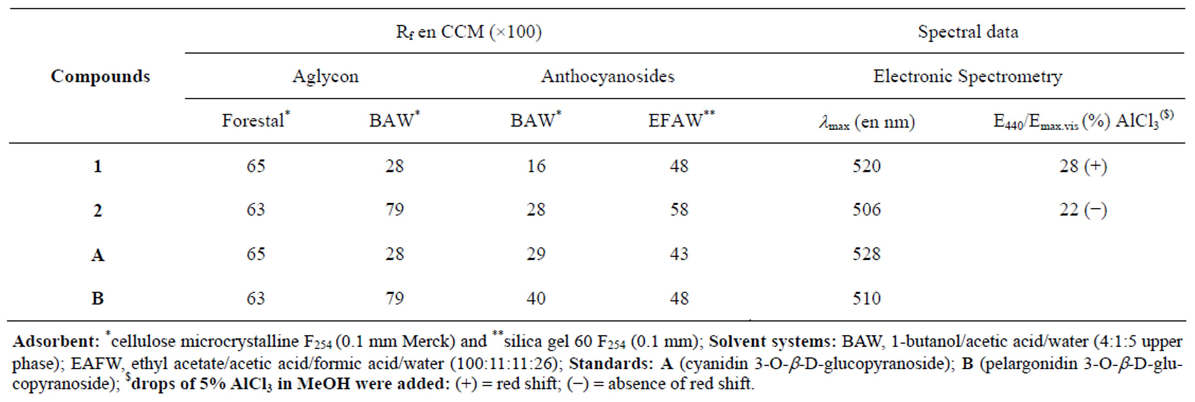
Table 1. Chromatographic and UV-Vis absorption data of anthocyanins 1 and 2 from C. paniculatum.
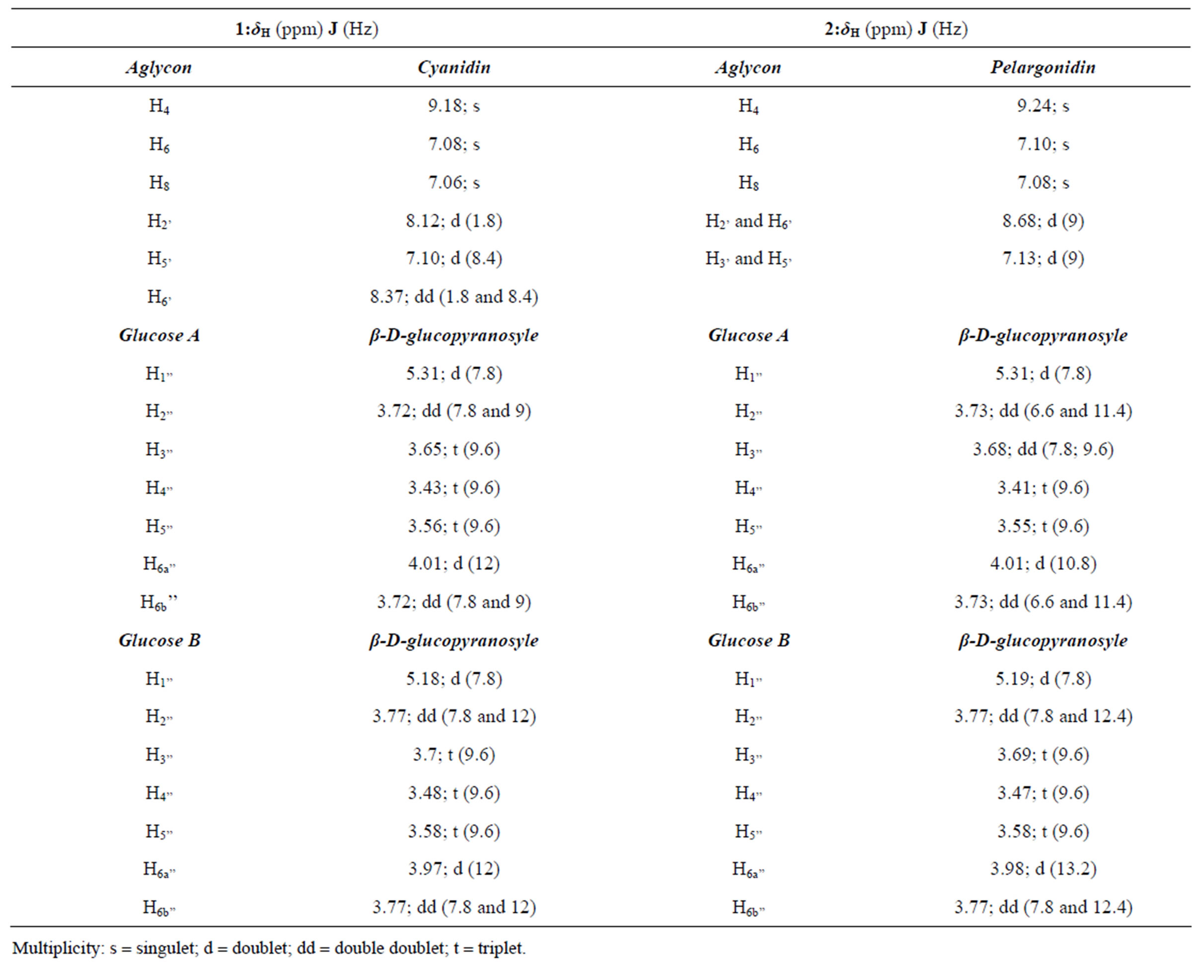
Table 2. 1H-NMR Spectral data of anthocyanins 1 and 2 [δ in CD3OD/TFA-d1 (5:1)].
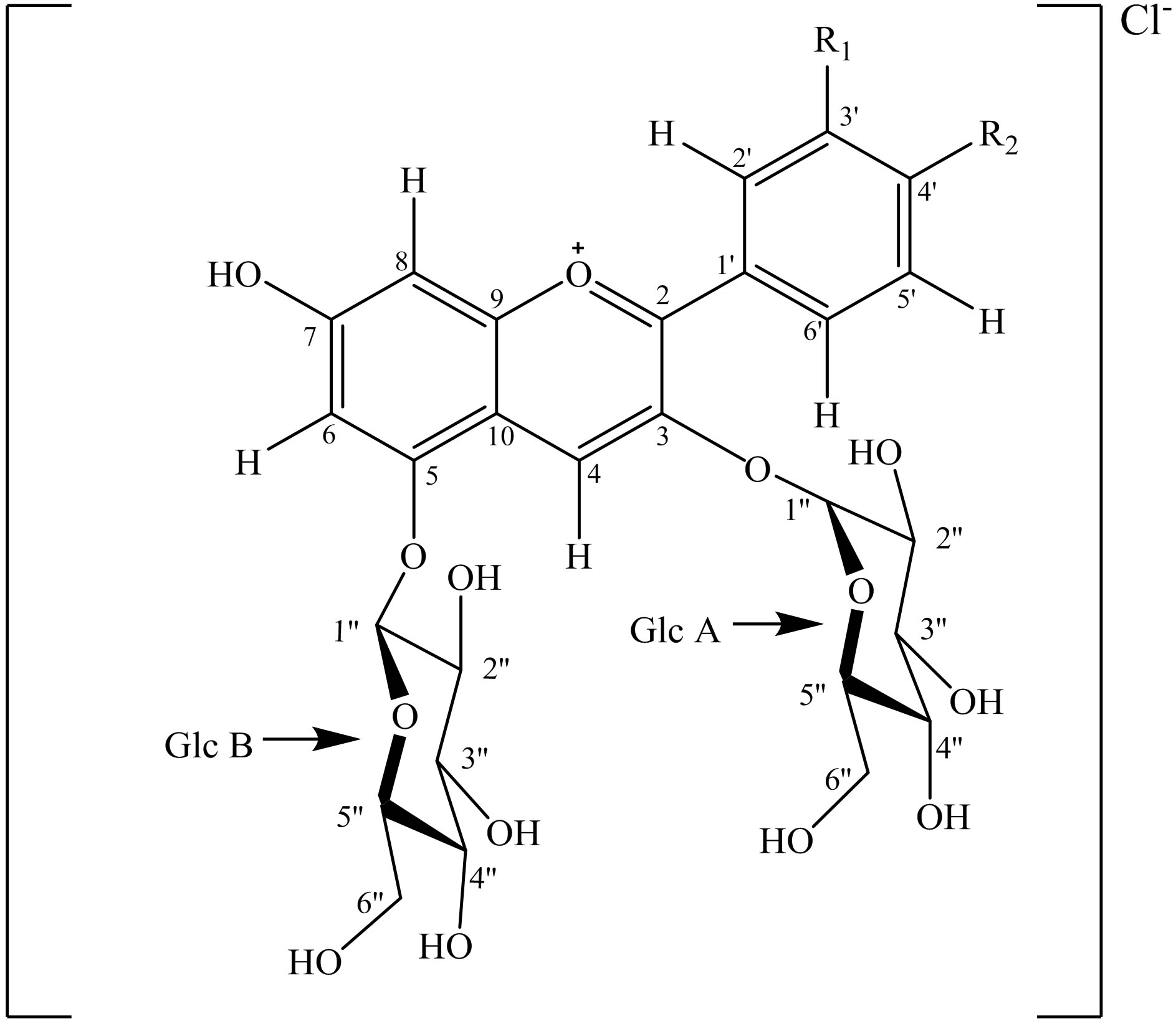
Figure 1. Complete structures of anthocyanins 1 and 2. Glc A: Glucose A; Glc B: Glucose B; 1: R1 = R2 = OH; 2: R1 = H and R2 = OH.
3.56 ppm. Signal at 4.01 ppm with a wide coupling constant (12 Hz) reveals a geminal coupling between two methylenic protons on 6’’ position. It characterized H6’’bA; the other proton (H6’’aA) also appears at 3.72 ppm.
By a similar reasoning based on spectra DQF-COSY 2, the glucoses A and B into 2 are identical to those detected in 1. Thus, isolated compounds from C. paniculatum flowers are identified as respectively as cyanidin and pelargonidin 3,5-O-β-D-diglucopyranoside (Figure 1).
4. ACKNOWLEDGEMENTS
The authors thanks the “Université Libre de Bruxelles”, Belgium and the University of Ouagadougou for their financial and technical supports.
REFERENCES
- Malvy, D. (2009) Antioxydants et alimentation. INSERM U330, Université Victor Segalen Bordeaux II, consulted on the 28th, December 2009. http://www.i-dietetique.com/?action=breves&id=4993
- Espinosa-Alonso, L.G., Lygin, A., Widholm, J.M., Valverde, M.E. and Paredes-Lopez, O.J. (2006). Polyphenols in wild and weedy mexican common beans (Phaseolus vulgaris L.). Journal of Agricultural and Food Chemistry, 54, 4436-4444. doi:10.1021/jf060185e
- Salinas-Moreno, Rojas-Herrera, Y., Sosa-Montes, L. and Pérez-Herrera, E. (2005). Anthocyanin composition in black bean varieties grown in Mexico. Agrociencia, 39, 385-394.
- Abbot B.J., Leiter, J., Hartwel, L., Caldwell, M.E., Beal, J.L., Perdue, R.E. and Schepartz, J.S.A. (1966). Screening data from the cancer chemotherapy national service center screening laboratories. Plant extracts. Cancer Research, 27, 364-527.
- Palé, E., Kouda-Bonafos, M., Nacro, M., Vanhaelen, M. and Vanhaelen-Fastré, R. (2003). Two triacylated and tetraglucosylated anthocyanins from Ipomoea asarifolia flowers. Phytochemistry, 64, 1395-1399. doi:10.1016/j.phytochem.2003.08.011
- Palé, E. (1998) Contribution à l’étude des composés anthocyaniques des plantes: Cas de Hibiscus sabdariffa, Lannea microcarpa, Vigna subterranea et Sorghum caudatum du Burkina Faso. Ph.D. Thesis, Université de Ouagadougou, Ouagadougou.
- Palé, E., Kouda-Bonafos, M., Nacro, M., Vanhaelen, M., and Vanhaelen-Fastré, R. (1997). Anthocyanins from Bambara Groundnut (Vigna subterranea). Journal of Agricultural and Food Chemistry, 45, 3359-3361. doi:10.1021/jf960897c