American Journal of Analytical Chemistry
Vol.3 No.11(2012), Article ID:24468,10 pages DOI:10.4236/ajac.2012.311097
Bioaccumulation of Some Heavy Metals in Fish Samples from River Benue in Vinikilang, Adamawa State, Nigeria
1Department of Chemistry, University of Maiduguri, Maiduguri, Nigeria
2Department of Chemistry, College of Education, Hong, Nigeria
3Department of Chemistry, College of Education, Waka Biu, Nigeria
Email: *joechemakan@yahoo.com
Received September 12, 2012; revised October 17, 2012; accepted October 25, 2012
Keywords: Bioaccumulation; Heavy Metals; Fish; Vinikilang; River Benue
ABSTRACT
This study was aim to determined the levels of some heavy metals in the gills, liver, stomach, kidney, bones and flesh of four fish species (Tilapia zilli, Clarias anguillaris, Synodentis budgetti and Oreochronmis niloticus) collected at River Benue in Vinikilang, Adamawa State, Nigeria for analysis of Cu, Zn, Co, Mn, Fe, Cr, Cd, Ni and Pb. These metals were chosen because at higher concentrations there might be toxic to the fish and by extension humans that depends on such fish as food. The concentrations of the metals were carried out using Flame Atomic Absorption Spectrophotometer (AAS, Unicam 969). Large differences in trace metal concentrations were observed between different tissues within each fish. The highest concentration of Fe (12.65 µg/g) was recorded in gill of Synodentis budgetti, while the lowest value of 0.68 µg/g was recorded in the flesh of Oreochronmis niloticus. The liver of Synodentis budgetti accumulates significant higher levels of Mn and Cd than other species; Fe and Zn was highest in the stomach of Tilapia zilli, while Clarias angullaris shows more of Cr, Pb, Cd and Co. The stomach of Synodentis budgetti accumulate significant higher levels of Fe than other species; Zn was highest in the stomach of Tilapia zilli, while Clarias angullaris shows more of Mn, Cr, Cu, Cd and Pb. Similarly, the bone of Synodentis budgetti accumulates significant higher levels of Mn and Cd than other species; Zn and Fe were highest in the bone of Tilapia zilli, while Clarias angullaris shows more of Cr, Pb, Ni, and Co. The highest levels of Fe (12.65 µg/g) observed in this study was recorded in the gill of Synodentis budgetti and it was below the high residue concentrations of Fe (34 - 107 ppm) in fish samples. Based on the above results, it can therefore be concluded that metals bioaccumulation in the entire fish species study did not exceeds the permissible limits set for heavy metals by FAO, FEPA and WHO.
1. Introduction
The bioaccumulation of heavy metals in living organisms and biomagnifications describes the processes and pathways of pollutants from one trophic level to another. Various species fish are mostly used as bio-indicators of heavy metals contamination [1]. The acidic conditions of aquatic environment might cause free divalent ions of many heavy metals to be absorbed by fish gills [2]. The concentrations of heavy metals in organs of fish show that the aquatic environment is polluted [3]. Heavy meals concentrations in the aquatic organism depict the past as well as the current pollution load in the environment in which the organism lives [4].
Pollution of the aquatic environment by inorganic chemicals has been considered a major threat to the aquatic organisms including fishes. The agricultural drainage water containing pesticides and fertilizers and effluents of industrial activities and runoffs in addition to sewage effluents supply the water bodies and sediment with huge quantities of inorganic anions and heavy metals [5]. Heavy metal can be incorporated into food chains and absorbed by aquatic organisms to a level that might affects their physiological state. Of the effective pollutants are the heavy metals which have drastic environmental impact on all organisms. Trace metals such as Zn, Cu and Fe play a biochemical role in the life processes of all aquatic plants and animals; therefore, they are essential in the aquatic environment in trace amounts. The results of many field studies of metal accumulation in fish living in polluted waters show that considerable amounts of various metals may be deposited in fish tissues without causing mortality. Various metals are accumulated in fish body in different amounts. These differences result from different affinity of metals to fish tissues, different uptake, deposition and excretion rates. Metals in natural waters occur in particulate or soluble form. Soluble species include labile and non-labile fractions. The labile metal compounds are the most dangerous to fish. They include various ionic forms of different to fish. Many data show that the amounts of metals in the labile fraction, and the share of various metal ions strongly depend on environmental conditions. Water temperature may cause the differences in metal deposition in various organs. Higher temperatures promote accumulation of cadmium especially in the most burdened organs: kidneys and liver [6]. Increased accumulation of metals by fish at higher temperatures probably results from higher metabolic rate, including higher rate of metal uptake and binding. Many data indicate that water acidification directly affects metal accumulation rates by the fish. Comparison of the data concerning metal levels in fish from various lakes indicates that the concentrations of cadmium and lead, but not zinc, are considerably higher in the fish from acidified lakes [7-10]. Accumulation of copper is also higher at lower pH [11]. Water hardness (mainly calcium concentration) considerably affects uptake of metals across the gill epithelium. According to [12], enrichment of water with calcium reduced copper accumulation in the gills. [13] reported that elevated dietary Ca2+ protected against both, dietary and waterborne Cd uptake. The results obtained by [14] indicate that calcium reduces zinc uptake by Oncorhynchus mykiss. Various species of fish from the same water body may accumulate different amounts of metals. Interspecies differences in metal accumulation may be related to living and feeding habits. [15] observed that predatory fish species accumulated more mercury but the benthivores contained more cadmium and zinc. Higher concentrations of mercury in the predatory fishes comparing to the non-predatory ones was also reported by [16]. [17] found that lead and zinc concentrations were higher in benthic fish. The results obtained by [18] indicate that predators accumulated more zinc and nickel than benthivores, while the latter contained more cadmium.
Fishes are most important organisms in the aquatic food chain, which are sensitive to heavy metals contamination. Most of the freshwater fishes are confined to specific microhabitat within inter connected river/stream system. If such system becomes contaminated by heavy metals, fish species either shift to less polluted segment of river/stream system or die off which ultimately disturb the food chains [19]. High level of heavy metals has apparent lethal and chronic effects on fishes [20]. Thus, fish not only indicates the pollution status of aquatic ecosystem but have significant impact on the food web [21]. It is one of the main sources of protein-enriched food all over the world [22]. Consumption of contaminated fish with heavy metals can result hazardous effects on human health [23]. Various pathways of metal accumulation in fish include such as ingestion of food, suspended particulate matter, metal ion exchange through gills and skin [24]. [24] also identified five routes through which heavy metals enter into fish viz; food, suspended particle, gills, intake of water and integuments. From these pathways, metals get absorbed into blood and transported to various organs for either storage or excretion. Level of trace metals in different organs of fish is used as an index of metal pollution in an ecosystem, which is considered as an important tool to highlight the role of elevated level of metals in aquatic organisms [25]. Concentration of heavy metals in different tissues/organs of fishes is directly influenced by contamination in aquatic environment, uptake, regulation and elimination inside the fish body [24]. Liver stores either heavy metals or excretes through bile. Other routes of heavy metal regulation are either kidneys or gills [24]. Accumulation of metals in various organs and tissues depends upon the way of exposure such as through diet or their elevated level in surrounding environment [24,26]. Morphological and behavioural abnormalities such as alteration in sensory reception, reduced responses to normal olfactory function (feeding, mating, selection or homing), reduction in swimming performance, gills purge, ventilation, coughs, learning impairment, loss of equilibrium that lapsed into paralysis, loss of reproductive efficiency and irregular metamorphosis appeared as symptom of toxic exposure of trace metals [22]. Concentration of metals becomes toxic to the fish when its level exceeds the permissible level [22].This threshold limit not only varies from metal to metal but also from one species to another [22]. Toxic effects of metals become more pronounced when various metabolic activities inside organism body fail to detoxify [24]. Heavy metals exhibit different accumulation pattern in organs [24]. Gills, liver and kidneys accumulate heavy metals in higher concentration in comparison to muscles, which exhibit lowest levels of metals accumulation [27]. Among different organs, liver accumulates higher concentrations of metals comparatively and has been used widely to investigate the process of bioaccumulation. Kidneys also play a vital role in excretion of trace metal ions [27]. Exchange of gases and absorption of heavy metals takes place from external aquatic to internal body environment through gills [27].
River Benue in Vinikilang, Adamawa State, received a wide variety of waste from agricultural activity within the Vinikilang area. The river is one of the main fish supply sources for this area. Most farmers within the Vinikilang area of Benue State use fertilizers and synthetic chemical pesticides to control pests on vegetables including a number of highly persistent organochlorine and organophosphurus pesticides. Pesticides are extensively used in agricultural production to check or control pestsdiseases weeds and other plant pathogens in an effort to reduce or eliminate yield losses and preserve high product quality. Lack of knowledge of the use and the effects of these pesticides and other agrochemicals among small and large scale farmers within this area of study has resulted in their misuse and consequently the waste generated flows into river Benue and may contaminate the river with a variety of heavy metals acting as point sources. Such contaminations might accumulation in the various organs of fishes; and such accumulation may affect humans and other species that depend on such fish as food. So the need for this study.
2. Materials and Methods
2.1. Sampling Area and Sample Collection
Sampling was from the River Benue in Vinikilang Area of Adamawa State, Nigeria. Fish samples (Tilapia zilli, Clarias anguillaris, Synodentis budgetti and Oreochronmis niloticus, were caught using gill nets from River Benue in Vinikilang Adamawa State, Nigeria; Fish samples of uniform size were collected in order to avoid the possible error due to size differences. The fish were labelled with an identification number. Samples of fishes were transported to the laboratory on the same day for Identified and dissection to remove the bone, liver, stomach, gill, flesh and kidney of each species of fish by an expert in the department of fisheries, University of Maiduguri, Nigeria.
2.2. Digestion of Fish Samples for Heavy Metal Determination
The bone, liver, stomach, gill, flesh and kidney of each fish samples (8.0 g) were dried at 105˚C until they reach a constant weight. Each dried sample was ground, using porcelain mortar and a pestle. The ground fish tissues were transferred to a porcelain basin and put into a Thermicon P muffle furnace at a temperature of 550˚C for 4 hrs. Samples were digested with tri-acid mixture (HNO3: HClO4·H2SO4 = 10:4:1) at a rate of 5 mL/per 0.5 g of sample and were placed on a hot plate at 100˚C temperature. Digestion was continued until the liquor becomes clear. All the digested liquors were filtered through Whatmann 541 filter paper and diluted to 25 mL with distilled water of the element in the sample solution times 20 as additional factor in µg/g dry weight. Determination of Cu, Zn, Co, Mn, Fe, Cr, Cd As, Ni and Pb were made directly on each final solution using PerkinElmer AAnalyst 300 Atomic Absorption Spectroscopy (AAS).
2.3. Calibration Solution
Standard solution of each sample Cu, Zn, Co, Mn, Fe, Cr, Cd, Ni and Pb were prepared according to Sc 2000 manufacturer procedure for Atomic absorption spectroscopy to be used. A known 1000 mg/l concentration of the metal solution was prepared from their salts.
2.4 Data Analysis
Data collected were subjected to one-way analysis of variance (ANOVA), and were used to assess whether samples varied significantly between species, possibilities less than 0.05 (p < 0.05) were be considered statisticcally significant.
3. Results and Discussion
3.1. Concentrations of Heavy Metals in Fish Samples
Figure 1 present the levels of heavy metals in the tissues of Tillabia zilli. Levels of Fe ranged from 1.08 to 9.23 µg/g; 0.33 to 3.45 µg/g Zn; 0.11 to 0.44 µg/g Mn; 0.05 to 0.32 µg/g Cr; 0.12 to 0.39 µg/g Cu; 0.11 to 0.96 µg/g Cd; 0.16 to 0.31 µg/g Pb; 0.11 to 0.69 µg/g Ni; 0.15 to 0.82 µg/g Co. The metal bioaccumulation in these tissue of Tilapia zilli are in the decreasing order of Fe > Zn > Cd > Co > Ni > Mn > Cu > Cr > Pb. The order of bioaccumulations of these metals might be as a result of the fact that different metals tend to accumulate differently in the tissues of different species of fish. In this study Fe was highest next is Zn, while Pb shows the least value.
The levels of heavy metals in the organs of Clarias Anguillaris are as presented in Figure 2. Fe ranged from 0.98 to 8.88 µg/g; 0.06 to 0.44 µg/g Zn; 0.14 to 0.38 µg/g Mn; 0.22 to 0.93 µg/g Cr; 0.08 to 0.29 µg/g Cu; 0.11 to 0.76 µg/g Cd; 0.13 to 0.45 µg/g Pb; 0.23 to 0.73 µg/g Ni; 0.26 to 0.89 µg/g Co. The order of metal bioaccumulation in these tissue are Fe > Cr >Co> Cd > Ni > Pb > Zn > Mn > Cu. The levels of heavy metals in the tissues of Synodentis budgetti is as presented in Figure 3. The levels of Fe ranged from 0.1.86 to 12.65 µg/g; 0.23 to 2.86 µg/g Zn; 0.21 to 2.33 µg/g Mn; 0.19 to 0.57 µg/g Cr; 0.11 to 0.31 µg/g Cu; 0.13 to 1.03 µg/g Cd; 0.04 to 0.38 µg/g Pb; 0.12 to 0.78 µg/g Ni; 0.08 to 0.34 µg/g Co. Figure 4 shows the concentrations of heavy metals in different tissues of Oreochronmis niloticus. Fe levels ranged from 0.68 to 8.92 µg/g; 0.08 to 0.21 µg/g Zn; 0.11 to 0.38 µg/g Mn; 0.33 to 0.85 µg/g Cr; 0.14 to 0.38 µg/g Cu; 0.18 to 0.85 µg/g Cd; 0.12 to 0.61 µg/g Pb; 0.23 to 0.95 µg/g Ni; 0.06 to 0.48 µg/g Co.
3.2. Comparison of Heavy Metals among Species of Fish
The comparison in the concentrations of heavy metals in the gills samples among the four species of fish are as presented in Figure 5. Fe ranged from 5.33 to 12.65 µg/g; 0.444 to 3.45 µg/g Zn; 0.44 to 2.33 µg/g Mn; 0.32 to

Figure 1. Mean concentrations of heavy metals in the gills, liver, stomach, kidney, bons and flesh of Tilapia zilli from River Benue in Vinikilang, Adamawa State, Nigeria.
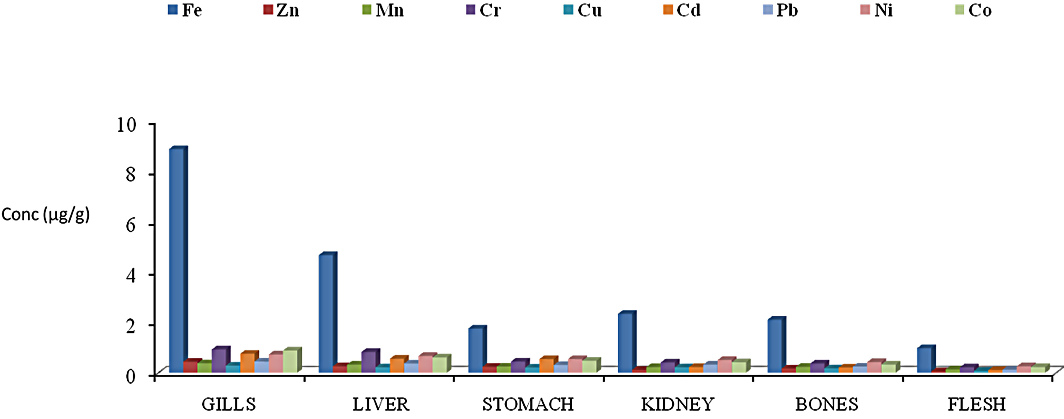
Figure 2. Mean concentrations of heavy metals in the gills, liver, stomach, kidney, bons and flesh of Clarias anaguillaris from River Benue in Vinikilang, Adamawa State, Nigeria.
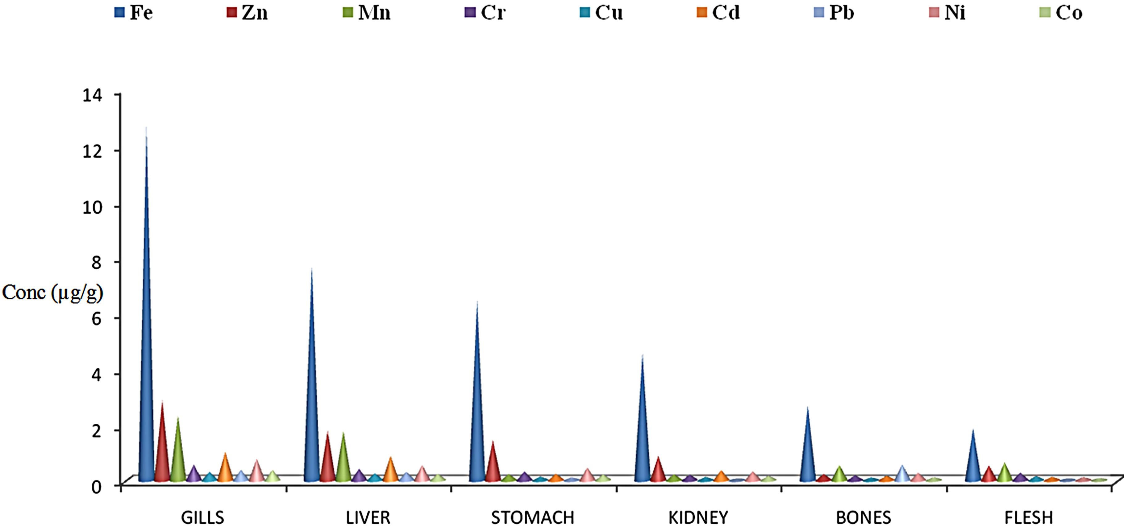
Figure 3. Mean concentrations of heavy metals in the gills, liver, stomach, kidney, bons and flesh of Synodentis budgetti from River Benue in Vinikilang, Adamawa State, Nigeria.
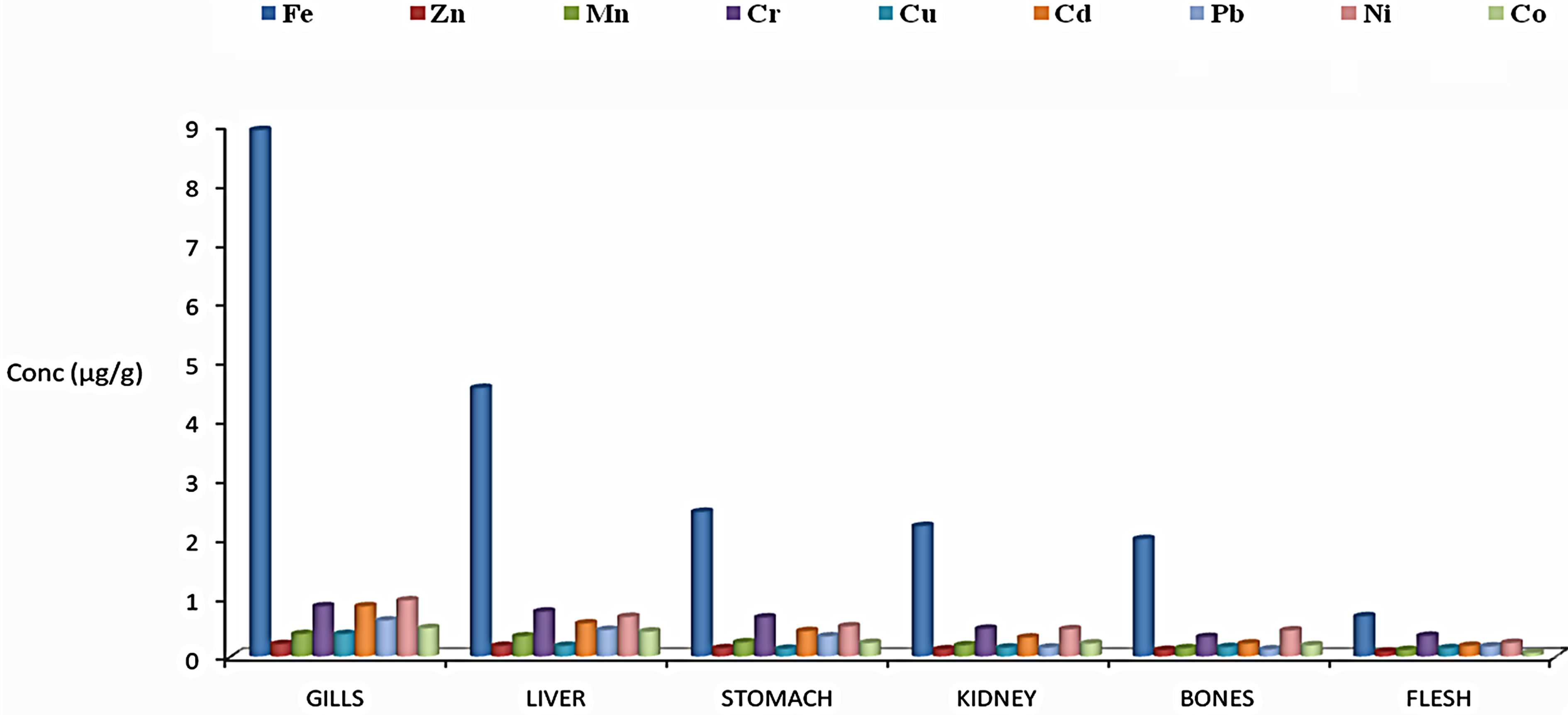
Figure 4. Mean concentrations of heavy metals in the gills, liver, stomach, kidney, bons and flesh of Oreochromis niloticus from River Benue in Vinikilang, Adamawa State, Nigeria.
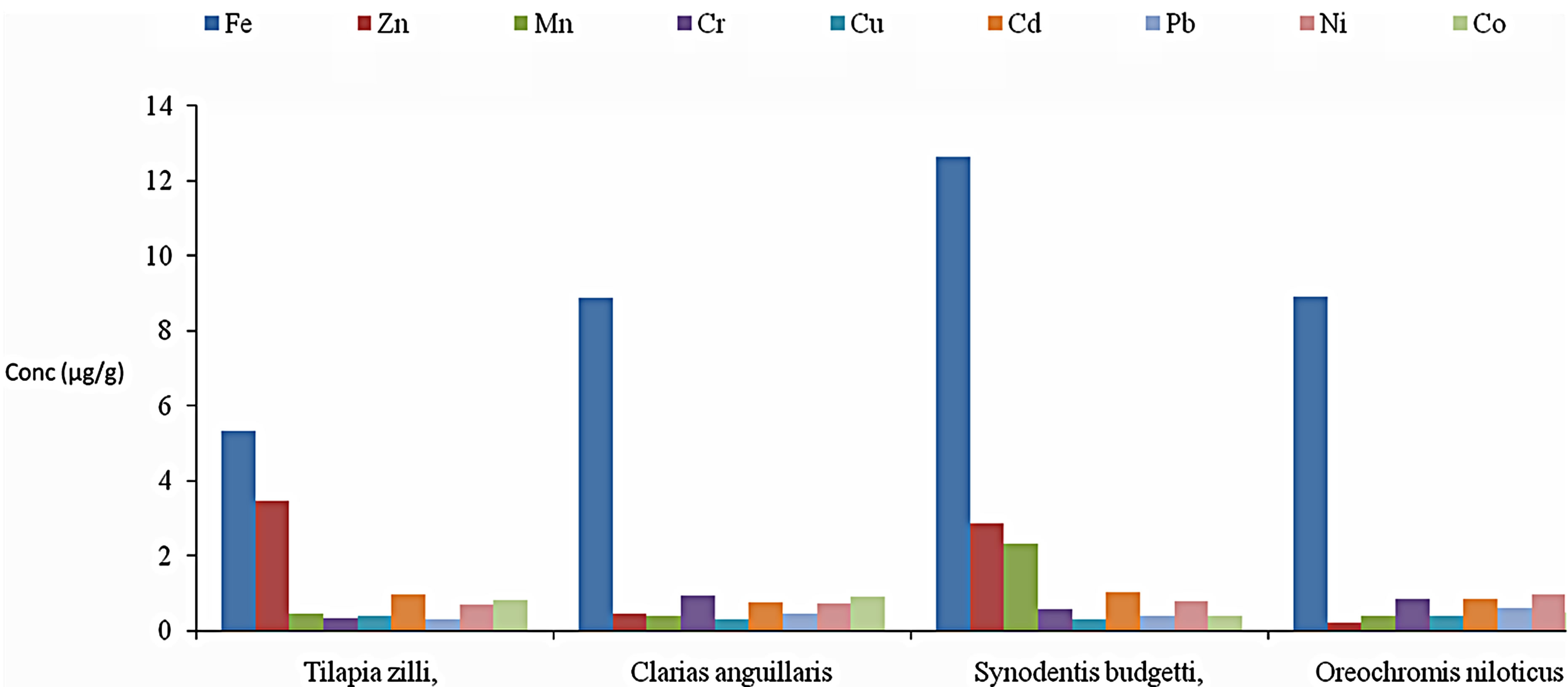
Figure 5. Comparison in the concentrations of heavy metals in the gills of different species of fishe from River Benue in Vinikilang, Adamawa State, Nigeria.
0.93 µg/g Cr; 0.31 to 0.39 µg/g Cu; 0.76 to 1.03 µg/g Cd; 0.31 to 0.61 µg/g Pb; 0.69 to 0.95 µg/g Ni; 0.38 to 0.82 µg/g Co. Gill surfaces are the first target of water-born metals (Spicer and Weber, 1991). The microenvironment of the gill surface consists of an epithelial membrane which primarily contains phospholipids covered by a mucous layer [28]. According to [29] the gill surface is negatively charged and thus provides a potential site for gill-metal interaction for positively charged metal. The gill of synodentis budgetti tends to accumulate the highest concentrations of all the metals, while Oreochronmis nilolitus showed the least concentrations. Laboratory experiments have indicated that in fishes which take up heavy metals from water, the gills generally show higher concentration than in the digestive tract. On the other hand, fish accumulating heavy metals from food show elevated metal levels in the digestive tract as compared to the gills [17,30]. The gills of all the fish tend to accumulate significant high levels of heavy metal than other tissues.
The comparison of heavy metals in liver tissues among the four species of fish is presented in Figure 6. Fe levels ranged from 4.55 to 9.23 µg/g; 0.26 to 1.78 µg/g Zn; 0.22 to 1.77 µg/g Mn; 0.31 to 0.83 µg/g Cr; 0.18 to 0.26 µg/g Cu; 0.58 to 0.88 µg/g Cd; 0.29 to 0.45 µg/g Pb; 0.29 to 0.67 µg/g Ni; 0.24 to 0.61 µg/g Co. The liver of Synodentis budgetti accumulates significant higher levels of Mn and Cd than other species; Fe and Zn was highest in
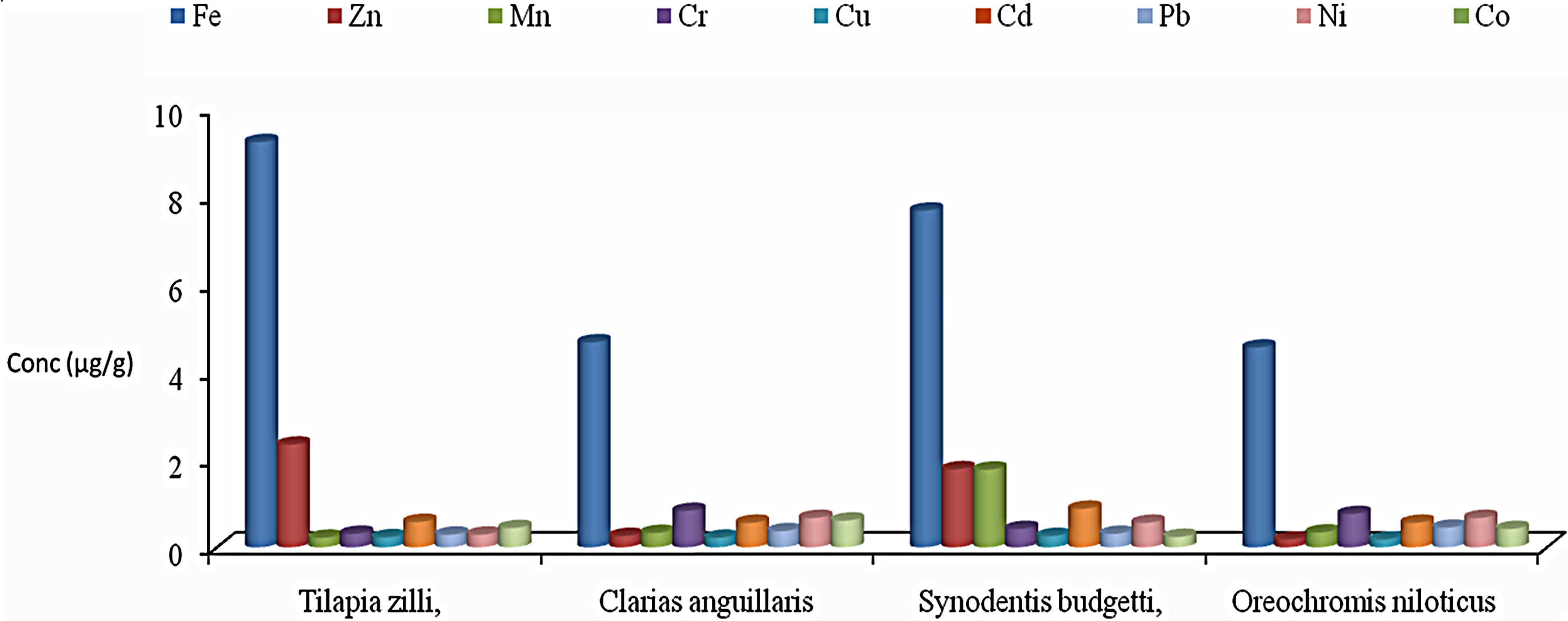
Figure 6. Comparison in the concentrations of heavy metals in the liver of different species of fishe from River Benue in Vinikilang, Adamawa State, Nigeria.
the stomach of Tilapia zilli, while Clarias angullaris shows more of Cr, Pb, Cd and Co. The liver plays an important role in accumulation and detoxification of heavy metals [31]. Exposure of fish to elevated levels of heavy metals induces the synthesis of metallothioneine proteins (MT), which are metal binding proteins [32,33]. Fishes are known to posses the metallothioneine proteins [34]. Metallothioneine proteins have high affinities for heavy metals and in doing so, concentrate and regulate these metals in the liver [35]. Metallothioneine proteins bind and detoxify the metal ion [35]. In the present study liver of synodentis budgetti accumulated more concentrations of the metals when compared to other metals. The liver tissue came second in terms of metals tissue accumulation after gills.
Figure 7 shows the comparison of heavy metals in stomach tissues among the four species of fish. Fe levels ranged from 1.51 to 6.44 µg/g; 0.14 to 1.56 µg/g Zn; 0.15 to 0.25 µg/g Mn; 0.23 to 0.66 µg/g Cr; 0.12 to 0.21 µg/g Cu; 0.24 to 0.55 µg/g Cd; 0.21 to 0.34 µg/g Pb; 0.17 to 0.56 µg/g Ni; 0.22 to 0.48 µg/g Co. The stomach of Synodentis budgetti accumulate significant higher levels of Fe than other species; Zn was highest in the stomach of Tilapia zilli, while Clarias angullaris shows more of Mn, Cr, Cu, Cd and Pb. Figure 8 presents the comparison of heavy metals in the kidney tissues among the four species of fish. The concentrations of Fe ranged from 1.22 to 24.56 µg/g; 0.12 to 1.55 µg/g Zn; 0.19 to 023 µg/g Mn; 0.17 to 0.47 µg/g Cr; 0.18 to 0.37 µg/g Cu; 0.04 to 0.33 µg/g Cd; 0.22 to 0.51 µg/g Pb; 0.17 to 0.42 µg/g Ni. Figure 9 present the comparison of heavy metals in bone tissues among species. The concentrations of Fe ranged from 1.99 to 4.22 µg/g; 0.11 to 0.33 µg/g Zn; 0.16 to 0.56 µg/g Mn; 0.19 to 0.37 µg/g Cr; 0.11 to 0.18 µg/g Cu; 0.11 to 0.22 µg/g Cd; 0.12 to 0.58 µg/g Pb; 0.26 to 0.44 µg/g Ni; 0.12 to 0.33 µg/g Co. The bone of Synodentis budgetti accumulates significant higher levels of Mn and Cd than other species; Zn and Fe were highest in the bone of Tilapia zilli, while Clarias angullaris shows more of Cr, Pb, Ni, and Co. The differences in metal accumulations could probably due to differences in feeding or metal sequestering habits between the fishes.
3.3. Comparison of Heavy Metals in Fish Samples with Standard Values
The concentration of Fe ranged from 0.68 to 12.65 µg/g. The highest concentration of 12.65 µg/g was recorded in gill of Synodentis budgetti, while the lowest value of 0.68 µg/g was recorded in the flesh of Oreochronmis niloticus. Fe is an essential element in human diet. It forms part of haemoglobin, which allows oxygen to be carried from the lungs to the tissues. Severe Fe deficiency causes anaemia in humans. The highest concentration observed in the present study was below the high residue concentrations of Fe (34 - 107 ppm) in fish samples on MNW Refuge [36].
Manganese was detected in the entire samples studied. The lowest concentration of 0.88 µg/g was observed in the flesh of Oreochronmis niloticus, while the highest concentration of 3.45 µg/g Mn was detected in gill of Tilapia zilli. Mn is an essential element for both animals and plants. Deficiencies of Mn result in severe skeletal and reproductive abnormalities in mammals. It is widely distributed throughout the body with little variation and does not accumulate with age [37]. The concentrations of Mn in all the fish samples exceeded the [38] limit of 0.01 ppm, thus pose a threat to the fishes with time.
Copper is an essential part of several enzymes and it is necessary for the synthesis of haemoglobin. Cu toxicity in fish is taken up directly from the water via gills, the
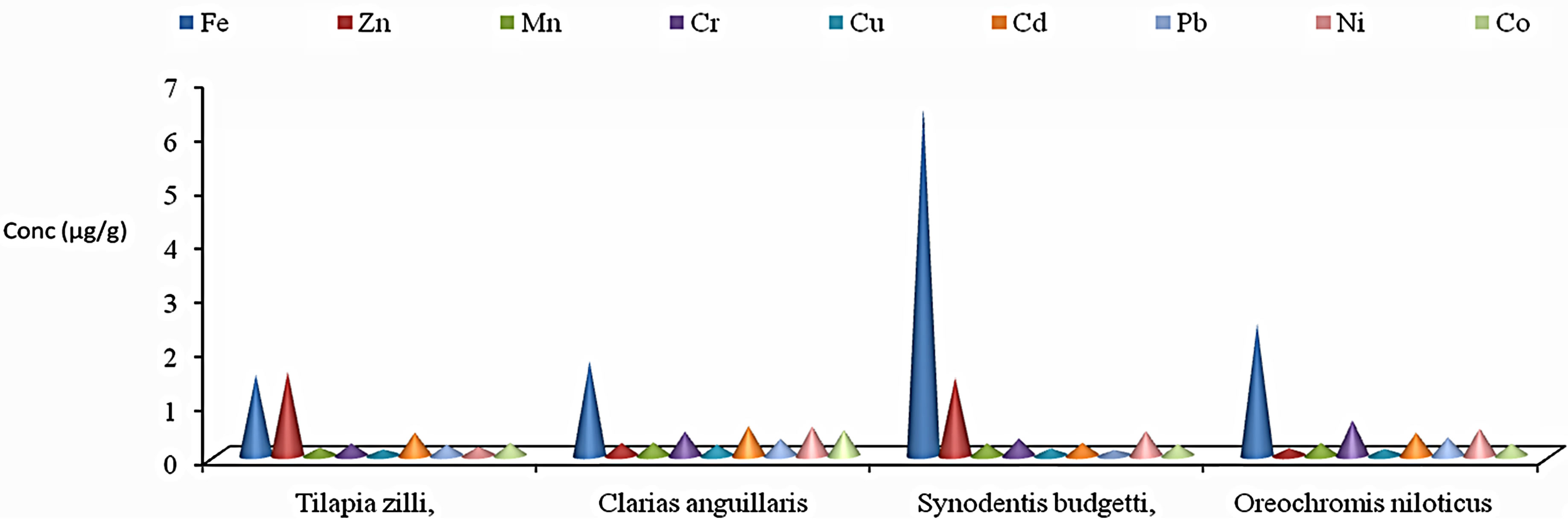
Figure 7. Comparison in the concentrations of heavy metals in the stomach of different species of fishe from River Benue in Vinikilang, Adamawa State, Nigeria.
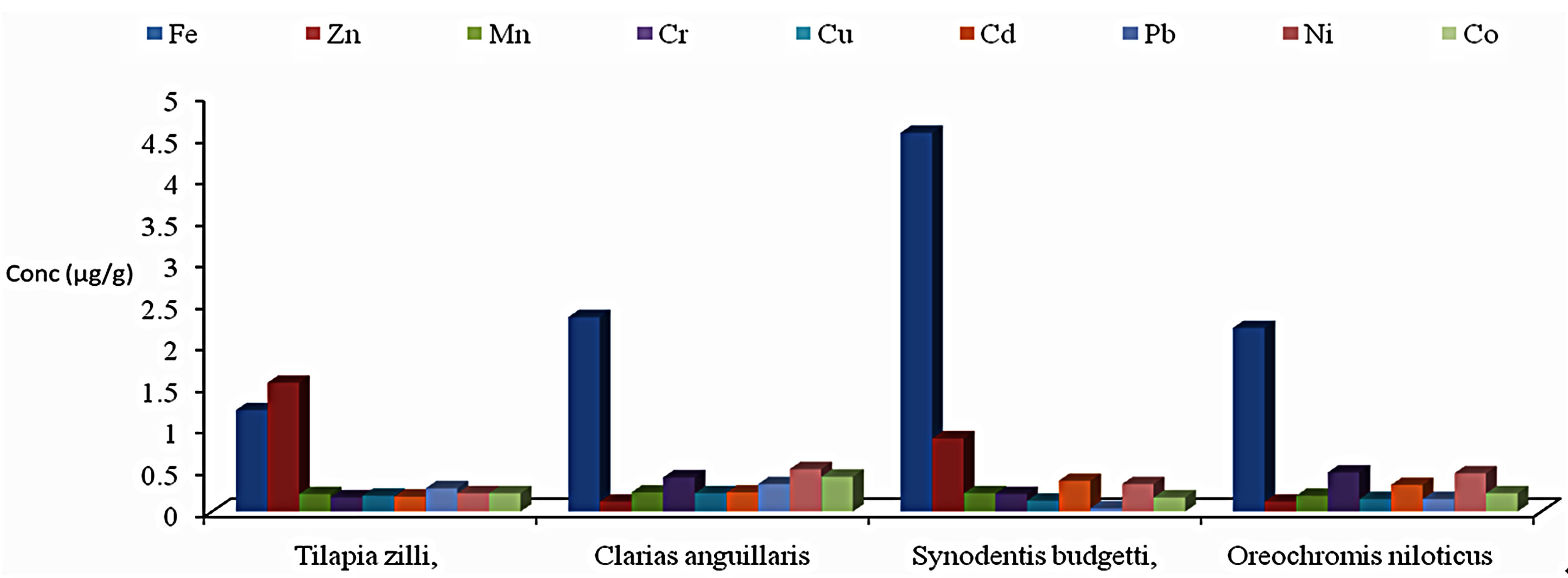
Figure 8. Comparison in the concentrations of heavy metals in the Kidney of different species of fishe from River Benue in Vinikilang, Adamawa State, Nigeria.
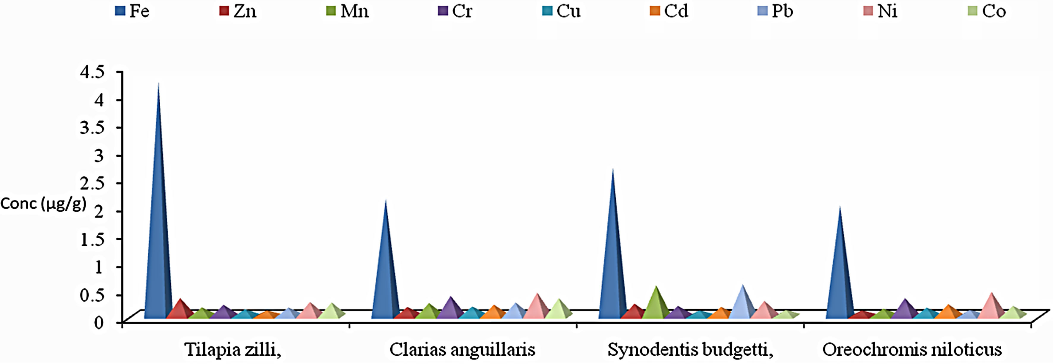
Figure 9. Comparison in the concentrations of heavy metals in the bones of different species of fishe from River Benue in Vinikilang, Adamawa State, Nigeria.
present study showed the similar accumulation of copper in the gills [39]. The concentration of Cu in the fish samples was highest (0.39 µg/g) in the gills of Tilapia zilli, while the lowest level of 0.06 µg/g was detected in the flesh of Oreochronmis niloticus. However, the highest value of 0.39 µg/g was below the FAO guideline of 30 µg/g. Thus the concentrations of Cu in the fish samples analysed were all below the FAO recommended guideline [40].
Zinc is an essential trace metal for both retarded growth, loss of taste and hypogonadism, leading to decreased fertility [37]. Zn toxicity is rare, but at concentrations in water up to 40 mg/kg, may induce toxicity, characterized by symptoms of irritability, muscular stiffness and pain, loss of appetite, and nausea [41]. The highest concentration of Zn (3.45 µg/g) was seen in the gill of Tilapia zilli, while the lowest value of 0.06 µg/g was measured in the flesh of Clarias angullaris. The FAO maximum guideline for Zn is 30 µg/g [40]. Thus, the concentrations of Zn in the fish samples were within the FAO guideline.
The major source of Ni for humans is food and uptake from natural sources, as well as food processing [42]. Increased incidence of cancer of the lung and nasal cavity caused by high intake of Ni has been also been reported in workers in Ni smelters. The highest concentration of Ni (0.95 µg/g) was measured in the gill of Oreochronmis niloticus, while the lowest detectable level of 0.11 µg/g was detected in the flesh of Clarias angullaris. The estimated maximum guideline [43] for Ni is 70 - 80 µg/g. Thus the concentrations of Ni in all the samples were far below the stipulated limit.
The lowest concentration of Cd, (0.11 µg/g) was detected in the flesh of Clarias angullaris, while the highest concentration of 1.03 µg/g was observed in the flesh of Synodentis budgetti. The source of Cd in humans is through food consumption. Severe toxic symptoms resulting from Cd ingestion are reported between 10 to 326 mg [37]. Fatal ingestions of Cd, producing shock and acute renal failure, occur from ingestions exceeding 350 mg/g [44]. The concentrations of Cd in all the fish samples, however, fell below the NCBP concentration of 2.1 μg/g threshold considered harmful to fish and predators [45,46].
Lead is classified as one of the most toxic heavy metals. The biological effects of sublethel concentrations of lead include delayed embryonic development, suppressed reproduction and inhalation of growth, increased mucous formation, neurological problems, enzyme inhalation and kidney disfunction [47]. The lowest concentration of 0.04 µg/g was observed in the flesh of Synodentis budgetti, while the highest concentration of 0.61 µg/g was detected in the gill of Oreochronmis niloticus. The highest concentration, 0.61 µg/g measured in the Oreochronmis niloticus was however within [40] guideline of 0.6 µg/g. All the other samples had Co concentrations below 0.61 µg/g detection limit.
The deficiency of Cr results in impaired growth and disturbances in glucose, lipid and protein metabolism [48]. Cr is an essential trace metal and the biologically usable form of Cr plays an essential role in glucose metabolism. The lowest detectable concentration of 0.05 µg/g was measured in the flesh of Synodentis budgetti, while the highest concentration of 0.93 was observed in the gill of Clarias angullaris. The maximum guideline of 12 - 13 µg/g stipulated by [49] was however, higher than the concentrations of Cr measured in all the fish samples, hence the samples are safe for human consumption.
4. Conclusion
Based on the result of this study, the levels of metals bioaccumulated in tissues of Tilapia zilli, Clarias anguillaris, Synodentis budgetti and Oreochronmis niloticus did not exceeds the permissible limits set for heavy metals by FAO, FEPA and WHO). The highest levels of all the metals in the present study were observed in gills and liver of the four fish samples, while flesh shows the lowest value. Therefore these fishes in this area of study did not pose any threat to human upon their consumption.
REFERENCES
- Z. Svobodova, O. Celechovska, J. Kolara, T. Randak and V. Zlabek, “Assessment of Metal Contamination in the Upper Reaches of the Ticha Orlice River,” Czech Journal of Animal Science, Vol. 49, No. 4, 2004, pp. 458-641.
- P. Part, O. Svanberg and A. Kiessling, “The Availability of Cadmium to Perfused Rainbow Troutgills in Different Water Qualities,” Water Research, Vol. 19, No. 2, 1985, pp. 427-434. doi:10.1016/0043-1354(85)90033-8
- A. Farkas, J. Salanki and I. Varanka, “Heavy Metal Concentrations in Fish of Lake Balaton, Lakes and Reservoirs,” Research and Management, Vol. 5, No. 4, 2000, pp. 271-279.
- R. C. Ravera, G. M. Beone, M. Dantas and P. Lodigiani, “Trace Element Concentrations in Freshwater Mussels and Macrophytes as Related to Those in Their Environment,” Journal of Limnology, Vol. 62, No. 1, 2003, pp. 61-70. doi:10.4081/jlimnol.2003.61
- ECDG, “European Commission DG ENV. E3 Project ENV. E.3/ETU/0058. Heavy Metals in Waste,” Final Report, 2002.
- H. N. Yang and H. C. Chen, “Uptake and Elimination of Cadmium by Japanese Eel, Anguilla japonica, at Various Temperatures,” Bulletin of Environmental Contamination and Toxicology, Vol. 56, No. 4, 1996, pp. 670-676. doi:10.1007/s001289900098
- T. M. Grieb, C. T. Driscoll, S. P. Gloss, C. L. Schofield, G. L. Bowie and D. B. Porcella, “Factors Affecting Mercury Accumulation in Fish in the Upper Michigan Peninsula,” Environmental Toxicology and Chemistry, Vol. 9, No. 7, 1990, pp. 919-930. doi:10.1002/etc.5620090710
- T. A. Haines and W. G. Brumbaugh, “Metal Concentration in the Gill, Gastrointestinal Tract, and Carcass of White Suckers (Catostomus commersoni) in Relation to lake Acidity,” Water, Air, and Soil Pollution, Vol. 73, No. 1, 1994, pp. 265-274. doi:10.1007/BF00477991
- J. G. Wiener, R. E. Martini, T. B. Sheffy and G. E. Glass, “Factors Influencing Mercury Concentrations in Walleyes in Northern Wisconsin Lakes,” Transactions of the American Fisheries Society, Vol. 119, No. 5, 1990, pp. 862-870. doi:10.1577/1548-8659(1990)119<0862:FIMCIW>2.3.CO;2
- R. J. Horwitz, B. Ruppel, S. Wisniewski, P. Kiry, M. Hermanson and C. Gilmour, “Mercury Concentrations in Freshwater Fishes in New Jersey,” Water, Air, and Soil Pollution, Vol. 80, No. 1-4, 1995, pp. 885-888. doi:10.1007/BF01189739
- H. Y. Cogun and F. Kargin, “Effects of pH on the Mortality and Accumulation of Copper in Tissues of Oreochromis niloticus,” Chemosphere, Vol. 55 No. 2, 2004, pp. 277-282. doi:10.1016/j.chemosphere.2003.10.007
- R. C. Playle, R. W. Gensemer and D. G. Dixon, “Copper Accumulation on Gills of Fathead Minnows: Influence of Water Hardness, Complexation and pH of the Gill Microenvironment,” Environmental Toxicology and Chemistry, Vol. 11, No. 3, 1992, pp. 381-391. doi:10.1002/etc.5620110312
- B. Baldisserotto, M. J. Chowdhury and C. M. Wood, “Effects of Dietary Calcium and Cadmium on Cadmium Accumulation, Calcium and Cadmium Uptake from the Water, and Their Interactions in Juvenile Rainbow Trout,” Aquatic Toxicology, Vol. 72, No. 1, 2005, pp. 99-117. doi:10.1016/j.aquatox.2004.11.019
- M. G. Barron and S. Albeke, “Calcium Control of Zinc Uptake in Rainbow Trout,” Aquatic Toxicology, Vol. 50, No. 3, 2000. pp. 257-264. doi:10.1016/S0166-445X(99)00099-5
- J. M. Kidwell, L. J. Phillips and G. F. Birchard, “Comparative Analyses of Contaminant Levels in Bottom Feeding and Predatory Fish Using the National Contaminant Biomonitoring Program Data,” Bulletin of Environmental Contamination and Toxicology, Vol. 54, No. 6, 1995, pp. 919-923. doi:10.1007/BF00197979
- H. R. Voigt, “Concentrations of Mercury (Hg) and Cadmium (Cd), and the Condition of Some Coastal Baltic Fishes,” Environmentalica Fennica, Vol. 21, No. 26, 2004, p. 8-13.
- J. J. Ney and J. H. Van Hassel, “Sources of Variability in Accumulation of Heavy Metals by Fishes in a Roadside Stream,” Archives of Environmental Contamination and Toxicology, Vol. 12, No. 6, 1983, pp. 701-706. doi:10.1007/BF01060754
- K. R. Campbell, “Concentrations of Heavy Metals Associated with Urban Runoff in Fish Living in Storm Water Treatment Ponds,” Archives of Environmental Contamination and Toxicology, Vol. 27, No. 3, 1994, pp. 352-356. doi:10.1007/BF00213171
- M. N. Rashed, “Monitory of Environmental Heavy Metals in Fish from Nasser Lake,” Environment International, Vol. 27, No.1, 2001, pp. 27-33.
- P. Kotze, H. H. du Preez and J. H. Van Vuren, “Bioaccumulation of Copper and Zinc in Oreochromis mossambicus and Clarias gariepinus, from the Olifants River, Mpumalanga, South Africa,” Water SA, Vol. 25, No. 1, 1999, pp. 99-110.
- Q.-Q. Chi, G.-W. Zhu and A. Langdon, “Bioaccumulation of Heavy Metals Infishes from Taihu Lake, China,” Journal of Environmental Sciences, Vol. 19, No. 12, 2007, pp. 1500-1504. doi:10.1016/S1001-0742(07)60244-7
- S. A. Mansour and M. M. Sidky, “Ecotoxicological Studies: Heavy Metals Contaminating Water and Fish from Fayum Governorate, Egypt,” Food Chemistry, Vol. 78, No. 1, 2002, pp. 15-22. doi:10.1016/S0308-8146(01)00197-2
- H. X, Mai, Y. W. Zhang, R. Si, Z. G. Yan, L. D. Sun, L. P. You and C. H. Yan, “High-Quality Sodium Rare-Earth Fluoride Nanocrystals: Controlled Synthesis and Optical Properties,” Journal of the American Chemical Society, Vol. 128, No. 19, 2006, pp. 6426-6436.
- G. Nussey, “Bioaccumulation of Chromium, Manganese, Nickel and Lead in the Tissues of the Moggel, Labeo umbratus (Cyprinidae), from Witbank Dam, Mpumalanga,” Water SA, Vol. 26, No. 2, 2000, pp. 269-284.
- J. Tarrio, M. Jaffor and M. Ashraf, “Levels of Selected Heavy Metals in Commercial Fish from Five Fresh Water Lake in Pakistan,” Toxicological & Environmental Chemistry, Vol. 33, No. 1-2, 1991, pp. 133-140. doi:10.1080/02772249109357755
- M. G. M. Alam, A. Tanaka, G. Allinson, L. J. B. Laurenson, F. Stagnitti and E. T. Snow, “A Comparison of Trace Element Concentrations in Cultured and Wild Carp (Cyprinus carpio) of Lake Kasumigaura, Japan,” Ecotoxicology and Environmental Safety, Vol. 53, No. 3, 2002, pp. 348-354. doi:10.1016/S0147-6513(02)00012-X
- W. Wepener, J. H. J. Vurenvan and H. H. Preezdu, “Uptake and Distribution of a Copper, Iron and Zinc Mixture in Gill, Live Rand Plasma of a Freshwater Teleost, Tilapia sparrmanii,” Water SA, Vol. 27, No. 1, 2001, pp. 99- 108.
- C. L. Bolis, A. Cambria and M. Famam, “Effects of Acid Stress on Fish Gills,” In: L. Zadunaisky and R. Gilles, Eds., Toxins, Drugs and Pollutants in Marine Mammals, Springer Verlag, Berlin, 1984, pp. 122-129. doi:10.1007/978-3-642-69903-0_10
- S. D. Reid and D. G. Mcdonald, “Metal Binding Activity of the Gills of the Rainbow Trout, Onchorhynchus mykiss,” Canadian Journal of Fisheries and Aquatic Sciences, Vol. 48, No. 6, 1991, pp. 1061-1068. doi:10.1139/f91-125
- A. G. Heath, “Water Psollution and Fish Physiology,” CRC Press, Boca Raton, 1990.
- A. M. Yousafzai, “Toxicological Effects of Industrial Effluents Dumped in River Kabul on Mahaseer, Tor Putitora at Aman Garh Industrial Area Nowshera, Peshawar, Pakistan,” Ph.D. Thesis, University of the Punjab, Lahore, 2004,
- G. C. Noel-Lambot and A. Disteche, “Distribution of Cd, Zn and Cu in Liver And Gills of The Eel Anguilla anguilla with Special Reference to Metallothioneins,” Comparative Biochemistry and Physiology Part C: Comparative Pharmacology, Vol. 61, No. 1, 1978, pp. 177-187.
- D. J. H. Phillips and P. S. Rainbow, “Strategies of Trace Metal Sequestration in Aquatic Organisms,” Marine Environmental Research, Vol. 28, No. 1-4, 1989, 207-210. doi:10.1016/0141-1136(89)90226-2
- L. Friberg, M. Piscator and G. Northberg, “Cadmuim in the Environment,” Chemical Rubber, Cleveland, 1971.
- E. Carpene and M. Vašák, “Hepatic Metallothioneins from Goldfish (Carassius auratus L),” Comparative Biochemistry and Physiology, Vol. 92B, No. 3, 1989, pp. 463- 468.
- C. S. Charbonneau and T. Nash, “Contaminants Program, Mingo National Wildlife Refuge (Region 3), Contaminants Survey Results,” US Fish and Wildlife Service, Columbia, 1993.
- P. Sivapermal, J. V. Sankar and P. G. Nair Viswanathan, “Heavy Metal Concentrations in Fish, Shellfish and Fish Products from Internal Markets of India visà-vis International Standards,” Food Chemistry, Vol. 102, No. 3, 2007, pp. 612-620. doi:10.1016/j.foodchem.2006.05.041
- WHO (World Health Organization), “Guidelines for Drinking Water Quality,” Recommendation WHO, Geneva, 1985.
- WHO, “Environmental Health Criteria 108: Nickel. International Programme on Chemical Safety,” World Health Organization, 1989. http:\\www.inchem.org/documents/ehc
- FAO (Food and Agriculture Organization), “Compilation of Legal Limits for Hazardous Substances in Fish and Fishery Products,” FAO Fisheries Circular No. 464, 1983, pp. 5-100.
- Nas-NRC, “National Academy of Sciences-National Research Council, Food and Nutrition Board, Recommended Dietary Allowances,” 8th Edition, National Academy Press, Washington DC,1974.
- Nas-NRC (National Academy of Sciences-National Research Council), “Division of Medical Sciences, Medical and Environmental Effects of Pollutants Nickel,” National Academic Press, Washington DC, 1975.
- USFDA, “Food and Drug Administration, Guidance Document for Nickel in Shell Fish,” DHHS/PHS/FDA/ CFSAN/ Office of Seafood, Washington DC, 1993.
- Nas-NRC (National Academy of Sciences-National Research Council), “Drinking Water and Health,” National Academic Press, Washington DC, 1982.
- C. J. Schmitt and W. G. Brumbaugh, “National Contaminant Biomonitoring Program: Concentrations of Arsenic, Cadmium, Copper, Lead, Mercury, Selenium and Zinc in US Freshwater Fish, 1976-1984,” Archives of Environmental Contamination and Toxicology, Vol. 19, No. 5, 1990, pp. 731-747. doi:10.1007/BF01183991
- S. M. Robertson, L. R. Gamble, and T. C. Maurer, “Contaminant Survey of La Sal Vieja, Willacy County, Texas, U.S. Fish Wild. Serv., Region 2, Contaminants Program. Fish and Wildlife Enhancement, Corpus Christi Field Office, Campus Box 338, 6300 Ocean Drive, Corpus Christi, Texas 78412,” Study Identifier 89-2-100, 1991.
- J. M. Rompala, F. W. Rutosky and D. J. Putnam, “Concentrations of Environmental Contaminants from Selected Waters in Pennsylvania,” US Fish and Wildlife Service. Special Scientific Report, State College, Pennsylvania, 1984.
- E. J. Calabrese, A. T. Canada and C. Sacco, “Trace Elements and Public Health,” Annual Review of Public Health, Vol. 6, No. 1, 1985, pp. 131-146. doi:10.1146/annurev.pu.06.050185.001023
- USFDA, “Food and Drug Administration, Guidance Document for Chromium in Shellfish,” DHHS/PHS/FDA/ CFSAN/Office of Seafood, Washington DC, 1993.
NOTES
*Corresponding author.