Modern Plastic Surgery, 2013, 3, 76-79
http://dx.doi.org/10.4236/mps.2013.32015 Published Online April 2013 (http://www.scirp.org/journal/mps)
The Preauricular Sinus: A Novel Approach for Complete
Bilateral Excision via a Modified F ace-Lift Incision*
Florian M. Lampert#, Georgios Koulaxouzidis, G. Björn Stark
Department of Plastic Surgery, University of Freiburg Medical Center, Freiburg, Germany.
Email: #florian.lampert@uniklinik-freiburg.de, georgios.koulaxouzidis@uniklinik-freiburg.de, bjoern.stark@uniklinik-freiburg.de
Received January 11th, 2013; revised February 15th, 2013; accepted February 24th, 2013
Copyright © 2013 Florian M. Lampert et al. This is an open access article distributed under the Creative Commons Attribution Li-
cense, which permits unrestricted use, distribution, and reproduction in any medium, provided the original work is properly cited.
ABSTRACT
Objectives/Hypothesis: To present a novel technique based on the combination of aesthetic and reconstructive tech-
niques for the radical and safe excision of preauricular sinus along with minimal cosmetic impairing. Study Design:
Case report, description of surgical method and brief overview of the literature. Methods: In a patient with bilateral
preauricular sinus, we used a modified face-lift incision and SMAS-fixation instead of solely excising the sinus with
skin ellipse. Results: Total removal of the bilateral fistulous systems could be achieved, along with a very aesthetically
pleasing result. Conclusions: The surgical approach shown here allows a radical excision with only minimal aesthetic
impairment using a shortened facelift-incision and SMAS-fixation.
Keywords: Preauricular Cyst; Preauricular Sinus; Face Lift Incision; SMAS Fixation; Aesthetic Reconstructive Surgery
1. Introduction
Using the example of a 28 years old male patient with a
long-lasting history of repetitive Infections of bilateral
preauricular sinuses, we describe a refinement of the op-
eration technique for the excision of this pathology. In-
stead of employing the standard operating technique,
which consists of an excision limited to the cyst along
with a skin spindle; we promote using a shortened pre-
tragal facelift incision. This approach provides an excel-
lent exposure of the operation site as a prerequisite for
complete removal of the affected tissue along with an
optimal cosmetic result of the inconspicuously placed
scar. The procedure can be carried out under local an-
aesthesia. These characteristics could contribute to an
optimized treatment of preauricular sinuses with further
reduction of the rate of rec urrence.
2. Case Report
A 28 years old male patient presented to us with a history
of repetitive painful swelling, redness and sebaceous and
purulent discharge from multiple bilateral preauricular
fistulae orifices from the level of the lobule up to the crus
of the helix (Figure 1) along with an oily skin type.
Apart from a previous pretragal incision and drainage
of an acute abscess on the more severly affected left side
several years ago, that had not led to any melioration of
the symptoms, patient history did not reveal any remark-
able occurrences. In particular, no other lesions of this
kind in further locations were found, also no alterations
indicative for embryonic developmental disorders such
as craniofacial dysmorphisms could be detected; family
history on this issue was unremarkable as well.
On the clinical tentative diagnosis of bilateral preauri-
cular sinuses, the indication for operative removal was
set. Unlike the customary recommendations from medi-
cal literature, we did not excise the lesions with an ellip se
of surrounding skin, but chose a surgical access via a
shortened pretragal facelift-incision (Figure 2(a)). By
doing so, we had the advantage of a superior exposure of
the operation site (Figure 2(b)), combined with the pos-
sibility of a radical removal of the complete system of
fistulous tracts along with the entire overlying skin, al-
most without any visible cosmetic impairment after
wound healing. Tension on the sutures could be avoided
by mobilisation of the superficial musculoaponeurotic
system (SMAS) and fixation to Loré’s fascia.
The operation, which was carried out under local an-
aesthesia, was uneventful, as was the postoperative
course. Sutures were removed 1 week postoperatively, at
this time, the patient was already free of discomfort.
*The authors have no funding, financial relationships, or conflicts o
f
interest to disclose.
#Corresponding author.
Copyright © 2013 SciRes. MPS
The Preauricular Sinus: A Novel Approach for Complete
Bilateral Excision via a Modified Face-Lift Incision 77
Figure 1. Preoperative view of the preauricular findings.
Histopathologic examination confirmed the in-sano-ex-
cision of multiple epidermal cysts with surround ing chro-
nic inflammation.
Until now (1 year postoperatively), there are no signs
of recurrence and almost invisible scarring (Figure 3; 3
months postoperativ ely).
3. Surgical Approach
The standard surgical access for facelift procedures is an
incision starting in the temporal region parallel to the
hairline towards the upper pole of the concha, then
threefold convexly curved preauricularly proximate to
the crus of the helix, the tragus and the earlobe, caudally
encircling the earlobe and ascending retroauricularly to
the superior aspect of the conchal bowl, then curving into
the scalp dorsally [1]. Especially in males, a retrotragal
incision should be avoided due to the risk of distorsion of
the hair-bearing skin onto the tragus. For the indication
presented here, we solely used the periauricular part of
the incision (Figures 2(a) and (b)).
After premedication with 7.5 mg Midazolame p.o. and
instillation of local anaesthesia (Mepivacain 1% with
Epinephrine 1:50,000; 3 ml per side), preparation was
carried out superficial to the capsula of the parotid gland
with mobilisation of the skin and the SMAS 3 cm ros-
trally (similar to a mini-Facelift-procedure). Having com-
(a)
(b)
Figure 2. (a) Visualization of the face lift incision line; (b)
Intraoperative view with excellent exposure of the whole
periauricular area.
plete exposure of the periauricular area, en-bloc-removal
of the fistulae-bearing tissue could be achieved by a
U-shaped excision, removal of cartilage was not neces-
sary in this case.
Due to the extended tissue mobilisation and underlying
fixation of the SMAS to Loré’s fascia (tympanoparotid
fascia), skin closure could be achieved free of tension,
there was no need for inserting a drainage.
4. Discussion
Preauricular sinuses, cysts or fistulae occur during auri-
Copyright © 2013 SciRes. MPS
The Preauricular Sinus: A Novel Approach for Complete
Bilateral Excision via a Modified Face-Lift Incision
78
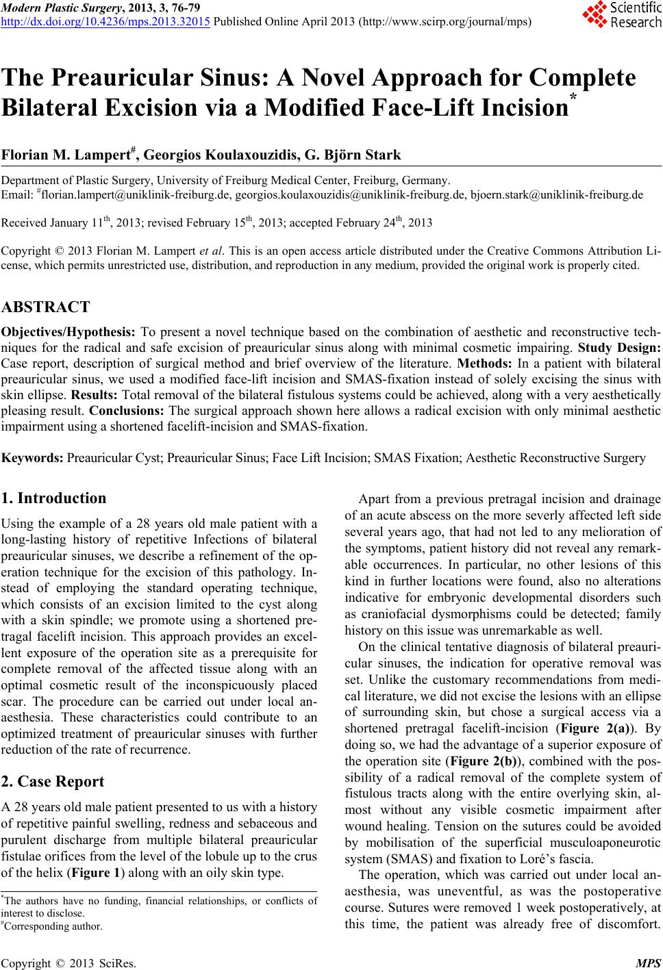
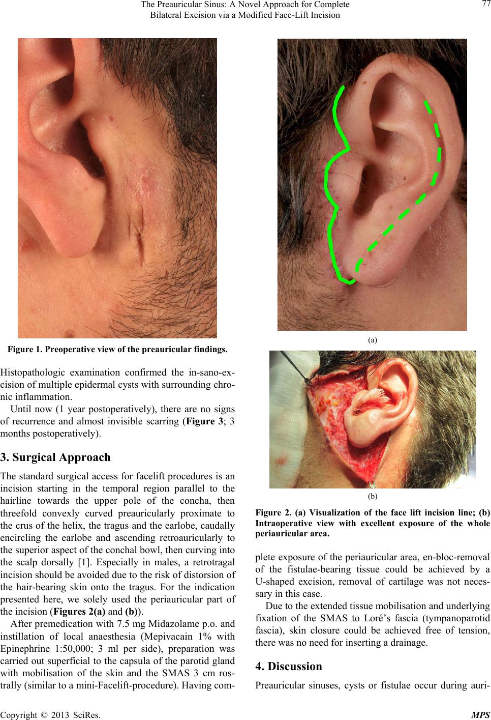
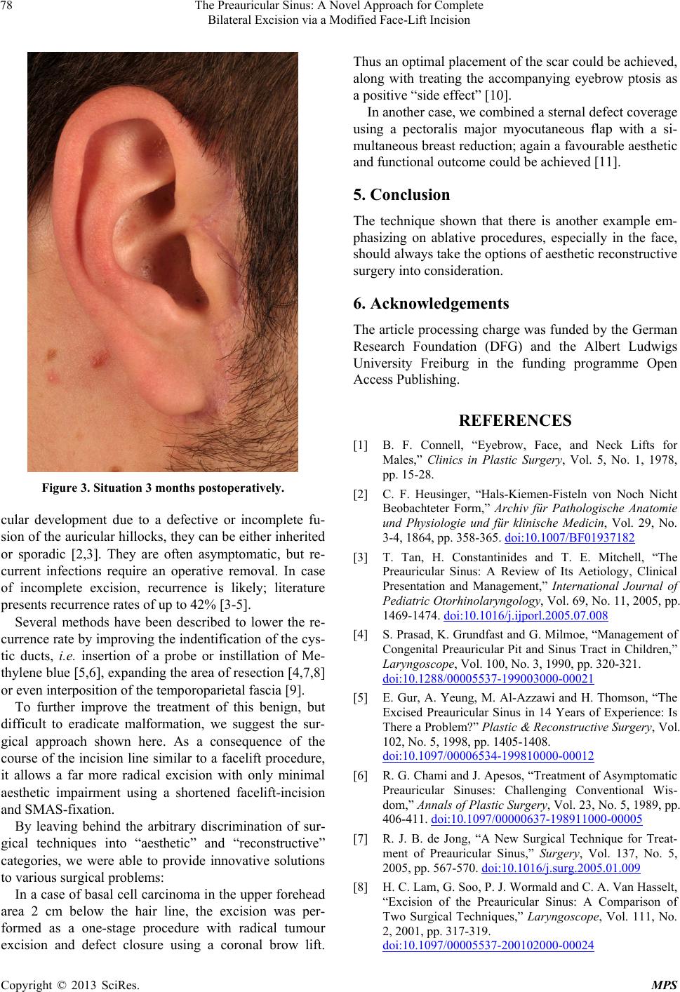
Figure 3. Situation 3 months postoperatively.
cular development due to a defective or incomplete fu-
sion of the auricular hillocks, they can be either inherited
or sporadic [2,3]. They are often asymptomatic, but re-
current infections require an operative removal. In case
of incomplete excision, recurrence is likely; literature
presents recurrence rates of up to 42% [3-5].
Several methods have been described to lower the re-
currence rate by improving the indentification of the cys-
tic ducts, i.e. insertion of a probe or instillation of Me-
thylene blue [5,6], expanding the area of resection [4,7,8]
or even interposition of the temporoparietal fascia [9].
To further improve the treatment of this benign, but
difficult to eradicate malformation, we suggest the sur-
gical approach shown here. As a consequence of the
course of the incision line similar to a facelift procedure,
it allows a far more radical excision with only minimal
aesthetic impairment using a shortened facelift-incision
and SMAS-fixation.
By leaving behind the arbitrary discrimination of sur-
gical techniques into “aesthetic” and “reconstructive”
categories, we were able to provide innovative solutions
to various surgical problems:
In a case of basal cell carcinoma in the upper forehead
area 2 cm below the hair line, the excision was per-
formed as a one-stage procedure with radical tumour
excision and defect closure using a coronal brow lift.
Thus an optimal placement of the scar could be achieved,
along with treating the accompanying eyebrow ptosis as
a positive “side effect” [10].
In another case, we combined a sternal defect coverage
using a pectoralis major myocutaneous flap with a si-
multaneous breast reduction; again a favourable aesthetic
and functional outcome could be achieved [11].
5. Conclusion
The technique shown that there is another example em-
phasizing on ablative procedures, especially in the face,
should always take the options of aesthetic reconstru ctive
surgery into consideration.
6. Acknowledgements
The article processing charge was funded by the German
Research Foundation (DFG) and the Albert Ludwigs
University Freiburg in the funding programme Open
Access Publishing .
REFERENCES
[1] B. F. Connell, “Eyebrow, Face, and Neck Lifts for
Males,” Clinics in Plastic Surgery, Vol. 5, No. 1, 1978,
pp. 15-28.
[2] C. F. Heusinger, “Hals-Kiemen-Fisteln von Noch Nicht
Beobachteter Form,” Archiv für Pathologische Anatomie
und Physiologie und für klinische Medicin, Vol. 29, No.
3-4, 1864, pp. 358-365. doi:10.1007/BF01937182
[3] T. Tan, H. Constantinides and T. E. Mitchell, “The
Preauricular Sinus: A Review of Its Aetiology, Clinical
Presentation and Management,” International Journal of
Pediatric Otorhinolaryngology, Vol. 69, No. 11, 2005, pp.
1469-1474. doi:10.1016/j.ijporl.2005.07.008
[4] S. Prasad, K. Grundfast and G. Milmoe, “Management of
Congenital Preauricular Pit and Sinus Tract in Children,”
Laryngoscope, Vol. 100, No. 3, 1990, pp. 320-321.
doi:10.1288/00005537-199003000-00021
[5] E. Gur, A. Yeung, M. Al-Azzawi and H. Thomson, “The
Excised Preauricular Sinus in 14 Years of Experience: Is
There a Problem?” Plastic & Reconstructive Surgery, Vol.
102, No. 5, 1998, pp. 1405-1408.
doi:10.1097/00006534-199810000-00012
[6] R. G. Chami and J. Apesos, “Treatment of Asymptomatic
Preauricular Sinuses: Challenging Conventional Wis-
dom,” Annals of Plastic Surgery, Vol. 23, No. 5, 1989, pp.
406-411. doi:10.1097/00000637-198911000-00005
[7] R. J. B. de Jong, “A New Surgical Technique for Treat-
ment of Preauricular Sinus,” Surgery, Vol. 137, No. 5,
2005, pp. 567-570. doi:10.1016/j.surg.2005.01.009
[8] H. C. Lam, G. Soo, P. J. Wormald and C. A. Van Hasselt,
“Excision of the Preauricular Sinus: A Comparison of
Two Surgical Techniques,” Laryngoscope, Vol. 111, No.
2, 2001, pp. 317-319.
doi:10.1097/00005537-200102000-00024
Copyright © 2013 SciRes. MPS
The Preauricular Sinus: A Novel Approach for Complete
Bilateral Excision via a Modified Face-Lift Incision
Copyright © 2013 SciRes. MPS
79
[9] C. I. Wu, C. C. Chen and S. C. Chang, “Treatment of
Recurrent Intractable Periauricular Fistula Using Tem-
poroparietal Fascia,” Journal of Craniofacial Surgery,
Vol. 21, No. 4, 2010, pp. 1224-1225.
doi:10.1097/SCS.0b013e3181e17b66
[10] N. Torio-Padron, H. Bannasch, S. U. Eisenhardt, A. S.
Dowlatshahi and G. B. Stark, “Surgical Treatment of a
Basal Cell Carcinoma in the Upper Forehead Area by
Combination of Reconstructive and Aesthetic Surgical
Techniques,” Journal of Plastic, Reconstructive & Aes-
thetic Surgery, Vol. 63, No. 2, 2010, pp. e151-e152.
doi:10.1016/j.bjps.2009.05.048
[11] N. Iblher, V. Penna, S. Krischak and G. B. Stark, “Simul-
taneous Pectoralis Major Myocutaneous Flap Combined
with Breast Reduction for Sternal Defect Coverage,”
Journal of Plastic, Reconstructive & Aesthetic Surgery,
Vol. 62, No. 8, 2009, pp. 1076-1080.
doi:10.1016/j.bjps.2008.04.023