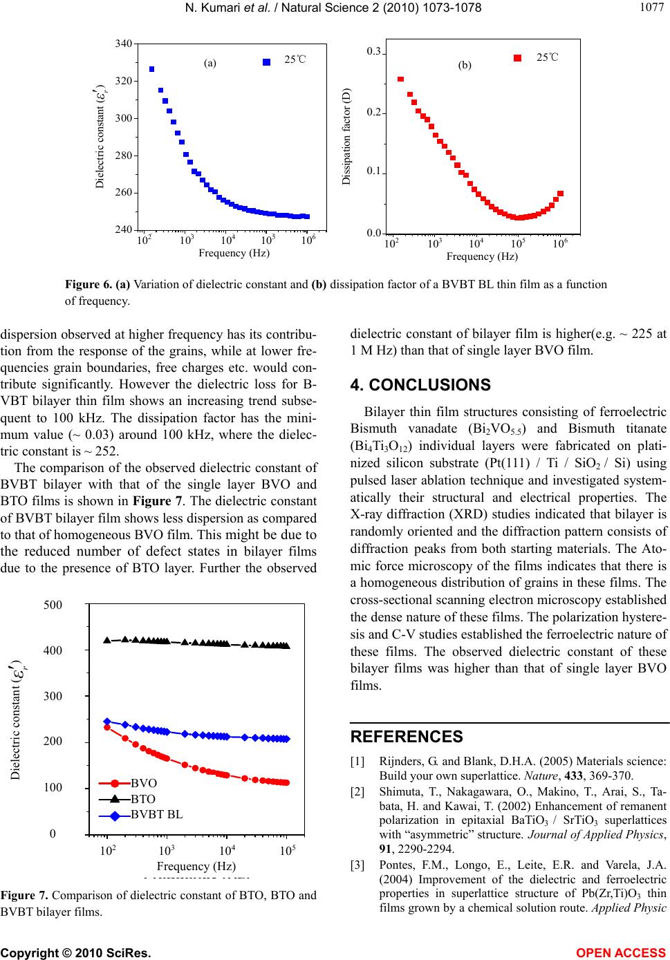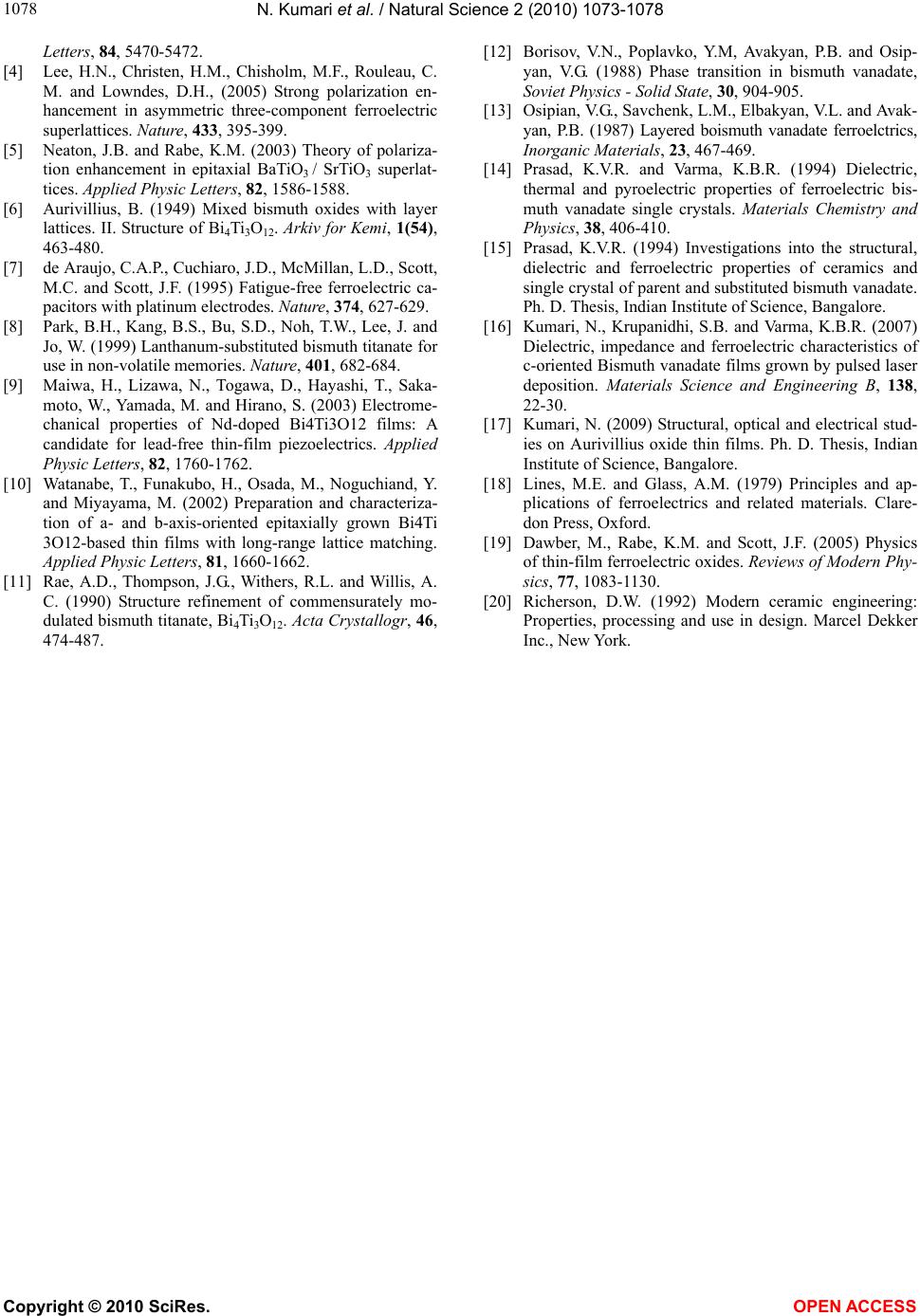
N. Kumari et al. / Natural Science 2 (2010) 1073-1078
Copyright © 2010 SciRes. OPEN ACCESS
1078
Letters, 84, 5470-5472.
[4] Lee, H.N., Christen, H.M., Chisholm, M.F., Rouleau, C.
M. and Lowndes, D.H., (2005) Strong polarization en-
hancement in asymmetric three-component ferroelectric
superlattices. Nature, 433, 395-399.
[5] Neaton, J.B. and Rabe, K.M. (2003) Theory of polariza-
tion enhancement in epitaxial BaTiO3 / SrTiO3 superlat-
tices. Applied Physic Letters, 82, 1586-1588.
[6] Aurivillius, B. (1949) Mixed bismuth oxides with layer
lattices. II. Structure of Bi4Ti3O12. Arkiv for Kemi, 1(54),
463-480.
[7] de Araujo, C.A.P., Cuchiaro, J.D., McMillan, L.D., Scott,
M.C. and Scott, J.F. (1995) Fatigue-free ferroelectric ca-
pacitors with platinum electrodes. Nature, 374, 627-629.
[8] Park, B.H., Kang, B.S., Bu, S.D., Noh, T.W., Lee, J. and
Jo, W. (1999) Lanthanum-substituted bismuth titanate for
use in non-volatile memories. Nature, 401, 682-684.
[9] Maiwa, H., Lizawa, N., Togawa, D., Hayashi, T., Saka-
moto, W., Yamada, M. and Hirano, S. (2003) Electrome-
chanical properties of Nd-doped Bi4Ti3O12 films: A
candidate for lead-free thin-film piezoelectrics. Applied
Physic Letters, 82, 1760-1762.
[10] Watanabe, T., Funakubo, H., Osada, M., Noguchiand, Y.
and Miyayama, M. (2002) Preparation and characteriza-
tion of a- and b-axis-oriented epitaxially grown Bi4Ti
3O12-based thin films with long-range lattice matching.
Applied Physic Letters, 81, 1660-1662.
[11] Rae, A.D., Thompson, J.G., Withers, R.L. and Willis, A.
C. (1990) Structure refinement of commensurately mo-
dulated bismuth titanate, Bi4Ti3O12. Acta Crystallogr, 46,
474-487.
[12] Borisov, V.N., Poplavko, Y.M, Avakyan, P.B. and Osip-
yan, V.G. (1988) Phase transition in bismuth vanadate,
Soviet Physics - Solid State, 30, 904-905.
[13] Osipian, V.G., Savchenk, L.M., Elbakyan, V.L. and Avak-
yan, P.B. (1987) Layered boismuth vanadate ferroelctrics,
Inorganic Materials, 23, 467-469.
[14] Prasad, K.V.R. and Varma, K.B.R. (1994) Dielectric,
thermal and pyroelectric properties of ferroelectric bis-
muth vanadate single crystals. Materials Chemistry and
Physics, 38, 406-410.
[15] Prasad, K.V.R. (1994) Investigations into the structural,
dielectric and ferroelectric properties of ceramics and
single crystal of parent and substituted bismuth vanadate.
Ph. D. Thesis, Indian Institute of Science, Bangalore.
[16] Kumari, N., Krupanidhi, S.B. and Varma, K.B.R. (2007)
Dielectric, impedance and ferroelectric characteristics of
c-oriented Bismuth vanadate films grown by pulsed laser
deposition. Materials Science and Engineering B, 138,
22-30.
[17] Kumari, N. (2009) Structural, optical and electrical stud-
ies on Aurivillius oxide thin films. Ph. D. Thesis, Indian
Institute of Science, Bangalore.
[18] Lines, M.E. and Glass, A.M. (1979) Principles and ap-
plications of ferroelectrics and related materials. Clare-
don Press, Oxford.
[19] Dawber, M., Rabe, K.M. and Scott, J.F. (2005) Physics
of thin-film ferroelectric oxides. Reviews of Modern Phy-
sics, 77, 1083-1130.
[20] Richerson, D.W. (1992) Modern ceramic engineering:
Properties, processing and use in design. Marcel Dekker
Inc., New York.