World Journal of AIDS, 2013, 3, 33-35
http://dx.doi.org/10.4236/wja.2013.31005 Published Online March 2013 (http://www.scirp.org/journal/wja) 33
Intracellular Transport of HIV-1 Matrix Protein
Associated with Viral RNA
Anatoliy I. Gozhenko1, Valentina A. Divocha1, Galina K. Vorkunova2, Alissa G. Bukrinskaya2,
Sergey I. Lupandin2
1School State Ukrainian Research Institute of the Medicine on Transport, Odessa, Ukraine; 2Ivanovsky Institute of Virology, Mos-
cow, Russia.
Email: bukrinskaya@yandex.ru
Received November 4th, 2012; revised December 23rd, 2012; accepted January 4th, 2013
ABSTRACT
HIV-1 matrix protein (MA) is a multifunctional structural protein localized on N terminus of Gag precursor p55. MA
participates in HIV-1 assembly as membranotropic part of Gag precursor as well as an individual protein spliced from
Gag early in infection. MA is found in th e nuclei of infected cells and in plasma membrane, the site of virus assembly,
in association with viral genome RNA. MA mutated variant M4 which contains two changed amino acids in N-terminal
regions is also associated with viral RNA, but it is localized in the nuclear and cytoskeleton fractions but not in the
plasma membrane suggesting that the mutant is deprived of membranotropic signal and “sticks” in the nuclei an d cy-
toskeleton, its previou s location sites. These data allow suggesting that MA involved into transmission of viral RNA is
transported to plasma membrane by cytos keleton.
Keywords: HIV-1 Matrix Protein; Gag Precursor P55; Cytoskeleton; Viral RNA; Transport of Viral Complex;
Plasma Membranes; Cell Fractionatiomn
1. Introduction
Matrix protein p17 is the smaller protein of HIV-1 but it
plays a lot of functions in viral life cycle. MA possessis
three transport signals and is involv ed in interaction with
Gag protein—the main protein in viral assembly. Its
presence in the nuclei could be connected with the for-
mation of the complex with the viral RNA as shown ear-
lier [1]. We studied this possibility by comparing the in-
tracellular localization of standart MA and mutant MA in
complex with viral RNA.
HIV-1 assembly process is driven and coordinated by
Gag precursor p55 which is generally sufficient for the
assembly of virus-like particles. An essential step of
HIV-1 assembly is the interaction of viral genome RNA
(gRNA) with Gag polyprotein, however, it is still un-
known when an d wh er e vir al ge nome RNA is tr anspo rted
from the nuclei to plasma membrane and is associated
with Gag precursor.
The membranotropic part of Gag on N-terminal is ma-
trix protein p17 ( MA). It po ssesses three transport signals
–membranotropic, nucleophy lic and the signal of nu-
clear export [2] and is regarded as a shuttling protein in
infected cells. MA could be spliced from Gag precursor
early in infection [3] but the authors believed that early
spliced viral proteins did not function in infected cells.
We suggested that early matrix protein could be involved
in viral life cycle [4] and particulary in virus assembly,
and tried to determine MA intracellular localization, it
association with viral RNA and the way of thear intra-
cellular transport.
Figure 1(a) shows the result of pulse-chase experi-
ment using HIV-1 infected MT4 cells. 14C leucine was
added 20 hours after infection for 20 minutes (pulse) and
the label was chased for 1, 2 and 3 hours. Cells were
fractionated as described [1] to get the fractions of the
cytosol, membranes and nuclei. It is seen that during
chase the amount of Gag precursor p55 in cytosol frac-
tion is diminished and after chase the protein is found in
membrane fraction (the place of virus assembly). Mean-
while, the amount of matrix protein in cytosol during the
chase is increasing suggesting that matrix protein is
spliced from Gag precursor soon after its synthesis. The
large amount of 120S glycoproteins show that they are
early synthesized and quickly transported to the mem-
branes.
It has been shown that during infection matrix protein
penetrates into the nuclei [5] and viral RNA is accumu-
lated in the nuclei following pulse-chase method and the
longer (for 3h) labeling of the infected cells with 14C
leucine (Figure 1(b)). MA has b een found in the nu clear
and membrane fractions of MT4 cells in association with
viral RNA including genome RNA using slot and blot
Copyright © 2013 SciRes. WJA
Intracellular Transport of HIV-1 Matrix Protein Associated with Viral RNA
34
(a)
(b)
F
igure 1. Localization o
f
vi
r
al p
r
otein
s
in cellula
r
fr
action
s
.
P
ul
s
e
-
cha
s
e expe
r
iment. (a) MT4 cells were infected with
HIV-1 virus (strain MVP-899) and 14C leucine was added
20 hours after infection for 20 min (pulse). Then the cells
were washed with PBS and the label was chased for 1, 2 and
3 hours. The cells were fractionated as described earlier [1]
to get cytosol, membrane and nuclear fractions. Lanes 1
and 6, protein markers, lanes 2 - 5, cytosol fraction, lanes 7
- 10, membrane fraction. Lanes 2 and 7, pulse labeling,
lanes 3 and 8, 4 and 9, 5 and 10, chase for 1, 2 and 3 hours
accordingly. The nuclear frac tion did not contain viral pro-
teins. (b) 14C leucine was added 40 h after infection for 30
min. (lanes 1, 3, 6) and the label was chased for 1 h (lanes 2,
4, 7) Lanes 5 and 8, the cells were labeled for 3 h. Lanes 1, 2,
cytosol fraction, 3 - 5, membrane fraction, 6 - 8, nuclear
fraction.
hybridization [1].
In 293T cells matrix protein has been revealed 40 h
after infection mainly in membrane fraction and in less
amount in nuclear fraction (Figure 2(a)). Viral RNA has
been also found by PCR in immunoprecipitates of mem-
brane fractions obtained after treatment with antiMA
antibody which do not interact with Gag precursor (the
primers used for LTR region of HIV-1 genome were L1
5’-g485ggagctctctggctaact-3’ and L2 5’-g931gattaactgc-
gaatcgttc-3’) (Figure 2(b)). These data suggest that MA
could be involved into viral RNA transmission to plasma
membrane.
To confirm the role of MA in viral RNA transmission,
we compared the intracellular localization and transport
of the complexes containing wild MA with that contain-
ing mutant MA. Plasmid containing mutant MA was
kindly presented by Dr. S. Dupont who also showed that
the virus containing mutant MA is not infectious [2]. The
mutant contained two changed amino acids in N-terminal
region: lysine in 18 position was chang ed on alanine and
arginine in 22 po sition on glycin e. 293T cells were trans-
fected by the plasmids expressing standart MA or mutant
MA, and after 24 hours of incubation the cells were frac-
tionated by the method described by Niederman [6] to
(a)
(b)
(c)
Figure 2. Intracellular localization of MA and gRNA 24 h
after 293T cells transfection. Lane 1, nuclear fraction, lane
2, cytoskeleton fraction, lane 3, membrane fraction, lane 4,
cytosol fraction. (c) Localization in cell fractions of MA-
gRNA complex after transfection of 293T cells with plas-
mids containing wild MA (lanes 1, 3, 5, 7) and mutant MA
(lanes 2, 4, 6, 8). 1, 2, nuclear fraction, 3, 4, cytoskeleton
fraction, 5, 6, membrane fraction, 7, 8, cytosol fraction. (a)
Immunoblot. (b) PCR using primers LTR-U5-gag to ge-
nome RNA. RNA was isolated from immunoprecipitate by
MA monoclonal antibody which does not react with Gag
precursor. Lanes 1 - 3, PCR using reverse transcription
reaction, lanes 4 - 6, control, PCR without preliminary re-
verse transcription.
Copyright © 2013 SciRes. WJA
Intracellular Transport of HIV-1 Matrix Protein Associated with Viral RNA
Copyright © 2013 SciRes. WJA
35
get the fractions of cytosol, membranes, nuclei and cy-
toskeleton. Localization of MA in cellular fractions was
determined using immunoblot.
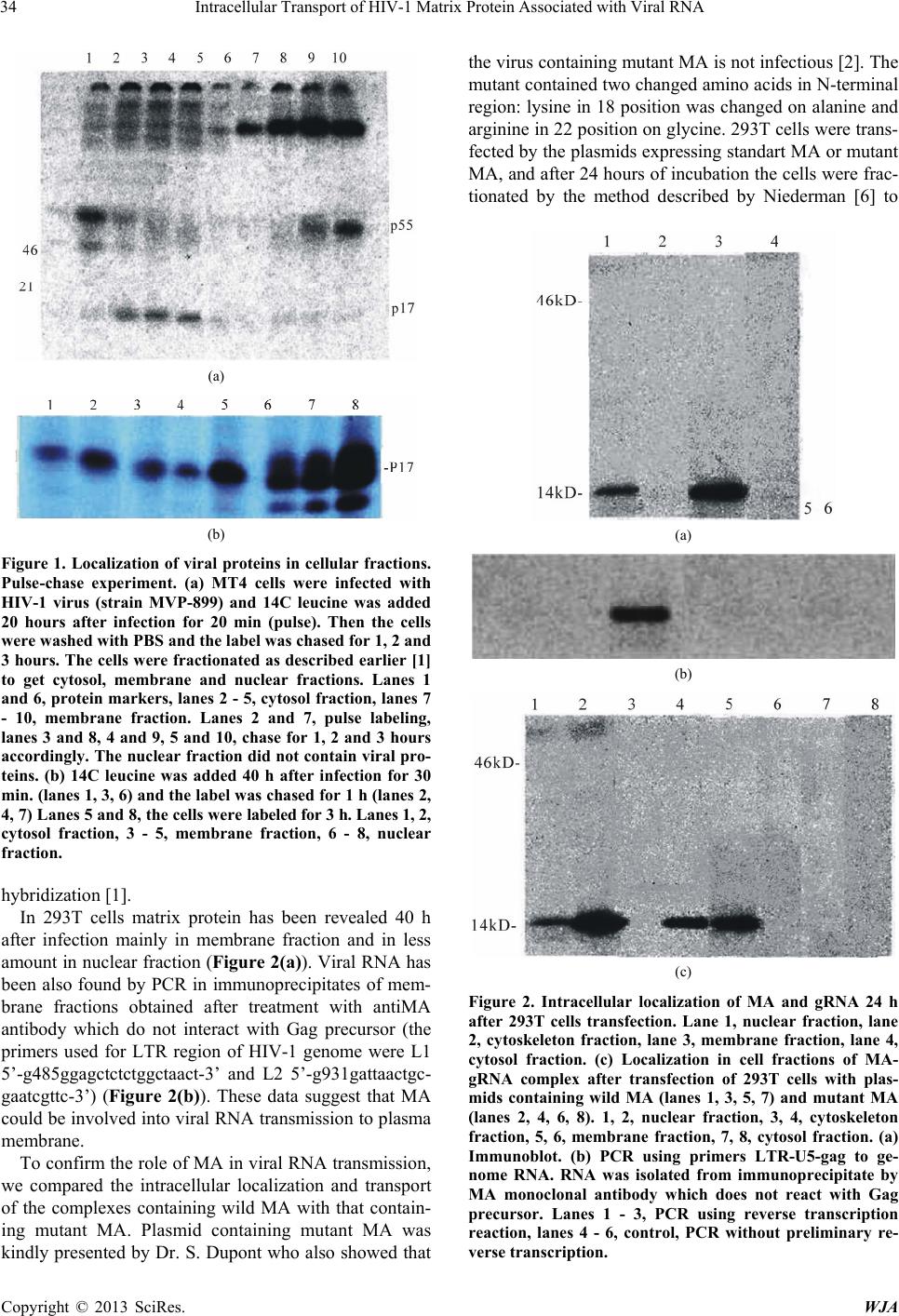
After transfection with plasmid expressing standard
MA, the small amount of MA was found in the nuclear
fraction while the main part of the protein was localized
in the membrane fraction. However, after transfection
with plasmid expressing mutant M4 matrix the protein
was not found in the membrane fraction but was local-
ized in the nuclear and cytoskeleton fractions (Figure
2(c)). These data show that the association of the mutant
MA with membranes is blocked, probably because mu-
tant MA lost its membranophylic signal (myristic acid)
on its N terminus, and the mutant MA protein associated
with viral RNA “sticks” in nuclei and cytoskeleton, the
fractions of its previous location. These results allow
suggesting that the complex MA-viral RNA is trans-
ported from the nuclei to membranes by cytoskeleton.
The reverse transport from membranes to nuclei was
described for viral reverse transcription and preintegra-
tion complexes [7] showing that the virus widely uses
cytoskeleton for its intracellular transport.
2. Conclusions
Despite the progress in defining determinates of Gag
participation in HIV assembly, Gag intracellular interac-
tions and trafficking to the assembly sites in the infected
cells are poorly understand. Our results indicate, that MA
is connected with viral RNA early after infection and the
complex is transported to the cellular membrane—the
place of viral assembly. When the mutant MA was used
instead of standard MA, the complex of MA and viral
RNA was not found in the cellular membrane, but sticks
in nucleus and cytoskeleton fractions. These results show
that MA-RNA complex is transported from the nuclei to
the cellular membrane by cytoskeleton.
The data also show that MA is cleaved from Gag pre-
cursor very early after Gag synthesis and thus could be
involved in to virus assembly as an individual protein.
The significant role of MA in HIV-1 replication shows
that this protein is the candidate antigen for therapeutic
vaccine against AIDS.
REFERENCES
[1] A. G. Bukrinskaya, G. K. Vorkunova and Y. Y. Tentsov,
“HIV-1 Matrix Protein P17 Resides in Cell Nuclei in As-
sociation with Genomic RNA,” AIDS Research and Hu-
man Retroviruses, Vol. 8, No. 10, 1992, pp. 1795-1801.
doi:10.1089/aid.1992.8.1795
[2] S. Dupont, N. Sharova, C. DeHoratius, C. M. A. Vir-
basius, X. C. Zhu, A. G. Bukrinskaya, M. Stevenson and
M. R. Green, “A Novel Nuclear Export Activity in HIV-1
Matrix Protein Required for Viral Replication,” Nature,
Vol. 402, No. 6762, 1999, pp. 681-685.
doi:10.1038/45272
[3] A. H. Kaplan and R. Swanstrom, “HIV-1 Gag Proteins
Are Processed in Two Cellular Compartments,” Procee-
dings of the National Academy of Sciences of USA, Vol.
88, No. 10, 1991, pp. 4528-4532.
doi:10.1073/pnas.88.10.4528
[4] A. Bukrinskaya, “HIV-1 Matrix Protein: A Mysterious
Regulator of the Viral Life Cycle,” Virus Research, Vol.
124, No. 1-2, 2007, pp. 1-11.
doi:10.1016/j.virusres.2006.07.001
[5] N. Sharova and A. Bukrinskaya, “P17 and P17-Con-
taining Gag Precursor of Input HIV Are Transported into
the Nuclei of Infected Cells,” AIDS Research and Human
Retroviruses, Vol. 7, No. 3, 1991, pp. 303-306.
doi:10.1089/aid.1991.7.303
[6] T. M. J. Niederman, W. R. Hastings and L. Ratner, “My-
ristoylation-Enhanced Binding of the HIV-1 Net Protein
to T Cell Skeletal Matrix,” Virology, Vol . 1 9 7 , N o . 1 , 1993,
pp. 420-425. doi:10.1006/viro.1993.1605
[7] A. Bukrinskaya, B. Brichacek, A. Mann and M. Steven-
son, “Establishment of a Functional HIV-1 Reverse Tran-
scription Complex Involves the Cytoskeleton,” The Jour-
nal of Experimental Medicine, Vol. 188, No. 11, 1998, pp.
2113-2125. doi:10.1084/jem.188.11.2113