Absolute Lymphocyte/Monocyte Ratio at Diagnosis and Interim Positron-Emission Tomography
Predict Survival in Classical Hodgkin Lymphoma
458
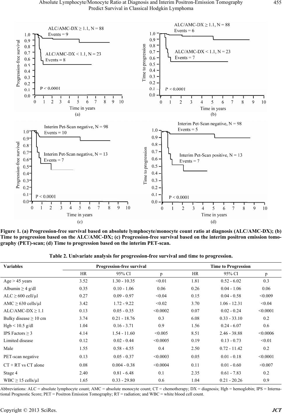
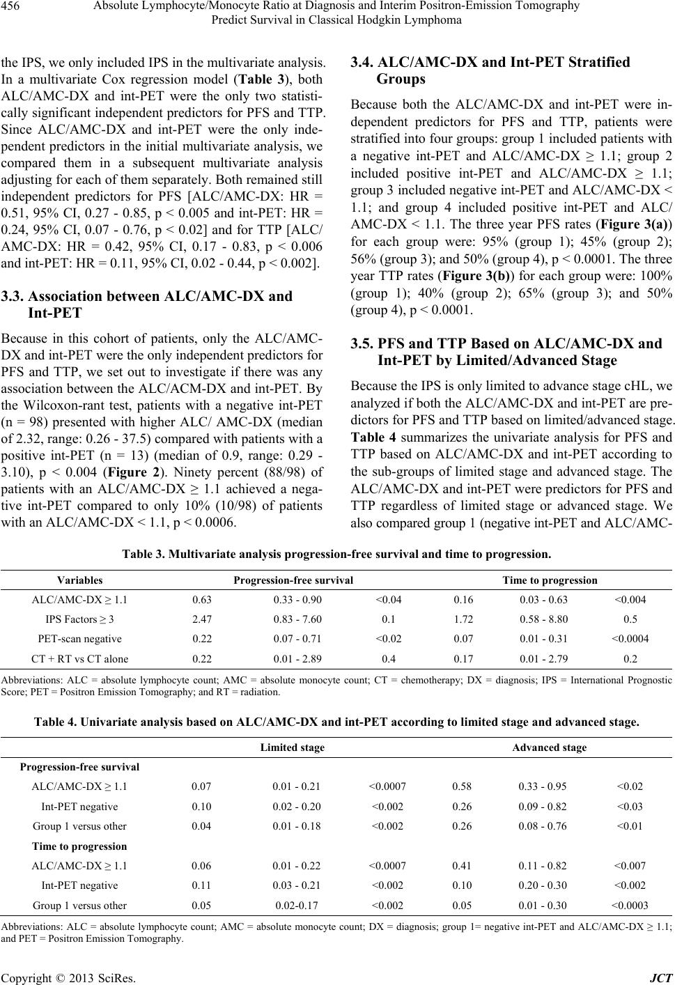
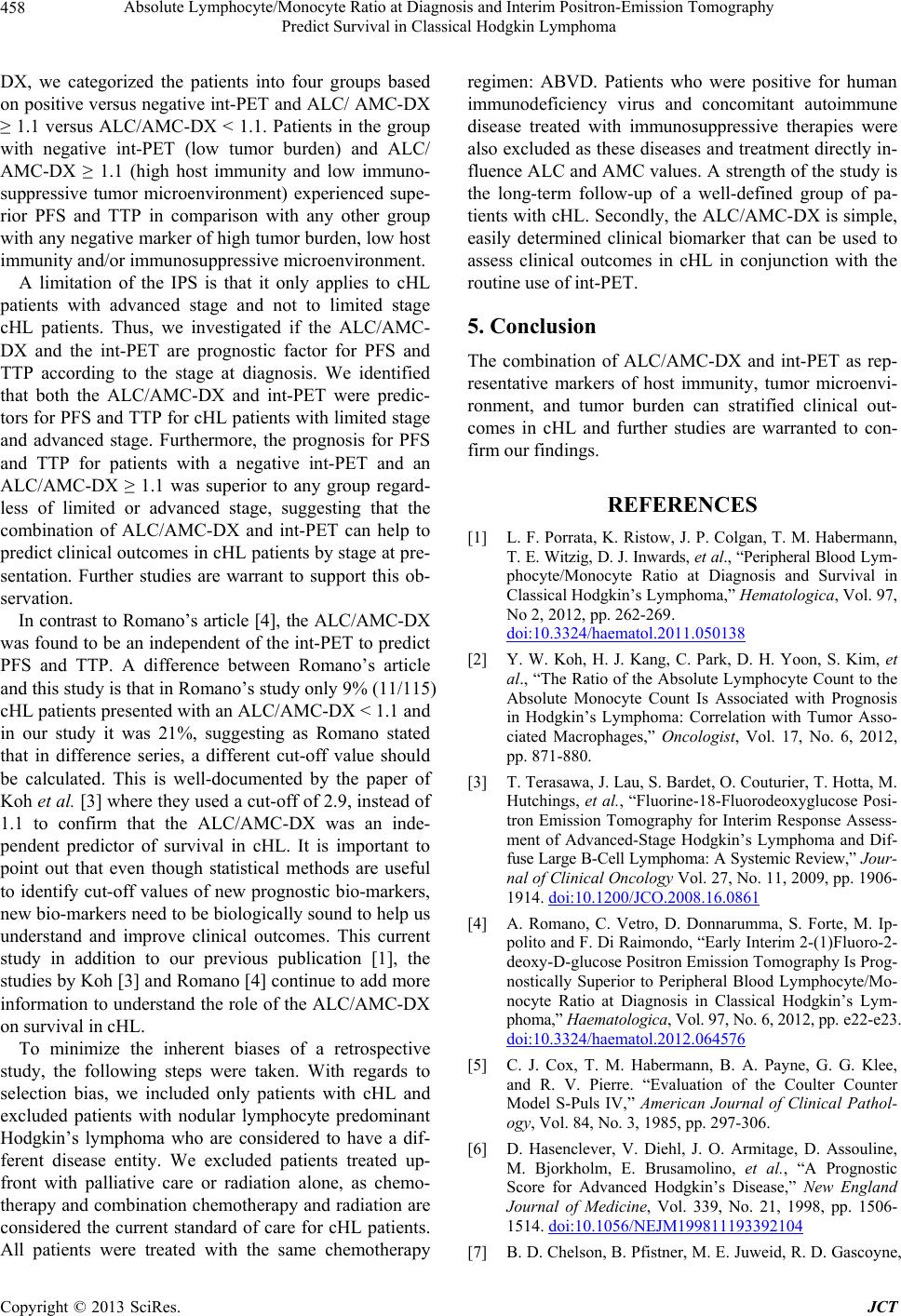
DX, we categorized the patients into four groups based
on positive versus negative int-PET and ALC/ AMC-DX
≥ 1.1 versus ALC/AMC-DX < 1.1. Patients in the group
with negative int-PET (low tumor burden) and ALC/
AMC-DX ≥ 1.1 (high host immunity and low immuno-
suppressive tumor microenvironment) experienced supe-
rior PFS and TTP in comparison with any other group
with any negative marker of high tumor burden, low host
immunity and/or immunosuppressive microenvironment.
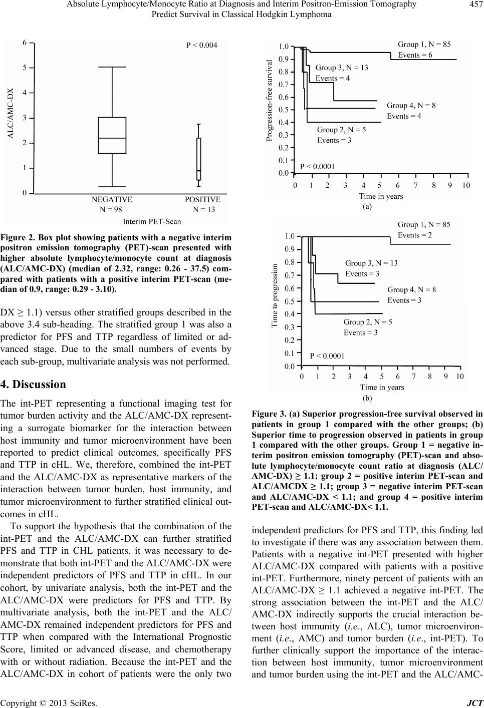
A limitation of the IPS is that it only applies to cHL
patients with advanced stage and not to limited stage
cHL patients. Thus, we investigated if the ALC/AMC-
DX and the int-PET are prognostic factor for PFS and
TTP according to the stage at diagnosis. We identified
that both the ALC/AMC-DX and int-PET were predic-
tors for PFS and TTP for cHL patients with limited stage
and advanced stage. Furthermore, the prognosis for PFS
and TTP for patients with a negative int-PET and an
ALC/AMC-DX ≥ 1.1 was superior to any group regard-
less of limited or advanced stage, suggesting that the
combination of ALC/AMC-DX and int-PET can help to
predict clinical outcomes in cHL patients by stage at pre-
sentation. Further studies are warrant to support this ob-
servation.
In contrast to Romano’s article [4], the ALC/AMC-DX
was found to be an independent of the int-PET to predict
PFS and TTP. A difference between Romano’s article
and this study is that in Romano’s study only 9% (11/115)
cHL patients presented with an ALC/AMC-DX < 1.1 and
in our study it was 21%, suggesting as Romano stated
that in difference series, a different cut-off value should
be calculated. This is well-documented by the paper of
Koh et al. [3] where they used a cut-off of 2.9, instead of
1.1 to confirm that the ALC/AMC-DX was an inde-
pendent predictor of survival in cHL. It is important to
point out that even though statistical methods are useful
to identify cut-off values of new prognostic bio-markers,
new bio-markers need to be biologically sound to help us
understand and improve clinical outcomes. This current
study in addition to our previous publication [1], the
studies by Koh [3] and Romano [4] continue to add more
information to understand the role of the ALC/AMC-DX
on survival in cHL.
To minimize the inherent biases of a retrospective
study, the following steps were taken. With regards to
selection bias, we included only patients with cHL and
excluded patients with nodular lymphocyte predominant
Hodgkin’s lymphoma who are considered to have a dif-
ferent disease entity. We excluded patients treated up-
front with palliative care or radiation alone, as chemo-
therapy and combination chemotherapy and radiation are
considered the current standard of care for cHL patients.
All patients were treated with the same chemotherapy
regimen: ABVD. Patients who were positive for human
immunodeficiency virus and concomitant autoimmune
disease treated with immunosuppressive therapies were
also excluded as these diseases and treatment directly in-
fluence ALC and AMC values. A strength of the study is
the long-term follow-up of a well-defined group of pa-
tients with cHL. Secondly, the ALC/AMC-DX is simple,
easily determined clinical biomarker that can be used to
assess clinical outcomes in cHL in conjunction with the
routine use of int-PET.
5. Conclusion
The combination of ALC/AMC-DX and int-PET as rep-
resentative markers of host immunity, tumor microenvi-
ronment, and tumor burden can stratified clinical out-
comes in cHL and further studies are warranted to con-
firm our findings.
REFERENCES
[1] L. F. Porrata, K. Ristow, J. P. Colgan, T. M. Habermann,
T. E. Witzig, D. J. Inwards, et al., “Peripheral Blood Lym-
phocyte/Monocyte Ratio at Diagnosis and Survival in
Classical Hodgkin’s Lymphoma,” Hematologica, Vol. 97,
No 2, 2012, pp. 262-269.
doi:10.3324/haematol.2011.050138
[2] Y. W. Koh, H. J. Kang, C. Park, D. H. Yoon, S. Kim, et
al., “The Ratio of the Absolute Lymphocyte Count to the
Absolute Monocyte Count Is Associated with Prognosis
in Hodgkin’s Lymphoma: Correlation with Tumor Asso-
ciated Macrophages,” Oncologist, Vol. 17, No. 6, 2012,
pp. 871-880.
[3] T. Terasawa, J. Lau, S. Bardet, O. Couturier, T. Hotta, M.
Hutchings, et al., “Fluorine-18-Fluorodeoxyglucose Posi-
tron Emission Tomography for Interim Response Assess-
ment of Advanced-Stage Hodgkin’s Lymphoma and Dif-
fuse Large B-Cell Lymphoma: A Systemic Review,” Jour-
nal of Clinical Oncology Vol. 27, No. 11, 2009, pp. 1906-
1914. doi:10.1200/JCO.2008.16.0861
[4] A. Romano, C. Vetro, D. Donnarumma, S. Forte, M. Ip-
polito and F. Di Raimondo, “Early Interim 2-(1)Fluoro-2-
deoxy-D-glucose Positron Emission Tomography Is Prog-
nostically Superior to Peripheral Blood Lymphocyte/Mo-
nocyte Ratio at Diagnosis in Classical Hodgkin’s Lym-
phoma,” Hae matologica, Vol. 97, No. 6, 2012, pp. e22-e23.
doi:10.3324/haematol.2012.064576
[5] C. J. Cox, T. M. Habermann, B. A. Payne, G. G. Klee,
and R. V. Pierre. “Evaluation of the Coulter Counter
Model S-Puls IV,” American Journal of Clinical Pathol-
ogy, Vol. 84, No. 3, 1985, pp. 297-306.
[6] D. Hasenclever, V. Diehl, J. O. Armitage, D. Assouline,
M. Bjorkholm, E. Brusamolino, et al., “A Prognostic
Score for Advanced Hodgkin’s Disease,” New England
Journal of Medicine, Vol. 339, No. 21, 1998, pp. 1506-
1514. doi:10.1056/NEJM199811193392104
[7] B. D. Chelson, B. Pfistner, M. E. Juweid, R. D. Gascoyne,
Copyright © 2013 SciRes. JCT