Journal of Cancer Therapy, 2013, 4, 441-447
http://dx.doi.org/10.4236/jct.2013.43A053 Published Online March 2013 (http://www.scirp.org/journal/jct)
441
Notch 1 and NF-κB Expression and Clinical Correlation in
Chinese Patients with Lymphoblastic Lymphoma
Lin Lin1,2, Xiaofei Sun1,2*, Juan Wang1,2, Zijun Zhen1,2, Suxia Lin1,3*, Gangling Tong1,2,
Yan Chen1,2
1State Key Laboratory of Oncology in South China, Guangzhou, China; 2Department of Pediatric Oncology, Sun Yat-Sen University
Cancer Center, Guangzhou, China; 3Department of Pathology, Sun Yat-Sen University Cancer Center, Guangzhou, China.
Email: *sunxf@sysucc.org.cn, *linsx@sysucc.org.cn
Received December 23rd, 2012; revised January 25th, 2013; accepted February 4th, 2013
ABSTRACT
T-cell acute lymphoblastic leukemia/lymphoma (T-ALL/T-LBL) is commonly associated with Notch 1 mutations.
There is limited data on the relationship between Notch l and NF-κB expression and clinical features in LBL. We
evaluated the expression of Notch l and NF-κB in LBL using immunohistochemistry and analyzed their relationship
with clinical characteristics, treatment results, and survival. From October 2000 to August 2008, 34 untreated patients
with LBL were enrolled in the study. Median age was 11.8 years (range, 1 - 25 years). Twenty-five patients were diag-
nosed with T-LBL and 9 patients with B-LBL. Most patients received chemotherapy consisting of modified ALL-BFM-
90. Notch l showed high expression in 68% of T-LBL and low expression in 100% of B-LBL (p = 0.015). High expres-
sion of Notch l positively correlated with presence of a mediastinal mass but not with 5-year event free survival (EFS)
in T-LBL. NF-κB showed high expression in 65% of all patients with LBL, with no difference between T- and B-LBL.
NF-κB expression was higher in T-LBL patients with bulky disease and B symptom; it did not correlate with 5-year
EFS in T-LBL. Expression of Notch 1 and NF-κB strongly correlated (p = 0.014) in T-LBL. Notch 1 is highly ex-
pressed in T-LBL. NF-κB is highly expressed in all patients with LBL with no difference between T-LBL and B-LBL.
Notch 1 expression was significantly associated with NF-κB expression in T-LBL. Notch l and NF-κB may play an im-
portant role in the development of T-LBL; further investigation is warranted.
Keywords: Lymphoblastic Lymphoma; Notch l; NF-κB
1. Introduction
Lymphoblastic lymphoma (LBL) is a highly invasive
malignancy originating from immature precursor lym-
phocytes. It is divided into precursor T cell lymphoblas-
tic lymphoma (T-LBL) and precursor B cell lymphoblas-
tic lymphoma (B-LBL). LBL and acute lymphoblastic
leukemia (ALL) share common morphologic and im-
munophenotypic features as well as a favorable outcome
after ALL-type chemotherapy; they are thought to repre-
sent a spectrum of a single disease entity, which is
termed T (or B) lymphoblastic leukemia/lymphoma in
the World Health Organization (WHO) [1] classification
schemes. Children and adolescents with LBL have better
survival when treated with intensive chemotherapeutic
regimens used in ALL, with a 5-year event free survival
of 70% - 90% [2,3]. However, 5-year event free survival
is less than 50% in high risk and refractory patients in
spite of intensive chemotherapy. New biological markers
and targeted therapies are need for these refractory LBL
patients.
In 1991, human Notch 1 was identified at the chromo-
somal breakpoint of a subset of T lymphoblastic neo-
plasm at the t (7;9) (q34;q34.3) chromosomal transloca-
tion [4]. Mammals have four Notch receptors (Notch 1 -
4) [5]. Notch l is a member of the Notch transmembrane
bound receptor family. Being a signaling pathway, Notch
1 not only plays an important part in differentiation and
development of normal cells, but also in the germination
and growth of neoplastic lesions, and as a suppressor
and/or promoter in various tumors [5,6]. Many studies
report Notch 1 mutations in greater than 50% of human
T-ALL cases [7-10]. G-secretase inhibitors may inhibit
the growth of T-ALL through blockage of the Notch 1
signal pathway [11]. Activation of Notch 1 leads to the
development of T-ALL in a mouse model [12].
NF-kappa B (NF-κB) was identified in B lymphocytes
in 1986. It acts as a regulator of κB light chain expres-
sion in B cells, and was thus termed nuclear transcription
factor-κB (NF-κB) [13]. NF-κB can promote the germi-
nation, proliferation, invasion, and metastasis of tumors
*Corresponding author.
Copyright © 2013 SciRes. JCT
Notch 1 and NF-κB Expression and Clinical Correlation in Chinese Patients with Lymphoblastic Lymphoma
442
through regulation of gene transfer [14]. The NF-κB path-
way is a major downstream target of Notch 1 in T-cell
leukemia and is highly active in established human T-
ALL. Inhibition of this pathway can efficiently restrict
tumor growth both in vitro and in vivo. Thus NF-κB is
one of the major mediators of Notch 1-induced transfor-
mation, suggesting that the NF-κB pathway is a potential
target for future T-ALL therapies [15].
The above mentioned studies focused on T-ALL and
less on LBL. Although ALL and LBL are the same dis-
ease entity, they show some differences in clinical char-
acteristics. The expression of Notch-1 and NF-kappa B in
precursor lymphoid neoplasms has been widely explored.
However, the data regarded mainly leukemic presenta-
tion while studies on lymphoblastic lymphoma are scarce.
Our research aimed to detect expression of Notch l and
NF-κB in tissues of LBL and normal lymph nodes using
immunohistochemistry. We also analyzed the relation-
ship between expression of these markers and clinical
characteristics and prognosis, aiming to provide evidence
for their potential use as biological markers.
2. Materials and Methods
2.1. Materials
From October 2000 to August 2008, the tumor samples
from 34 untreated patients with lymphoblastic lymphoma
at Sun Yat-Sen University Cancer Center were obtained.
Patients included 30 males and 4 females with a median
age of 11.8 years (range, 1 - 25 years; 1 - 18 years: 29
cases; 18 - 25 years: 5 cases). Twenty-five patients were
diagnosed with T-LBL and 9 with B-LBL. According to
the St. Jude staging system, our patient sample include 1
stage I, 4 stage II, 12 stage III, and 17 stage IV patients.
Nine patients had B symptoms, 9 had large masses, 17
had bone marrow involvement, 22 had an anterior medi-
astinal mass, 9 had extranodal lesions, and 20 had in-
creased serum lactate dehydrogenase (LDH) levels.
Thirty-one patients were treated according to the modi-
fied ALL-BFM-90 protocol, 2 patients received CHOAP,
and 1 patient abandoned treatment. Five normal lymph
nodes were obtained from October 2007 to October 2008
to serve as a control group. The informed consent of pa-
tients or their guardian had been obtained.
2.2. Immunohistochemistry
The tissue was stained by a two-step immunohistochem-
istry staining method. The first antibody for Notch 1
staining was goat anti-human antibody from Santa Cruz
(working concentration 1:100) and the first antibody for
NF-κB staining was mouse anti-human antibody from
Santa Cruz (working concentration 1:150). The second
antibody for both stains was derived from an instant de-
tection kit (PV-6003) from Zhongshan Goldenbridge Bio-
technology Co., Ltd.
2.3. Microscopic Evaluation
Using the semi-quantitative evaluation method by Car-
cangiu et al. [16], membrane, plasma, and nuclear details
of tumor cell were observed microscopically and evalu-
ated for staining degree and number of positive cells. The
staining degree of cells was defined as colorless, light tan,
tan, or brown and was assigned a score of 0, 1, 2, or 3,
respectively. The number of positive cells was defined as
<5%, 5% - 35%, 36% - 70%, or >70% and was assigned
a score of 0, 1, 2, or 3, respectively. Results were as-
sessed by the product of the two scores as follows: 1 as
negative (), 2 - 3 as weak positive (+), 4 - 5 as median
positive (++), and 6 as strong positive (+++). Negative
and weak positive (+) were considered low expression,
and median positive (++) and strong positive (+++) were
considered high expression.
2.4. Follow-Up
Follow-up studies were performed at three-month inter-
vals after ending maintenance therapy during the first
year, six-month intervals during the second year, and
one-year intervals during the following three years.
Event free survival (EFS) was calculated from the first
day of chemotherapy to death due to any cause, relapse,
progression, second malignancy, or the date of the last
follow-up contact for patients who were alive.
2.5. Statistical Analysis
Differences between two sample rates were analyzed
with the Fisher exact test two-sided for discrete variables.
The correlation between two nonparametric samples was
analyzed with spearman correlation test. Kaplan-Meier
test was implemented in survival analysis. The statistical
analysis was performed using SPSS statistics (version
16.0; SPSS). P values were considered significant at less
than 0.05.
3. Results
3.1. Expression of Notch-1 and NF-Kappa B in
T-LBL Compared to B-LBL
Notch 1 protein was expressed in the cytomembrane and
cytoplasm. High expression of Notch-1 was found in
50% (17/34) of all LBL cases. In T-LBL, high expression
was seen in 68% (17/25) and low expression in 32%
(8/25). In B-LBL, 100% (9/9) showed low expression.
Five normal lymph nodes also showed low expression.
Expression of Notch l was significantly different between
T-LBL and B-LBL (p = 0.001); expression was also sig-
Copyright © 2013 SciRes. JCT
Notch 1 and NF-κB Expression and Clinical Correlation in Chinese Patients with Lymphoblastic Lymphoma
Copyright © 2013 SciRes. JCT
443
nificantly different when comparing T-LBL and normal
lymph nodes (p = 0.009). B-LBL and normal lymph
nodes all showed low expression of Notch 1 (Table 1
and Figure 1).
NF-κB protein was expressed in the cytoplasm and
nucleus. Low expression was seen in 35% (12/34) and
high expression in 65% (22/34) of all LBL cases. In
T-LBL, low expression occurred in 32% (8/25) and high
expression in 68% (17/25). In B-LBL, low expression
was seen in 44% (4/9) and high expression in 56% (5/9).
There was no difference in NF-κB expression between
T-LBL and B-LBL, p = 0.687 (Table 2 and Figure 2).
We analyzed correlation of Notch-1 and NF-κB ex-
pression in T-LBL and found 14 cases with high expres-
sion of Notch1 also presented with high expression of
NF-κB. Notch 1 expression correlated significantly with
NF-κB expression in T-LBL, p = 0.014, no correlated
was seen in B-LBL (Table 3).
3.2. Association of Notch-1 and NF-κB
Expression with Clinical Characteristics of
T-LBL
We analyzed T-LBL by one factor correlation analysis
and found that high Notch l expression correlated sig-
nificantly with the presence of a mediastinal mass (p =
0.046). However, Notch l expression did not significantly
correlate with gender, age, extranodal lesions, bulky dis-
ease, LDH level, or presence of B symptoms (p > 0.05).
NF-κB expression was positively related to bulky disease
and B symptom (p = 0.013 and 0.000, respectively).
NF-κB expression did not correlate with other clinical
Table 1. Expression of Notch 1 in LBL and normal lymph nodes.
Low expression High expression
Samples Case (n)
Case (n) Percent Case (n) Percent p-value
T-LBL 25 8 32 17 68
B-LBL 9 9 100 0 0
0.001
T-LBL 25 8 32 17 68
Normal lymph nodes 5 5 100 0 0 0.009
B-LBL 9 9 100 0 0
Normal lymph nodes 5 5 100 0 0 No
Table 2. Expression of NF-κB in LBL and normal lymph nodes.
Low expression High expression
Case (n)
Case (n) Percent Case (n) Percent p-value
T-LBL 25 8 32 17 68
B-LBL 9 4 44 5 56
0.687
T-LBL 25 8 32 17 68
Normal lymph nodes 5 5 100 0 0 0.009
B-LBL 9 4 44 5 56
Normal lymph nodes 5 5 100 0 0 0.086
Table 3. Correlation of Notch-1 and NF-κB expression in T-LBL and B-LBL.
NF-κB expression
Notch 1 expression + ++ +++
R value p value
T-LBL 0 2 2 0
+ 1 2 1 0
++ 3 0 1 3
+++ 0 0 6 4
0.485 0.014
B-LBL 2 0 3 0
+ 1 1 0 2
++ 0 0 0 0
+++ 0 0 0 0
6.3
0.098
Notch 1 and NF-κB Expression and Clinical Correlation in Chinese Patients with Lymphoblastic Lymphoma
444
Figure 1. Notch 1 expression in lymphoblastic lymphoma
(400×). (A) Low expression of Notch1; (B) High expression
of Notch1.
Figure 2. NF-κB expression in lymphoblastic lymphoma
(400×). (A) Low expression of NF-κB; (B) High expression
of NF-κB.
characteristics (Table 4).
3.3. Relationship between Notch 1 and NF-κB
Expression and Survival in T-LBL
In the 25 patients with T-LBL, median follow up was 55
months (range, 11 - 142 months). Twenty-one patients
were alive and 4 had died from disease. Five-year event
free survival was 82% ± 8.1%. In the Notch l high ex-
pression and low expression groups, 5-year event free
survival rates were 80.2% ± 10% and 87.5% ± 11%, re-
spectively (p = 0.80) (Figure 3). The 5-year event free
survival in the NF-κB high expression and low expres-
sion groups were 86.3% ± 9% and 75.0% ± 15%, respec-
tively (p = 0.32) (Figure 4).
4. Discussion
Deregulated Notch signaling has been implicated in
many diseases, but the clearest example of a pathogenic
role is found in T-cell lymphoblastic leukemia/lym-
phoma, where the majority of human and murine tumors
have acquired mutations that lead to aberrant increase in
Notch 1 signaling. Many studies report Notch 1 muta-
tions in greater than 50% of human T-ALL/T-LBL cases
[7-10]. Most of these studies detected Notch 1 mutation
by gene sequencing. Notch 1 gene mutation may result in
overexpression of Notch 1 protein [17]. Immuohisto-
chemical detection of Notch 1 protein is a very common
and simple method in clinical practice. Notch 1 is a trans-
membrane bound receptor expressed in the cytomem-
brane and cytoplasm. Notch 1 antibody, which targets the
Notch 1 protein, can be used to detect expression of
Notch 1 by immunohistochemistry. Franziska identified
Notch 1 protein expression in 90 cases of lymphoma by
immunohistochemistry and found a 100% positivity rate
in classic Hodgkin lymphoma and anaplastic large cell
lymphoma but no expression in B cell lymphoma [18].
Kamstrup also detected the Notch1 expression in cuta-
neous T-cell lymphoma by using immunohistochemistry
and found the prominent expression in 21/40 cases, the
expression of Notch increased with the more advanced
stage [19]. In the present study we found high expression
of Notch l in 68% of T-LBL and low expression in 100%
of B-LBL. Although we did not evaluate for Notch 1
gene mutation in our patients, our finding of high Notch
1 protein expression in T-LBL is similar to the results of
Notch 1 mutation analysis in many studies reporting mu-
tations in more than 50% of T-ALL/LBL cases [7-10].
These results confirm that Notch l may play an important
role in the germination and proliferation of T-LBL but
not B-LBL. Detection of Notch 1 expression by immu-
nohistochemistry may be supplementary to understand-
ing deregulated Notch 1 signaling in T-LBL.
We analyzed the relationship between Notch 1 expres-
sion and clinical features in patients with T-LBL and
found that Notch l expression positively correlated with
the presence of a mediastinal mass. Most T-LBL patients
with an anterior mediastinal mass showed high expres-
sion of Notch 1. Zhu [20] reported that Notch l mutations
were significantly associated with higher blast counts in
the peripheral blood at diagnosis in T-ALL. In spite of
the similarities between T-ALL and T-LBL, there are
also obvious differences in their clinical features. It is
interesting that the predominant manifestation in T-LBL
patients is an anterior mediastinal mass, while in patients
with T-ALL bone marrow involvement is the predomi-
nant site of disease. Thus both findings, i.e. Notch 1 mu-
tation correlating with higher blast counts in the periph-
eral blood in T-ALL and with mediastinal mass in T-
LBL, show that Notch 1 signaling may play an important
role in development of T-ALL/T-LBL.
Multiple clinical series confirm that Notch 1 mutations
in T-ALL/LBL are frequent in all genetic and clinical
subtypes of human T-ALL/LBL, leading investigators to
ask if mutational status is a useful biomarker. However,
associations between mutation status and outcome are in-
consistent. While a few series suggest that Notch 1 muta-
tions are associated with worse outcomes [20], some
studies have shown no association [21,22], and the BFM
groups studies reported that Notch 1 mutations correlate
with better treatment response and overall survival in
T-ALL [8,10]. Callens reported that Notch 1 and/or
FBXW7 mutations were associated with improved event-
free survival and overall survival in pediatric T-lympho-
blastic lymphoma, patients with NOTCH1 mutation de-
monstrated a significantly better outcome than NOTCH1
wild type patients with 96% (SE, 0.04) versus 53% (SE,
Copyright © 2013 SciRes. JCT
Notch 1 and NF-κB Expression and Clinical Correlation in Chinese Patients with Lymphoblastic Lymphoma 445
Table 4. Notch 1 and NF-κB expression and clinical characteristics in T-LBL.
Notch 1 Expression NF-κB Expression
n
Low High
p value Low High
p value
Male 22 4 (18%) 18 (82%) 4 (18%) 18 (82%)
Sex Female 3 0 (0%) 3 (75%) 0.420 0 (0%) 3 (100%) 0.420
18 21 4 (23%) 17 (77%) 4 (18%) 17 (82%)
Age >18 4 0 (0%) 4 (100.0%) 0.341 0 (0%) 4 (100%) 0.341
No 5 3 (60%) 2 (40%) 0 (0%) 5 (100%)
Mediastinal
mass Yes 20 1 (5%) 19 (95%) 0.046 4 (20%) 16 (80%) 0.275
Normal 8 0 (0%) 8 (100%) 2 (25%) 6 (75%)
LDH High 17 4 (23%) 13 (77%) 0.134 2 (12%) 15 (88%) 0.400
0 20 4 (20%) 16 (80%) 4 (20%) 16 (80%)
Extralymph node 1 5 0 (0%) 5 (100%) 0.275 0 (0%) 5 (100%) 0.275
III 11 2 (18%) 9 (82%) 4 (36%) 7 (64%)
Stage IV 13 5 (39%) 8 (62%)
0.386 3 (23%) 10 (77%) 0.659
No 20 4 (20%) 16 (80%) 0 (0%) 20 (100%)
B symptoms Yes 5 0 (0%) 5 (100%) 0.275 4 (80%) 1 (20%) 0.000
No 17 2 (12%) 15 (88%) 0 (0%) 17 (100%)
Bulky Yes 8 2 (25%) 6 (75%) 0.088 4 (50%) 4 (50%) 0.013
Figure 3. Survival curve and expression of Notch 1 in T-
LBL.
Figure 4. Survival curve and expression of NF-κB in T-
LBL.
0.11) 5-year EFS, respectively, (p = 0.002) [23], all these
patients received the BFM protocol treatment. It showed
the Notch1 mutation status were associated with survival
in T-ALL/LBL patients who received BFM protocol. So
far the BFM protocol is one of best protocols in treat-
ment of pediatric ALL/LBL. In our study, we only de-
tected the expression of Notch 1 in T-LBL, no detecting
the Notch1 mutation, and found that Notch l expression
did not relate to 5-year event free survival in T-LBL.
Through high expression of Notch1 were found in our
patients, but the sample was small and it did not get a
well evaluation when comparing the association between
the expression and survival. Moreover, the relationship
between Notch l mutation and expression needs to fur-
ther study.
Notch-related T-ALL/LBL is also associated with con-
stitutive activation of NF-κB [24,25]. NF-κB is one of
the important nuclear transcript regulating factors and
plays a role in the onset of T-ALL as a downstream re-
gulator of Notch 1. Blocking this pathway can suppress
carcinoma growth, implying that NF-κB may be a future
treatment target in T-ALL [26]. To produce these thera-
peutic agents, the relationship between NF-κB and Notch
1 expression in LBL should be understood. Up to now,
only a few reports on the expression of NF-κB in LBL
exist. In our research, we found that high expression of
NF-κB occurred in 65% of all LBL cases. There was no
difference in expression of NF-κB between T-LBL and
B-LBL. High expression of NF-κB was significantly re-
lated to bulky disease and B symptom. These findings
show that NF-κB is highly activated in LBL and closely
related to the germination and proliferation of LBL. We
Copyright © 2013 SciRes. JCT
Notch 1 and NF-κB Expression and Clinical Correlation in Chinese Patients with Lymphoblastic Lymphoma
446
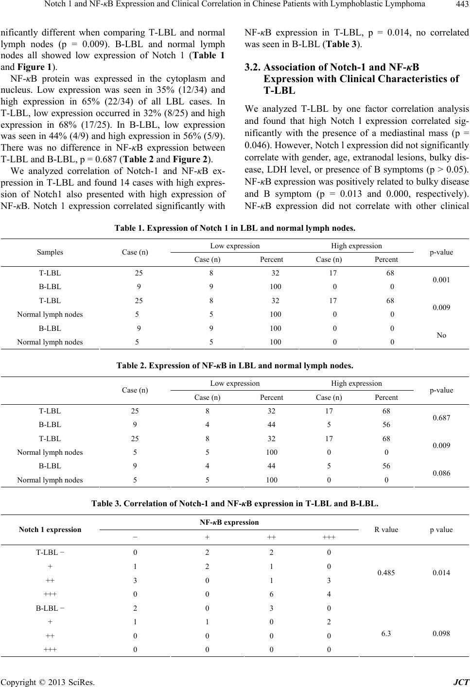
analyzed the relationship between expression of NF-κB
and Notch 1 in LBL and found there was a significant
positive correlation between the expression of Notch 1
and NF-κB in T-LBL (p = 0.014). Our results showed
that high expression Notch 1 was accompanied by high
expression of NF-κB in T-LBL, and both have well cor-
relation. This supports the hypothesis that the Notch 1
signaling pathway is associated with constitutive activa-
tion of NF-κB and demonstrates that NF-κB may be a
signal transduction factor downstream of the Notch 1
pathway active in the germination of T-LBL. In contrast,
we found low expression of Notch1 and high expression
of NF-κB in B-LBL, and no correlation between the ex-
pression of Notch 1 and NF-κB in B-LBL, It suggesting
that NF-κB might be activated via other pathways in B-
LBL to result in tumor proliferation.
When analyzing the relationship between NF-κB ex-
pression and clinical characteristics of T-LBL, we found
NF-κB expression to be higher in T-LBL patients with
bulky disease and B symptoms. On the other hand, no
correlation was found between NF-κB expression and
sex, age, or extranodal disease. Bavi detected expression
of NF-κB in 203 cases of diffuse large B cell lymphoma
(DLBCL) using immunohistochemistry and reported that
NF-κB expression occurred in 25.6% (52/203) of DL-
BCL tumors, was associated with activated B cell (ABC)
phenotype, and showed a significantly poorer overall
survival as compared to those without NF-κB expression
[27]. A study in laryngeal squamous cell carcinoma also
showed that overexpression of NF-κB was associated
with worse overall survival and was an independent prog-
nostic factor [28]. In our study we found that NF-κB ex-
pression did not correlate with 5-year event free survival
in T-LBL. However, our sample size was small and fur-
ther investigation is warranted.
This study showed that Notch 1 is highly expressed in
T-LBL and weakly expression in B-LBL. NF-κB was
highly expressed in LBL with no difference between T-
LBL and B-LBL. Notch 1 expression was significantly
associated with NF-κB expression in T-LBL. Notch l and
NF-κB may play important roles in the germination and
development of T-LBL and are potential therapeutic tar-
gets worthy of additional investigation.
REFERENCES
[1] H. S. Steven, C. Elias, L. H. Nancy, et al., “WHO Classi-
fication of Tumours of Haematopoietic and Lymphoid
Tissues,” IARC, France, 2008.
[2] A. Reiter, M. Schrappe, W. D. Ludwig, et al., “Intensive
ALL-Type Therapy without Local Radiotherapy Provides
a 90% Event-Free Survival for Children with T-Cell Lym-
phoblastic Lymphoma: A BFM Group Report,” Blood,
Vol. 95, No. 2, 2000, pp. 416-421.
[3] B. Burkhardt, W. Woessmann, M. Zimmermann, et al.,
“Impact of Cranial Radiotherapy on Central Nervous Sys-
tem Prophylaxis in Children and Adolescents with Cen-
tral Nervous System-Negative Stage III or IV Lympho-
blastic Lymphoma,” Journal of Clinical Oncology, Vol.
24, No. 3, 2006, pp. 491-499.
doi:10.1200/JCO.2005.02.2707
[4] L. W. Ellisen, J. Bird, D. C. West, et al., “TAN21, the
Human Homolog of the Drosophila Notch Gene, Is Bro-
ken by Chromosomal Translocations in T Lymphoblastic
Neoplasms,” Cell, Vol. 66, No. 4, 1991, pp. 649-661.
doi:10.1016/0092-8674(91)90111-B
[5] S. Artavanis-Tsakonas, K. Matsuno and M. E. Fortini,
“Notch Signaling,” Science, Vol. 268, Vol. 5208, 1995,
pp. 225-232.
[6] U. Koch and F. Radtke, “Notch and Cancer: A Double-
Edged Sword,” Cellular and Molecular Life Sciences,
Vol. 64, No. 21, 2007, pp. 2746-2762.
doi:10.1007/s00018-007-7164-1
[7] A. P. Weng, A. A. Ferrando, W. Lee, et al., “Activating
Mutations of NOTCH1 in Human T Cell Acute Lym-
phoblastic Leukemia,” Science, Vol. 306, No. 5694, 2004,
pp. 269-271. doi:10.1126/science.1102160
[8] V. Asnafi, A. Buzyn, S. Le Noir, et al., “NOTCH1/
FBXW7 Mutation Identifies a Large Subgroup with Fa-
vorable Outcome in Adult T-Cell Acute Lymphoblastic
Leukemia (T-ALL): A Group for Research on Adult
Acute Lymphoblastic Leukemia (GRAALL) Study,” Blood,
Vol. 113, No. 17, 2009, pp. 3918-3924.
doi:10.1182/blood-2008-10-184069
[9] S. Breit, M. Stanulla, T. Flohr, et al., “Activating NO-
TCH1 Mutations Predict Favorable Early Treatment Re-
sponse and Long-Term Outcome in Childhood Precursor
T-Cell Lymphoblastic Leukemia,” Blood, Vol. 108, No. 4,
2006, pp. 1151-1157. doi:10.1182/blood-2005-12-4956
[10] A. Larson Gedman, Q. Chen, S. Kugel Desmoulin, et al.,
“The Impact of NOTCH1, FBW7 and PTEN Mutations
on Prognosis and Downstream Signaling in Pediatric T-
Cell Acute Lymphoblastic Leukemia: A Report from the
Children’s Oncology Group,” Leukemia, Vol. 23, No. 8,
2009, pp. 1417-1425. doi:10.1038/leu.2009.64
[11] T. Palomero, K. C. Barnes, P. J. Real, et al., “CUTLL1, a
Novel Human T-Cell Lymphoma Cell Line with t(7;9)
Rearrangement, Aberrant NOTCH1 Activation and High
Sensitivity to Gamma-Secretase Inhibitors,” Leukemia,
Vol. 20, No. 7, 2006, pp. 1279-1287.
doi:10.1038/sj.leu.2404258
[12] W. S. Pear, J. C. Aster, M. L. Scott, et al., “Exclusive
Development of T Cell Neoplasms in Mice Transplanted
with Bone Marrow Expressing Activated Notch Alleles,”
The Journal of Experimental Medicine, Vol. 183, No. 5,
1996, pp. 2283-2291. doi:10.1084/jem.183.5.2283
[13] D. Paris, A. Quadros and N. Patel, “Inhibition of Angio-
genesis and Tumor Growth by Beta and Gamma-Secret-
ase Inhibitors,” European Journal of Pharmacology, Vol.
514, No. 1, 2005, pp. 1-15.
doi:10.1016/j.ejphar.2005.02.050
[14] M. L. Bernal, C. M. Lovly and L. Ratner, “The Role of
NF-{Kappa} B-1 and NF-{kappa} B-2-Mediated Resis-
Copyright © 2013 SciRes. JCT
Notch 1 and NF-κB Expression and Clinical Correlation in Chinese Patients with Lymphoblastic Lymphoma
Copyright © 2013 SciRes. JCT
447
tance to Apoptosis in Lymphomas,” Proceedings of the
National Academy of Sciences, Vol. 103, No. 24, 2006,
pp. 9220-9222. doi:10.1073/pnas.0507809103
[15] T. ilimas, J. Mascarenhas and T. Palomero, “Targeting
the NF-kB Signaling Pathway in Notch1-Induced T-Cell
Leukemia,” Nature Medicine, Vol. 13, No. 1, 2007, pp.
70-77. doi:10.1038/nm1524
[16] M. L. Carcangiu, J. T. Chambers and I. M. Voynick, “Im-
munohistochemical Evaluation of Estrogen and Proges-
terone Receptor Content in 183 Patients with Endometrial
Carcinoma. Part I: Clinical and Histologic Correlations,”
American Journal of Clinical Pathology, Vol. 94, No. 3,
1990, pp. 247-254.
[17] X. Y. Li, F. Gounari, A. Protopopov, et al., “Oncogenesis
of T-ALL and Nonmalignant Consequences of Overex-
pressing Intracellular NOTCH1,” Journal of Experimen-
tal Medicine, Vol. 205, No. 12, 2008, pp. 2851-2861.
doi:10.1084/jem.20081561
[18] J. Franziska, A. Ioannis, F. Reinhold, et al., “Activated
Notch1 Signaling Promotes Tumor Cell Proliferation and
Survival in Hodgkin and Anaplastic Large Cell Lym-
phoma,” Blood, Vol. 99, No. 9, 2002, pp. 3398-3403.
doi:10.1182/blood.V99.9.3398
[19] M. R. Kamstrup, L. M. Gjerdrum, E. Biskup, et al.,
“Notch1 as a Potential Therapeutic Target in Cutaneous
T-Cell Lymphoma,” Blood, Vol. 116, No. 14, 2010, pp.
2504-2512. doi:10.1182/blood-2009-12-260216
[20] Y.-M. Zhu, W.-L. Zhao and J.-F. Fu, “NOTCH1 Muta-
tions in T-Cell Acute Lymphoblastic Leukemia: Prognos-
tic Significance and Implication in Multifactorial Leuke-
mogenesis,” Clinical Cancer Research, Vol. 15, No.12,
2006, pp. 3043-3049.
[21] M. R. Mansour, M. L. Sulis, V. Duke, et al., “Prognostic
Implications of NOTCH1 and FBXW7 Mutations in
Adults with T-Cell Acute Lymphoblastic Leukemia Treated
on the MRC UKALLXII/ECOG E2993 Protocol,” Jour-
nal of Clinical Oncology, Vol. 27, No. 26, 2009, pp.
4352-4356. doi:10.1200/JCO.2009.22.0996
[22] M. van Grotel, J. P. Meijerink, E. R. van Wering, et al.,
“Prognostic Significance of Molecular-Cytogenetic Ab-
normalities in Pediatric T-ALL Is Not Explained by Im-
munophenotypic Differences,” Leukemia, Vol. 22, No. 1,
2008, pp. 124-131. doi:10.1038/sj.leu.2404957
[23] C. Callens, F. Baleydier, E. Lengline, et al., “Clinical Im-
pact of NOTCH1 and/or FBXW7 Mutations, FLASH De-
letion, and TCR Status in Pediatric T-Cell Lymphoblastic
Lymphoma,” Journal of Clinical Oncology, Vol. 30, No.
16, 2012, pp. 1966-1973. doi:10.1200/JCO.2011.39.7661
[24] D. Bellavia, A. F. Campese, E. Alesse, et al., “Constitu-
tive Activation of NF-kappaB and T-Cell Leukemia/Lym-
phoma in Notch3 Transgenic Mice,” The EMBO Journal,
Vol. 19, No. 13, 2000, pp. 3337-3348.
doi:10.1093/emboj/19.13.3337
[25] T. Vilimas, J. Mascarenhas, T. Palomero, et al., “Target-
ing the NF-kappaB Signaling Pathway in Notch1-Induced
T-Cell Leukemia,” Nature Medicine, Vol. 13, No. 1, 2007,
pp. 70-77. doi:10.1038/nm1524
[26] I. Aifantis, T. Vilimas and S. Buonamici, “Notches, NF
Kappa Bs and the Making of T Cell Leukemia,” Cell Cy-
cle, Vol. 6, No. 4, 2007, pp. 403-406.
doi:10.4161/cc.6.4.3858
[27] P. Bavi, S. Uddin, R. Bu, et al., “The Biological and
Clinical Impact of Inhibition of NF-κB-Initiated Apop-
tosis in Diffuse Large B Cell Lymphoma (DLBCL),” The
Journal of Pathology, Vol. 224, No. 3, 2011, pp. 355-366.
doi:10.1002/path.2864
[28] L. Z. Jiang, P. Wang, B. Deng, et al., “Overexpression of
Forkhead Box M1 Transcription Factor and Nuclear Fac-
tor-κB in Laryngeal Squamous Cell Carcinoma: A Poten-
tial Indicator for Poor Prognosis,” Human Pathology, Vol
42, No. 8, 2011, pp. 1185-1193.
doi:10.1016/j.humpath.2010.06.017