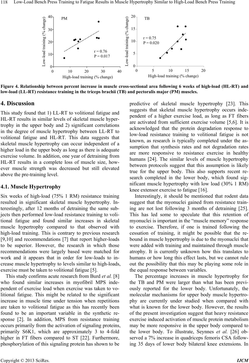
Low-Load Bench Press Training to Fatigue Results in Muscle Hypertrophy Similar to High-Load Bench Press Training
120
2011, pp. 748-752. doi:10.1016/j.mehy.2011.07.029
[6] S. M. Phillips, “Physiologic and Molecular Bases of Mus-
cle Hypertrophy and Atrophy: Impact of Resistance Exer-
cise on Human Skeletal Muscle (Protein and Exercise
Dose Effects),” Applied Physiology, Nutrition, and Me-
tabolism, Vol. 34, No. 3, 2009, pp. 403-410.
doi:10.1139/H09-042
[7] American College of Sports Medicine (ACSM) Position
Stand, “Progression Models in Resistance Training for
Healthy Adults,” Medicine and Science in Sports and Ex-
ercise, Vol. 41, No. 3, 2009, pp. 687-708.
doi:10.1249/MSS.0b013e3181915670
[8] N. A. Burd, D. W. West, A. W. Staples, P. J. Atherton, J.
M. Baker, D. R. Moore, A. M. Holwerda, G. Parise, M. J.
Rennie, S. K. Baker and S. M. Phillips, “Low-Load High
Volume Resistance Exercise Stimulates Muscle Protein
Synthesis More than High-Load Low Volume Resistance
Exercise in Young Men,” PLoS One, Vol. 5, No. 8, 2010,
e12033. doi:10.1371/journal.pone.0012033
[9] J. M. Willardson, “The Application of Training to Failure
in Periodized Multiple-Set Resist ance Exercise Programs,”
Journal of Strength and Conditioning Research, Vol. 21,
No. 2, 2007, pp. 628-631.
[10] G. E. Campos, T. J. Luecke, H. K. Wendeln, K. Toma, F.
C. Hagerman, T. F. Murray, K. E. Ragg, N. A. Ratamess,
W. J. Kraemer and R. S. Staron, “Muscular Adaptations
in Response to Three Different Resistance-Training Re-
gimens: Specificity of Repetition Maximum Training
Zones,” European Journal of Applied Physiology, Vol. 88,
No. 1-2, 2002, pp. 50-60.
doi:10.1007/s00421-002-0681-6
[11] B. Leger, R. Cartoni, M. Praz, S. Lamon, O. Deriaz, A.
Crettenand, C. Gobelet, P. Rohmer, M. Konzelmann, F.
Luthi and A. P. Russell, “Akt Signalling through GSK-
3Beta, mTOR and Foxo1 Is Involved in Human Skeletal
Muscle Hypertrophy and Atrophy,” Journal of Physiol-
ogy, Vol. 576, No. 3, 2006, pp. 923-933.
doi:10.1113/jphysiol.2006.116715
[12] D. W. West, N. A. Burd, J. E. Tang, D. R. Moore, A. W.
Staples, A. M. Holwerda, S. K. Baker and S. M. Phillips,
“Elevations in Ostensibly Anabolic Hormones with Re-
sistance Exercise Enhance neither Training-Induced Mus-
cle Hypertrophy nor Strength of the Elbow Flexors,”
Journal of Applied Physiology, Vol. 108, No. 1, 2010, pp.
60-67. doi:10.1152/japplphysiol.01147.2009
[13] D. W. West and S. M. Phillips, “Associations of Exer-
cise-Induced Hormone Profiles and Gains in Strength and
Hypertrophy in a Large Cohort after Weight Training,”
European Journal of Applied Physiology, Vol. 112, No. 7,
2012, pp. 2693-2702. doi:10.1007/s00421-011-2246-z
[14] V. G. Coffey, Z. Zhong, A. Shield, B. J. Canny, A. V.
Chibalin, J. R. Zierath and J. A. Hawley, “Early Signaling
Responses to Divergent Exercise Stimuli in Skeletal Mus-
cle from Well-Trained Humans,” FASEB Journal, Vol. 20,
No. 1, 2006, pp. 190-192.
[15] S. B. Wilkinson, M. A. Tarnopolsky, E. J. Grant, C. E.
Correia and S. M. Phillips, “Hypertrophy with Unilateral
Resistance Exercise Occurs without Increases in Endo-
genous Anabolic Hormone Concentration,” European
Journal of Applied Physiology, Vol. 98, No. 6, 2006, pp.
546-555. doi:10.1007/s00421-006-0300-z
[16] C. J. Mitchell, T. A. Churchward-Venne, D. W. West, N.
A. Burd, L. Breen, S. K. Baker and S. M. Phillips, “Resis-
tance Exercise Load Does Not Determine Training-Me-
diated Hypertrophic Gains in Young Men,” Journal of
Applied Physiology, Vol. 113, No. 1, 2012, pp. 71-77.
doi:10.1152/japplphysiol.00307.2012
[17] R. Ogasawara, R. S. Thiebaud, J. P. Loenneke and T. Abe,
“Time Course for Arm and Chest Muscle Thickness
Changes Following Bench Press Trai ning, ” Interventional
Medicine and Applied Science, Vol. 4, No. 4, 2012, pp.
217-220. doi:10.1556/IMAS.4.2012.4.7
[18] R. Ogasawara, T. Yasuda, N. Ishii and T. Abe, “Com-
parison of Muscle Hypertrophy Following 6-Month of
Continuous and Periodic Strength Training,” European
Journal of Applied Physiology, in Pr es s.
doi:10.10007/s00421-012-2511-9
[19] P. Kannus, D. Alosa, L. Cook, R. J. Johnson, P. Renstrom,
M. Pope, B. Beynnon, K. Yasuda, C. Nichols and M.
Kaplan, “Effect of One-Legged Exercise on the Strength,
Power and Endurance of the Contralateral Leg. A Ran-
domized, Controlled Study Using Isometric and Concen-
tric Isokinetic Training,” European Journal of Applied
Physiology and Occupational Physiology, Vol. 64, No. 2,
1992, pp. 117-126. doi:10.1007/BF00717948
[20] T. Abe, D. V. DeHoyos, M. L. Pollock and L. Garzarella,
“Time Course for Strength and Muscle Thickne ss Changes
Following Upper and Lower Body Resistance Training in
Men and Women,” European Journal of Applied Physi-
ology, Vol. 81, No. 3, 2000, pp. 174-180.
doi:10.1007/s004210050027
[21] T. Yasuda, R. Ogasawara, M. Sakamaki, H. Ozaki, Y.
Sato and T. Abe, “Combined Effects of Low-Intensity
Blood Flow Restriction Training and High-Intensity Re-
sistance Training on Muscle Strength and Size,” Euro-
pean Journal of Applied Physiology, Vol. 111, No. 10,
2011, pp. 2525-2533. doi:10.1007/s00421-011-1873-8
[22] J. Tannerstedt, W. Apro and E. Blomstrand, “Maximal
Lengthening Contractions Induce Different Signaling Re-
sponses in the Type I and Type II Fibers of Human Ske-
letal Muscle,” Journal of Applied Physiology, Vol. 106,
No. 4, 2009, pp. 1412-1418.
doi:10.1152/japplphysiol.91243.2008
[23] G. Terzis, G. Georgiadis, G. Stratakos, I. Vogiatzis, S.
Kavouras, P. Manta, H. Mascher and E. Blomstrand,
“Resistance Exercise-Induced Increase in Muscle Mass
Correlates with p70S6 Kinase Phosphorylation in Human
Subjects,” European Journal of Applied Physiology, Vol.
102, No. 2, 2008, pp. 145-152.
doi:10.1007/s00421-007-0564-y
[24] M. J. Rennie, H. Wackerhage, E. E. Spangenburg and F.
W. Booth, “Control of the Size of the Human Muscle
Mass,” Annual Review of Physiology, Vol. 66, 2004, pp.
799-828. doi:10.1146/annurev.physiol.66.052102.134444
[25] J. C. Bruusgaard, I. B. Johansen, I. M. Egner, Z. A. Rana
and K. Gundersen, “Myonuclei Acquired by Overload
Exercise Precede Hypertrophy and are Not Lost on De-
training,” Proceedings of the National Academy of Sci-
Copyright © 2013 SciRes. IJCM