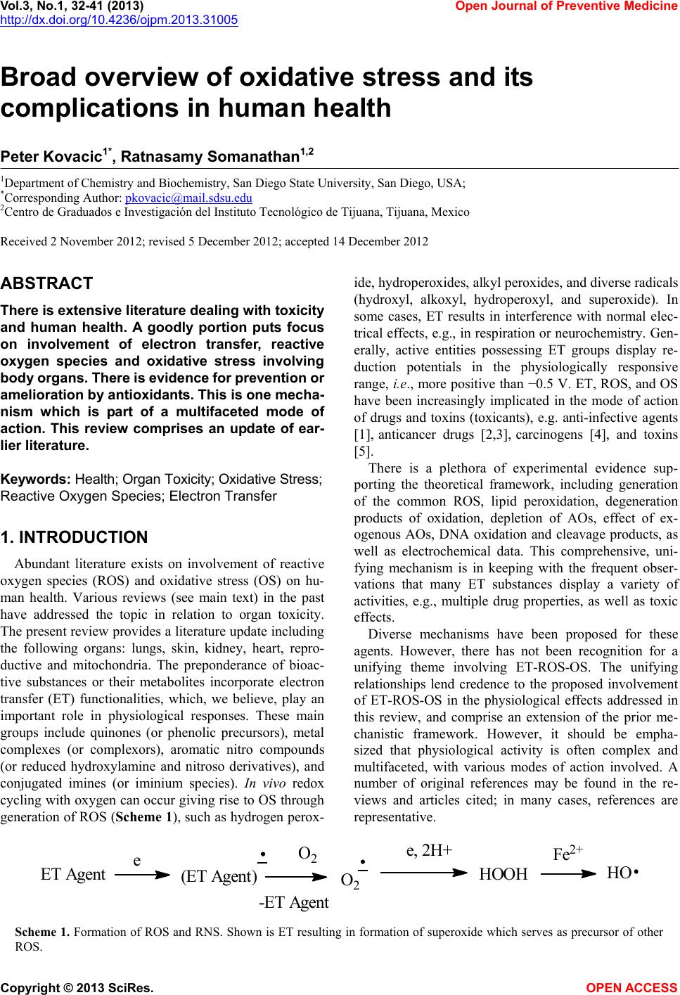
 Vol.3, No.1, 32-41 (2013) Open Journal of Preventive Medicine http://dx.doi.org/10.4236/ojpm.2013.31005 Broad overview of oxidative stress and its complications in human health Peter Kovacic1*, Ratnasamy Somanathan1,2 1Department of Chemistry and Biochemistry, San Diego State University, San Diego, USA; *Corresponding Author: pkovacic@mail.sdsu.edu 2Centro de Graduados e Investigación del Instituto Tecnológico de Tijuana, Tijuana, Mexico Received 2 November 2012; revised 5 December 2012; accepted 14 December 2012 ABSTRACT There is extensive literature dealing with toxicity and human health. A goodly portion puts focus on involvement of electron transfer, reactive oxygen species and oxidative stress involving body organs. Th ere is evidence for pr evention or amelioration by antioxidants. This is one mecha- nism which is part of a multifaceted mode of action. This review comprises an update of ear- lier literature. Keywords: Health; Organ Toxicity; Oxidative Stress; Reactive Oxygen Species; Electron Transfer 1. INTRODUCTION Abundant literature exists on involvement of reactive oxygen species (ROS) and oxidative stress (OS) on hu- man health. Various reviews (see main text) in the past have addressed the topic in relation to organ toxicity. The present review provides a literature update including the following organs: lungs, skin, kidney, heart, repro- ductive and mitochondria. The preponderance of bioac- tive substances or their metabolites incorporate electron transfer (ET) functionalities, which, we believe, play an important role in physiological responses. These main groups include quinones (or phenolic precursors), metal complexes (or complexors), aromatic nitro compounds (or reduced hydroxylamine and nitroso derivatives), and conjugated imines (or iminium species). In vivo redox cycling with oxygen can occur giving rise to OS through generation of ROS (Scheme 1), such as hydrogen perox- ide, hydroperoxides, alkyl peroxides, and diverse radicals (hydroxyl, alkoxyl, hydroperoxyl, and superoxide). In some cases, ET results in interference with normal elec- trical effects, e.g., in respiration or neurochemistry. Gen- erally, active entities possessing ET groups display re- duction potentials in the physiologically responsive range, i. e., more positive than −0.5 V. ET, ROS, and OS have been increasingly implicated in the mode of action of drugs and toxins (toxicants), e.g. anti-infective agents [1], anticancer drugs [2,3], carcinogens [4], and toxins [5]. There is a plethora of experimental evidence sup- porting the theoretical framework, including generation of the common ROS, lipid peroxidation, degeneration products of oxidation, depletion of AOs, effect of ex- ogenous AOs, DNA oxidation and cleavage products, as well as electrochemical data. This comprehensive, uni- fying mechanism is in keeping with the frequent obser- vations that many ET substances display a variety of activities, e.g., multiple drug properties, as well as toxic effects. Diverse mechanisms have been proposed for these agents. However, there has not been recognition for a unifying theme involving ET-ROS-OS. The unifying relationships lend credence to the proposed involvement of ET-ROS-OS in the physiological effects addressed in this review, and comprise an extension of the prior me- chanistic framework. However, it should be empha- sized that physiological activity is often complex and multifaceted, with various modes of action involved. A number of original references may be found in the re- views and articles cited; in many cases, references are representative. ET Agent(ETAgent) O2 -ET Agent O2 ,2H HOOH HO Fe2+ e Scheme 1. Formation of ROS and RNS. Shown is ET resulting in formation of superoxide which serves as precursor of other ROS. Copyright © 2013 SciRes. OPEN ACCE SS  P. Kovacic, R. Somanathan / Open Journal of Preventive Medicine 3 (2013) 32-41 33 2. DISCUSSION 2.1. Neurodegenerative Diseases There has been treatment of neurotoxicity and neu- rodegenerative diseases involving ROS and OS. A re- cent review represents an update of neurodegenerative diseases based on extensive literature [6]. The redox ap- proach comprises a unifying theme which can be applied to a large number of illnesses in this class, including Parkinson’s, Huntington’s, Alzheimer’s, prions, Down’s syndrome, ataxia, multiple sclerosis, Creutzfeldt-Jacob disease, amyotrophic lateral sclerosis, schizophrenia, and tardive dyskinesia. An earlier review addressed neuro- degeneration from a similar mechanistic viewpoint based on ROS-OS [7]. The brain consumes more oxygen under physiological conditions than other organs, thereby increasing its sus- ceptibility to OS since generation of higher levels of ROS can lead to pathological changes when these are in excess of the buffering capacity of endogenous antioxi- dant systems [8]. Extensive data on OS, signaling path- ways, cell death and neuroprotection have been gener- ated in many studies. Hyperoxia produces toxicity, including that of the nervous system. The mammalian brain appears to be particularly sensitive to oxidative damage, one reason being the high oxygen consumption. Rises in calcium interfere with mitochondrial function (including neural), increasing formation of superoxide which can react with nitric oxide (NO) to form the potent oxidant peroxyni- trite (ONOO-), accompanied by lipid peroxidation. Sev- eral neurotransmitters, including dopamine, L-DOPA, serotonin and norepinephrine, can produce ROS, evi- dently via quinone/semiquinone metabolites. Iron is found throughout the brain as complexes with various proteins (see Role of Iron below). Neural membrane lip- ids are replete with polyunsaturated fatty acids whose oxidation products, such as 4-hydroxynonenal and 4- oxononenal [9], can act as sources of ROS, and are espe- cially cytotoxic to neurons. Brain metabolism generates an abundance of hydrogen peroxide via SODs (superox- ide dismutases) and other enzymes [7]. AO defenses are modest, such as catalase levels. Brain microglia can be- come activated to produce superoxide, hydrogen per- oxide and cytokines. Microglia and astrocytes are major players in brain inflammation which is associated with ROS [10]. Some cytochromes leak electrons during the catalytic redox cycle, thus providing ROS [7]. An- other source of brain ROS is NADPH oxidase enzymes. He- moglobin, a neurotoxin, can release heme which is a powerful promoter of lipid peroxidation. The complex of hemoglobin with NO can also generate OS [11]. The Halliwell review presents various means for defense against neurotoxins [7]. There is also reference to earlier treatment of neurodegenerative diseases. A broad overview of neurotoxins was presented based on electron transfer (ET), reactive oxygen species (ROS), and oxidative stress (OS) [12]. It is relevant that metabo- lites from toxins generally posses ET functionalities which can participate in redox cycling. Toxic effects at the molecular level include lipid peroxidation, DNA at- tack, adduction, enzyme inhibition, oxidative attack on the CNS, and cell signaling. The toxins fall into many categories. Beneficial effects of AOs are documented. A related update was reported in 2012 [13]. A similar arti- cle deals with nitric oxide (NO), catecholamine and glu- tamate [14]. The review treats the mechanism of these agents as important neurotransmitters and as neurotoxins, based on involvement of ET-ROS-OS. Role of Iron A recent review presents a unifying theme for cellular death and neurotoxicity by iron agents [6,15]. The basic theme involves continuing and autocatalytic generation of hydroxyl radicals by way of the Fenton reaction in- volving poorly liganded iron. 3. PULMONARY TOXICITY The pulmonary system is one of the main targets for toxicity. In the industrial age, there has been a large in- crease in atmospheric pollutants. Many adverse reactions can occur, some of the principal ones being asthma, COPD and cancer. It is often unclear what role natural components play in the mechanism of pathogenesis. However, a common factor appears to be the upregulation of ROS in lung cells upon exposure. In vivo redox cycling with oxygen can occur giving rise to OS through generation of ROS. ROS can arise from diverse sources, both endogenous and exogenous [16]. Reduction of O2 to ROS, e.g., su- peroxide, occurs as a by-product of metabolism. When cellular injury occurs, release of species, such as iron, into extracellular space can lead to generation of delete- rious ROS. Neutrophils and macrophages are adept at transforming oxygen into ROS which eliminate foreign organisms, accompanied by the undesirable effect of OS on normal cells. The lung is especially susceptible to injury by this gas. For example, hyperoxia damages en- dothelial and alveolar epithelial cells [17]. A recent re- view deals with the consequence of hyperoxia and toxic- ity of oxygen in the lung [18]. Exposure results in in- creased intracellular generation of superoxide and, hence, of other ROS, with ensuing autoxidation reactions. A dramatic example of lung toxicity involving ROS is adult respiratory distress syndrome (ARDS) which is brought about by trauma, shock, sepsis, vomiting, and inhalation of toxins. Inflammation, a common result of Copyright © 2013 SciRes. OPEN ACCE SS  P. Kovacic, R. Somanathan / Open Journal of Preventive Medici ne 3 (2013) 32-41 34 lung insult by toxic substances, is a precursor of subse- quent events triggered by ROS. There is considerable evidence that OS is a contributing factor in ARDS [17]. We wish to emphasize the following quote: “In general, free radicals represent an important component in the pathogenesis of lung disease” [16]. The role of ROS in lung damage is further buttressed by the increased ac- tiveity of free radical-scavenging enzymes in lungs chal- lenged by a variety of toxins. There is appreciable literature on ROS, OS and pul- monary toxicity. In our 2009 review, we surveyed a number of pulmonary toxicants [16]. In this review only those toxicants from more recent years will be cited. 3.1. Role of Nanomaterials In recent years, a wide range of nanomaterials have been developed for various applications. Increasing evi- dence suggests that the special physicochemical proper- ties of these nanomaterials pose potential risk to human health. Recent reviews in this area deal with the biome- chanism and toxicity of nanoparticles, including pulmo- nary insults [19,20]. Data indicate that the composition and size of nano- materials, as well as the target cell type, are critical de- terminants of intracellular responses, degree of cytotox- icity and potential mechanism of toxicity [21]. ROS plays a major role in its toxicity. Alveolar epithelial cells exposed to manganese (II,III)oxide nanoparticles gener- ated ROS leading to OS and apoptosis [22]. Copper ox- ide nanoparticles induce OS and cytotoxicity in airway epithelial cells [23]. A study correlated the conduction band energy with cellular redox potential of Co3O4, Cr2O3, Ni2O3, Mn2O3 and CoO nanoparticles and its ability to induce oxygen radicals, OS, and inflammation [24]. A review deals with the mammalian toxicity of ZnO nanoparticles, through inhalation results in ROS generation which plays an im- portant role in inflammatory response [25]. A 2012 re- view covers toxicity induced by nanoparticles [26]. Mul- tiwalled carbon nanotubes induce OS, with increased ROS production and depletion of intracellular GSH. Multiwalled carbon nanotubes also induce a fibrogenic response by stimulating ROS production [27]. Data showed that exposure of cultured RAW 264.7 cells and A549 human lung cells to multiwalled carbon nanotubes led to OS induced cytotoxicity [28]. 3.2. Role of Organic Toxins OS and oxidative effects on DNA are increased in mice exposed to styrene or styrene oxide, and these may play a role in the lung tumorigenesis [29]. In a related study N-acetylcysteine and GSH were shown to act as AOs in preventing OS in mice exposed to styrene [30]. Chlorobenzene and 1,2-dichlorobenzene cause OS and induce apoptosis in lung epithelial cells at non-acute toxic concentrations [31]. 2-Chloroethyl ethyl sulfide, a sulfur mustard, causes a significant increase in mito- chondrial dysfunction, involving increase in ROS in lung cell injury [32]. Metalloporphyrin acts as an AO in de- creasing mitochondrial ROS, DNA oxidation and the increasing intracellular GSH. A study revealed diallyl trisulfide, a major constituent of garlic oil, induces apop- tosis of U937 human leukemia cells by generation of ROS [33]. Research showed that wood dust from pine, birch and oak is cytotoxic, being able to increase the production of ROS [34]. 4. DERMAL TOXICITY Insults to the skin may be mild, serious or lethal. Various constituents of the skin may be affected by der- mal toxicants. Cutaneous damage may also result from inhalation or ingestion of toxins, in addition to direct skin contact. Similarly, substances that induce toxicity through absorption by the skin can also migrate to and adversely affect other organs. In this review, we draw lines of evidence to support the concept that the ET-ROS-OS unifying theme, which has been successful in describing the means by which many other classes of toxins induce their effects, can also be applied to dermatotoxins [35]. Such toxin classes in- clude a variety of structurally diverse substances. Exposure to the chemical warfare agent sulfur mustard is reported to cause depletion of GSH, which plays an important role in sulfur mustard-linked OS and skin in- jury. Cultured skin epidermal cells and SKH-1 mouse skin when exposed to 2-chloroethyl ethyl sulfide, an analog of mustard gas, led to amelioration by GSH and induction of toxicity [36]. N-Acetyl cysteine, a GSH analog, acts as an AO and shows both protective and therapeutic effects. The sulfur mustard analog 2- chloethyl ethyl sulfide induced oxidative DNA damage in skin epidermal cells and fibroblasts [37]. A related study also revealed that the sulfur mustard analog in- duces OS and activates transcription factors AP-1 and NF-κB via upstream signaling pathways including MAPKs and Akt in SKH-I hairless mouse skin [38]. Cr(IV) induced apoptosis with involvement of reactive oxidants [39]. Data indicated that topical exposure to unpurified single walled carbon nanotubes induced free radical generation, OS, and inflammation, depletion of glutathione, oxidation of protein thiols and carbonyls, elevated myeloperoxidase activity and skin thickening [40]. Amorphous nanosilica induces endocytosis-de- pendent ROS generation and DNA damage in human keratinocytes [41]. Cytotoxicity of uranium, has been in the spotlight in recent decades. Uranyl acetate induces Copyright © 2013 SciRes. OPEN ACCE SS  P. Kovacic, R. Somanathan / Open Journal of Preventive Medici ne 3 (2013) 32-41 35 OS [42]. Carotenoids are known to be potent quenchers of singlet molecular oxygen. Solar light-induced photooxi- dative stress causes skin photoaging by accelerating the generation of ROS. Intake of carotene lowered the met- alloproteinase family which is responsible for wrinkles and sagging of skin, and helps in slowing down ultravio- let-A-induced photoaging in human skin by acting as a singlet oxygen quencher [43]. A 2012 review deals with protective mechanisms of green tea AOs polyphenols in skin [44]. Another article deals with the ROS-mediated skin damage and skin protection by AOs [45]. 5. NEPHROTOXICITY Toxic processes have been implicated in the patho- genesis of several systemic diseases including kidney, which induces OS in the kidney [46]. A 2012 review summarizes the induction of OS in kidney [46]. Much of the nephrotoxicity can be attributed to OS-induced by drugs, and reports address the role of prescription drug- induced OS and toxicity [47,48]. Drug-induced OS is implicated as a mechanism of toxicity in numerous organs, including liver. Well char- acterized drugs associated with OS, include cancer therapies, anti-inflammatory drugs, antiviral agents, an- tipsychotics and analgesics [48]. Metabolism of a drug may generate a reactive intermediate that can induce ROS generation, leading to OS in various organs. Cis- platin is an example of a drug that exhibits multi organ toxicity, acting as an antineoplastic agent. Clinically, renal injury has been described. The kidney accumulates cisplatin to a greater degree than other organs [48]. The disproportionate accumulation of cisplatin in kidney tis- sue contributes to free radical generation, depletion of antioxidants leading to OS and nephrotoxicity [48,49]. Research also indicated that cisplatin nephrotoxicity is associated with mutual mitochondrial/lysosomal poten- tiation (cross talk) of OS in renal proximal tubular cells [50]. This cross-talk results in release of lysosomal di- gestive protease and phospholipases and mitochondrial permeability transition pore opening leading to cyto- chrome c release and activation of caspase cascade, which signals apoptosis. Several reports exist for AO action against cisplatin neph- rotoxicity. Selenium nanoparticles functionalized with 11- mercapto-1-undecanol inhibit ROS-mediated apoptosis [51]. Captopril, an angiotensin-converting enzyme inhibitor containing the sufhydryl group can protect against cis- platin-induced nephrotoxicity in rats [52]. Canabidiol attenuates cisplatin-induced nephrotoxicity by decreasing OS, inflammation and cell death [53]. Data demonstrate a key role of lysosomal iron and early ROS production in gentiomicin-induced lysosomal membrane premeabilization and apoptosis [54]. Salicylic acid was found to attenuate the nephrotoxicity in rats [55]. Ifosfamide, an antineoplastic drug, causes severe nephrotoxicity. The metabolite chloroactaldehyde is be- lieved to be the chemical responsible for the nephrotox- icity [56]. N-Acetyl cysteine acts as an AO in decreasing the nephrotoxicity. A study showed that ROS, OS, and MAPK signaling is involved in promoting cyclosporine- induced glomerular dysfunction and subsequent nephro- toxicity [57]. Increased production of ROS by anticancer drugs trichostatin and 5-aza-deoxycytidine, has been described in patients with various malignancies, which is attributed to their nephrotoxicity. A study showed uric acid attenuates toxicity by pre- venting systemic and renal oxidative stress and tissue damage induced by mercuric chloride in rats [58]. Cad- mium (Cd) is a well known human carcinogen and potent nephrotoxin. Cd caused renal toxicity by inducing lipid peroxidation and morphological alterations [59]. Curcu- min, a natural product from turmeric protects Cd-induced nephrotoxicity in rats. Colistin (polymyxin E), a cationic polypeptide antibiotic, causes OS-induced nephrotoxicity. Melatonin was found to attenuate colistin-induced neph- rotoxicity in rats [60]. Zinc oxide and cadmium sulfide nanoparticles generate ROS that leads to OS-induced nephrotoxicity [61]. 6. CARDIOVASCULAR TO XICITY As described in drug-induced OS and nephrotoxicity, many antibiotic drugs also induce cardiotoxicity [48,62]. The antineoplastic activity of doxorubicin is mediated by interaction with DNA [48]. Reduction of doxorubicin by one electron via mitochondrial reductase may generate anthracycline semiquinone free radicals that produce ROS [48]. Reaction between iron and doxorubicin may also generate ROS. Extensive data have been generated in numerous model systems showing that administration of AOs protects cardiomycetes from doxorubicin-in- duced damage. The range of molecules explored is di- verse, including plant extracts, vitamins C and E, N- acetylcycteine, L-carnitine, beta-blocker carvedilol, co- enzyme Q10, and dexrazonxane [48,63]. Adriamycin is another antineoplastic anthracycline used to treat solid tumors and various forms of cancer, but it displays cardiac toxicity [64]. Adriamycin is well known to produce large amounts of ROS, which may be lethal to cancerous cells. However, unchecked ROS gen- eration typically leads to OS. Adriamycin has been docu- mented to cause oxidative damage in several organs, including heart [64]. Gamma-glutamycysteine ethyl ester was found to suppress the OS induced by adriamycin. Several reviews deal with the mechanism and protection Copyright © 2013 SciRes. OPEN ACCE SS  P. Kovacic, R. Somanathan / Open Journal of Preventive Medici ne 3 (2013) 32-41 36 in anthracycline-induced cardiotoxicity [65-68]. Platinum-based compounds are commonly used cyto- toxic agents in the treatment of several solid tumors. However, their application is still limited in elderly pa- tients, due to the risk in cardiovascular toxicity. The in- creased risk is mainly due to ROS production [69]. AOs have been involved in cancer treatment by their property to suppress the oxidant injury [69,70]. Administration of glucose degradation products in- creased cardiovascular damage in rat models [71]. A related study showed that glucose degradation products and advanced glycation end (AGEs) products play a role in the pathophysiolgy of cardiotoxicity [72]. Mechanistic aspects of AGEs have been addressed [73]. Cocaine is one of the most common illicitly used drugs in the world and causes the most frequent drug- related deaths in young adults. Chronic cocaine con- sumption is associated with serious cardiovascular com- plications. Chronic cocaine administration causes severe myocardial OS through increased ROS production [74]. Cocaine cardiotoxicity may be mediated indirectly through its sympathomimetic effect, inhibiting reuptake and in- creasing the levels of neuronal catecholamines. Exposure to nanoparticles significantly impairs endo- thelium-dependent vasoreactivity in coronary arterioles, and this may be due in large part to increases in mi- crovascular ROS [75]. Another study showed that expo- sure to ambient particulate pollution induces arrhythmia via OS and calcium calmodulin kinase II activation [76]. In recent years two reviews have appeared dealing with heavy metal (Ar, Pb, Cd, Hg) poisoning and car- diovascular disease. The reviews implicate ROS and OS as major players in the pathophysiology of atherosclero- sis [77,78]. 7. REPRODUCTIVE TOXICITY Several earlier reviews have addressed this topic [79- 81]. The present contribution provides recent develop- ments. There is evidence that several teratogens affect the developing embryo by increasing OS resulting in severe embryonic damage. This mechanism seems to operate in diabetic-induced embryonic damage, as well as in the mechanism of teratogenicity caused by ionizing radiation, hypoxia, alcohol and cocaine use and cigarette smoking. Under diabetic conditions, there was a signifi- cant decrease in the activity of endogenous AO enzymes and of vitamins C and E in the embryos. Human and animal studies show that the main mechanism of fetal damage induced by high levels of ionizing irradiation, cocaine and alcohol abuse, hypoxia and cigarette smok- ing is also by increased embryonic OS. Similarly, several drugs exert their teratogenic activity via embryonic OS. Abnormal placentation may also cause enhanced placen- tal OS, resulting in embryonic death. Animal studies also show that a variety of AOs are effective in decreasing the damaging effects of hightened OS induced by teratogens. Concurrent administration of chloroamphenicol (CAP) with multivitamin-haematinics complex (MHC) is a common practice to cushion anticipated anaemic effects in repro- ductive toxicity [82]. Alone, MHC treatment markedly decreased catalase (CAT) and glutathione S-transferase (GST) activities, whereas it resulted in significant in- crease in superoxide dismutase (SOD) activity. Signifi- cant increase in testicular lipid peroxidation and sperm abnormalities were accompanied by reduction in sperm number, sperm motility and live-dead ratio in all treat- ment groups. MHC-induced testicular toxicity occurred vis OS. Mequindox (MEQ) is a synthetic antimicrobial che- mical [83]. A study was designed to investigate the hy- pothesis that MEQ exerts testicular toxicity by causing OS. Superoxide dismutase (SOD), reduced glutathione (GSH) and 8-hydroxydeoxyguananosine (8-OHdG) lev- els were elevated, whereas the malondialdehyde (MDA) level was slightly increased. The findings provide evi- dence in vivo for the formation of free radicals. Reports document the protective effect of AOs, such as vitamin E [84] and Ginko biloba [85]. Various studies on reproductive toxicity deal with the role of metals: Pb [86], Cr [87,88], and Se [89]. 8. MITOCHONDRIA Mitochondrial OS has long been implicated in normal aging, and a host of human diseases, including cancer and neurodegenerative disorders. Mitochondria are the major source of oxygen free radicals in most cell types. Low concentrations of ROS can serve as signaling func- tions, triggering the activation of specific pathways [90]. However, high concentrations of ROS cause lipid per- oxidation, damage to cell membranes, proteins, and DNA. Mitochondria are, therefore, a major source of ROS and a major target of ROS-induced OS and damage. A high rate of mitochondrial DNA mutation eventually exacer- bates mitochondrial dysfunction and reduces mitochon- drial energy production [90]. Under OS conditions, mitochondria release various pro-apoptoic factors. This release is caused by the per- meabilization of the mitochondrial outer membrane, which accompanies the depolarization of the mitochon- drial intermembrane potential. Aldehydes are generated during numerous physiological processes. Aldehydes are highly reactive agents, which form adducts with lipids, proteins, and DNA, affecting the function of these mac- romolecules. Aldehyde dehydrogenases are important enzymes that eliminate toxic aldehydes by catalyzing their oxidation to non-reactive acids. A review discusses the mitochobdrial aldehyde dehydrogenase and cardiac diseases [91]. Inhibition of aldehyde dehydrogenase 2 by Copyright © 2013 SciRes. OPEN ACCE SS  P. Kovacic, R. Somanathan / Open Journal of Preventive Medici ne 3 (2013) 32-41 37 OS is associated with cardiac dysfunction in diabetic rats [92]. A study demonstrated that TNF-α-induced OS al- ters redox homeostasis by impairing the membrane per- meability transition pore opening proteins adenine nu- cleotide translocator and voltage-dependent anion chan- nel, thereby resulting in the pore opening, causing mito- chondrial dysfunction and attenuated cardiac function [93]. A recent review deals with the mitochondria death/ survival signaling pathways in cardiotoxicity induced by anthracyclines [94]. Data shows that inhibition of mito- chondrial transition permeability prevents doxorubicin- induced cardiotoxicity. OS and mitochondrial dysfunc- tion have been implicated in atherosclerosis [95]. Au- thors suggest mitochondria-targeted AOs as potential therapy. Alzheimer’s disease, the most common form of dementia with a progressive course, evidences neuronal damage in specific vulnerable brain regions and circuits involved in memory and language [96]. Two recent re- views deal with mitochondrial- and endoplasmic reticu- lum-associated OS in Alzheimer’s disease [96,97]. There is an emerging consensus that aging is a multi- factorial process, which is genetically determined and influenced epigenetically by environment. OS induced DNA damage, oxidation of proteins, lipid peroxidation and ROS have been implicated as causative factors of aging [98]. Two reviews deal with the OS, mitochondrial dysfunction and aging [98,99]. 9. CONCLUSION This review presents recent reports dealing with elec- tron transfer, reactive oxygen species and oxidative stress as part of the mechanism of toxicity involving or- gans in human health. Various portions of the organ cells may be involved in the insults. Antioxidants may play a role in prevention. 10. ACKNOWLEDGMENTS Editorial assistance by Thelma Chavez is acknowledged. REFERENCES [1] Kovacic, P. and Becvar L.E. (2000) Mode of action of anti-infective agents: Emphasis on oxidative stress and electron transfer. Current Pharmaceutical Design, 6, 143- 167. doi:10.2174/1381612810006020143 [2] Kovacic, P. and Osuna J.A. (2000) Mechanisms of anti- cancer agents: Emphasis on oxidative stress and electron transfer. Current Pharmaceutical Design, 6, 277-309. doi:10.2174/1381612003401046 [3] Kovacic, P. (2007) Unifying mechanism for anticancer agents involving electron transfer and oxidative stress: Clinical implications. Medical Hypotheses, 69, 510-516. doi:10.1016/j.mehy.2006.08.046 [4] Kovacic, P. and Jacintho, J.D. (2001) Mechanism of car- cinogenesis: Focus on oxidative stress and electron trans- fer. Current medicinal chemistry, 8, 773-796. doi:10.2174/0929867013373084 [5] Kovacic, P. and Somanathan, R. (2008) Unifying mecha- nism for eye toxicity: Electron transfer, reactive oxygen species, antioxidant benefits, cell signaling and cell mem- branes. Cell Membranes and Free Radical Research, 1, 56-69 (and references therein). [6] Kovacic, P. and Somanathan, R. (2012) Redox process in neurodegerative disease involving reactive oxygen spe- cies. Current Neuropharmacology, 10, in Press. [7] Halliwell, B. (2006) Oxidative stress and neurodegenera- tion: Where are we now? Journal of Neurochemistry, 97, 1634-1658. doi:10.1111/j.1471-4159.2006.03907.x [8] Robert, R., Smith, R.A., Safe, S., Szabo, C., Tjalkens, R. B. and Robertson, F.M. (2010) Toxicological and path- physiological roles of reactive oxygen and nitrogen spe- cies. Toxicology, 276, 85-94. doi:10.1016/j.tox.2010.07.009 [9] Kovacic, P. (2006) Novel electrochemical approach to enhanced toxicitry of 4-oxo-2-nonenal vs. 4-hydroxy-2- nonenal (role of imine): Oxidative stress and therapeutic modalities. Medical Hypotheses, 67, 151-156. doi:10.1016/j.mehy.2005.10.034 [10] Min, K.J., Yang, M.S., Kim, S.U. and Joe, E.H. (2006) Astrocytes induce hemeoxygenase-1 expression in mi- croglia: A feasible mechanism for preventing excessive brain inflammation. The Journal of Neuroscience, 26, 1880- 1887. doi:10.1523/JNEUROSCI.3696-05.2006 [11] Chiu, D.T., van den Berg, J., Kuypers, F.A., Hung, I.-J., Wei, J.-S. and Liu, T.-Z. (1996)Correlation of membrane lipid peroxidation with oxidation of hemoglobin variants: Possibly related to the rates of hemin release. Free Radi- cal Biology & Medicine, 21, 89-95. doi:10.1016/0891-5849(96)00035-4 [12] Kovacic, P. and Somanathan, R. (2012) Nervous about developments in electron transfer-ROS-oxidative stress: mechanisms of neurotoxicity? In: Laher, J, Ed., Systems Biology of Free Radicals and Antioxidants, Springer Ver- lag, New York, in Press. [13] Kovacic, P. and Somanathan, R. (2005) Neurotoxicity: the broad framework of electron transfer, oxidative stress and protection by antioxidants. Current Medicinal Chem- istry—Central Nervous System Agents, 5, 249-258. doi:10.2174/156801505774913044 [14] Jacintho, J.D. and Kovacic, P. (2003) Neurotransmission and neurotoxicity by nitric oxide, catecholamines, and glutamate: Unifying themes of reactive oxygen species and electron transfer. Current Medicinal Chemistry, 10, 2693-2703. doi:10.2174/0929867033456404 [15] Kell, D.B. (2010) Towards a unifying, systems biology understanding of large-scale cellular death and destruc- tion caused by poorly liganded iron: Parkinson’s, Hunt- ington’s, Alzheimer’s. prions, bactericides, chemical toxi- cology and others as examples. Archives of Toxicology, 84, 825-889. doi:10.1007/s00204-010-0577-x [16] Kovacic, P. and Somanathan, R. (2009) Pulmonary toxic- Copyright © 2013 SciRes. OPEN ACCE SS  P. Kovacic, R. Somanathan / Open Journal of Preventive Medici ne 3 (2013) 32-41 38 ity and environmental contamination: Radicals, electron transfer, and protection by antioxidants. Reviews of Envi- ronmental Contamination and Toxicology, 201, 41-69. doi:10.1007/978-1-4419-0032-6_2 [17] Halliwell, B. and Gutteridge, J.M.C. (1999) Free Radicals in Biology and Medicine. Oxford University Press, New York, 1-897. [18] Mach, W.J., Thimmesch, A.R., Pierce, J.T. and Pierce, J.D. (2011) Consequence of hyperoxia and the toxicity of oxygen in the lung. Nursing Research and Practice, 2011, Article ID: 260482, 7 Pages. doi:10.1155/2011/260482 [19] Kovacic, P. and Somanathan, R. (2010) Biomechanisms of nanoparticles (toxicants, antioxidants and therapeutics): Electron transfer and reactive oxygen species. Journal of Nanoscience and Nanotechnology, 10, 1-12. doi:10.1166/jnn.2010.3028 [20] Jia’en Li, J., Muralikrishnan, S., Ng, C.-T., Yung, L.Y.L. and Bay, B.-H. (2010) Nanoparticle-induced pulmonary toxicity. Experimental Biology and Medicine, 235, 1025- 1033. doi:10.1258/ebm.2010.010021 [21] Sohaebuddin, S.K., Thevenot, P., Baker, D., Eaton, J.W. and Tang, L. (2012) Nanomaterial cytotoxicity is compo- sition, size, and cell type dependent. Particle and Fibre Toxicology, 7, 1-17. [22] Frick, R., Müller-Edenborn, B., Sclicker, A., Rothen- Rutishauser, B., Raemy, D.O., Günther, D., Hattendorf, B., Stark, W. and Beck-Schimmer, B. (2011) Comparison of manganese oxide nanoparticles and manganese sulfate with regard to oxidative stress, uptake and apoptosis in alveolar epithelial cells. Toxicology Letters, 205, 163-172. doi:10.1016/j.toxlet.2011.05.1037 [23] Fahmy, B. and Cormier, S.A. (2009) Copper oxide nanoparticles induce oxidative stress and cytotoxicity in airway epithelial cells. Toxicology in Vitro, 23, 1365-1371. doi:10.1016/j.tiv.2009.08.005 [24] Zhang, H., Ji, Z., Xia, T., Meng, H., Low-Kam, C., Liu, R., Pokhrel, S., Lin, S., Wang, X., Liao, Y.-P., Wang, M., Li, L., Rallo, R., Damoiseaux, R., Telesca, D., Mädler, L., Cohen, Y., Zink, J. I. and Nel, A.E. (2012) Use of metal oxide nanoparticle band gap to develop a predictive para- digm for oxidative stress and acute pulmonary inflamma- tion. ACS Nano, 6, 4349-4368. doi:10.1021/nn3010087 [25] Vanderbriel, R.J. and De Jong, W.H. (2012) A review of mammalian toxicity of ZnO nanoparticles. Journal of Nanotechnology, Science and Applications, 5, 61-71. doi:10.2147/NSA.S23932 [26] Shinde S.K., Grampurohit, N.D., Gaikwad, D.D., Jadhav, S.L., Gadhave, M.V. and Shelke, P.K. (2012) Toxicity induced by nanoparticles. Asian Pacific Journal of Tro- pical Disease, 2, 331-334. doi:10.1016/S2222-1808(12)60072-3 [27] He, X., Young, S.H., Schwegler-Berry, D., Chisholm, W.P., Fernback, J.E. and Ma, Q. (2011) Multiwalled car- bon nanotubes induce a fibrogenic response by stimulat- ing reactive oxygen species production, activating NF- κB signaling, and promoting fibroblast-to-myofibroblast transformation. Chemical Research in Toxicology, 24, 2237- 2248. doi:10.1021/tx200351d [28] Chen, B., Liu, Y., Song, W.M., Hayashi, Y., Ding, X.C. and Li, W.H. (2011) In vitro evaluation of cytotoxicity and oxidative stress induced by multiwalled carbon nano- tubes in murine RAW 264.7 macrophages and human A549 lung cells. Biomedical and Environmental Sciences, 24, 593-601. [29] Harvilchuck, J.A., Pu, X., Klaunig, J.E. and Carlson, G.P. (2009) Indicators of oxidative stress and apoptosis in mouse whole lung and Clara cells following exposure to styrene and its metabolites. Toxicology, 264, 171-178. doi:10.1016/j.tox.2009.08.001 [30] Meszka-Jordan, A., Mahlapuu, R., Soomets, U. and Carl- son, G. P. (2009) Oxidative stress due to (R)-styrene oxide exposure and the role of antioxidants in non-Swiss albino (NSA) mice. Journal of Toxicology and Environmental Health, Part A, 72, 642-650. doi:10.1080/15287390902769436 [31] Mörbt, N., Tomm, J., Feltens, R., Mögel, I., Kalkhof, S., Murugesan, K., Wirth, H., Vogt, C., Binder, H., Lehmann, I. and von Bergen, M. (2011) Chlorinated benzenes cause concomitantly oxidative stress and induction of apoptotic markers in lung epithelial cells (A549) at non-acute toxic concentrations. Journal of Proteome Research, 10, 363- 378. doi:10.1021/pr1005718 [32] Gould, N.S., White, C.W. and Day, B.J. (2009) A role for mitochondrial oxidative stress in sulfur mustard analog 2-chloroethyl ethyl sulfide-induced lung injury and anti- oxidant protection. Journal of Pharmacology and Ex- perimental Therapeutics, 328, 732-739. doi:10.1124/jpet.108.145037 [33] Choi, Y.H. and Park, H.S. (2012) Apoptosis induction of U937 human leukemia cells by diallyl trisulfide induces through generation of reactive oxygen species. Journal of Biomedical Science, 19, 50. doi:10.1186/1423-0127-19-50 [34] Pylkkänen, L., Stockmann-Javala, H., Alenius, H., Hus- gafvel-Pursiainen, K. and Savolainen, K. (2009) Wood dusts induce the production of reactive oxygen species and caspase-3 activity in human bronchial epithelial cells. Toxicology, 262, 265-270. doi:10.1016/j.tox.2009.06.019 [35] Kovacic, P. and Somanathan, R. (2010) Dermal toxicity and environmental contamination: Electron transfer, reac- tive oxygen species, oxidative stress, cell signaling, and protection by antioxidants. Reviews of Environmental Contamination and Toxicology, 203, 119-138. doi:10.1007/978-1-4419-1352-4_4 [36] Tewari-Singh, N., Agarwal, C., Huang, J., Day, B.J., White, C.W. and Agarwal, R. (2011) Efficacy of glu- tathione in ameliorating sulfur mustard analog-induced toxicity in cultutred skin epidermal cells and in SKH-1 mouse skin in vivo. Journal of Pharmacology and Ex- perimental Therapeutics, 336, 450-459. doi:10.1124/jpet.110.173708 [37] Tewari-Singh, N, Gu, M., Ahotriya, S., Gomnez, J., Agarwal, C., White, C.W. and Agarwal, R. (2011) Mecha- nisms of sulfur mustard analog 2-chloeroethyl ethyl sul- fide-induced DNA damage in skin epidermal cells and fi- broblasts. Free Radical Biology & Medicine, 51, 2272- 2280. doi:10.1016/j.freeradbiomed.2011.08.020 [38] Pal, A., Twari-Singh, N., Gu, M., Agarwal, C., Huang, J., Copyright © 2013 SciRes. OPEN ACCE SS  P. Kovacic, R. Somanathan / Open Journal of Preventive Medici ne 3 (2013) 32-41 39 Day, B. J., White, C. W. and Agarwal, R. (2009) Sulfur mustard analog induces oxidative stress and activates sig- naling cascades in the skin of SKH-1 hairless mice. Free Radical Biology & Medicine, 47, 1640-1651. doi:10.1016/j.freeradbiomed.2009.09.011 [39] Son, Y.-O., Hitron, J.A., Wang, X., Chang, Q., Pan, J., Zhang, Z., Liu, J., Wang, S., Lee, J.-C. and Shi, X. (2010) Cr(IV) induces mitochondrial-mediated and caspase-de- pendent apoptosis through reactive oxygen species-me- diated p53 activation in JB6 C141 cells. Toxicology and Applied Pharmacology, 245, 226-235. doi:10.1016/j.taap.2010.03.004 [40] Murray, A.R., Kisin, E., Leonard, S.S., Young, S.H., Kommineni, C., Kagan, V.E., Castranova, V. and Shve- dova, A.A. (2009) Oxidative stress and inflammatory re- sponse in dermal toxicity of single walled carbon nano- tubes. Toxicology, 257, 161-171. doi:10.1016/j.tox.2008.12.023 [41] Nabeshi, H., Yoshikawa, T., Matsuyama, K., Nakazato, Y., Tochigi,S., Kondoh, S., Hirai, T., Akase, T., Nagano, K., Abe, Y., Yoshioka, Y., Kamada, H., Itoh, N., Tsunoda, S.-I, and Tsutsumi, Y. (2011) Amorphous nanosilica induce endocytosis-dependent ROS generation and DNA damage in human keratinocytes. Particle and Fibre Toxicology, 8, 1. doi:10.1186/1743-8977-8-1 [42] Daraei, B., Pourahmad, J., Hamidi-Pour, N., Hosseneini, M.-J., Shaki, F. and Solleimani, M. (2012) Uranyl acetate induces oxidative stress and mitochondrial membrane potential collapse in the human dermal fibroblast primary cells. Iranian Pharmaceutical Research, 11, 495-501. [43] Terao, J., Minami, Y. and Bando, N. (2011) Singlet mo- lecular oxygen-quenching activity of carotenoids: Rele- vance to protection of the skin from photoaging. Journal of Clinical Biochemistry and Nutrition, 48, 57-62. doi:10.3164/jcbn.11-008FR [44] White, P.O., Tribout, H. and Baron, E. (2012) Protective mechanisms of green tea polyphenols in skin. Oxidative Medicine and Cellular Longevity, 201 2, Article ID: 560682, 8 Pages. doi:10.1155/2012/560682 [45] Ascenso, A., Ribeiro, H.M., Marques, H.C. and Simoes, S. (2011) Topical delivery of antioxidants. Current Drug De- livery, 8, 640-660. doi:10.2174/156720111797635487 [46] Ozbek, E. (2012) Induction of oxidative stress in kidney. International Journal of Nephrology, 2012, Article ID: 465897, 9 Pages. doi:10.1155/2012/465897 [47] Ramatillah, D.L., Gillani, S.W. and Suardi, M. (2012) Effect of cytotoxic medications (MTX, cisplatin, 5-FU and cyclophosphamide) against creatinine clearance. Pa- tient relationships and creatinine clearance urea with cancer patients. International Journal of Pharmacy Edu- cation, 3, 240-244. [48] Deavall, D.G., Mertin, E.A., Horner, J.M. and Roberts, R. (2012) Drug-induced oxidative stress and toxicity. Jour- nal of Toxicology, 2012, Article ID: 645460, 13 Pages. doi:10.1155/2012/645460 [49] Yao, X., Panichpisal, K., Kurtzman, N. and Nugent, K. (2007) Cisplatin nephrotoxicity; a review. The American Journal of the Medical Sciences, 334, 115-124. doi:10.1097/MAJ.0b013e31812dfe1e [50] Pourahmad, J., Hosseini M.J., Eskandari, M.R., Shekarabi, S.M. and Daraei, B. (2010) Mitochondrial/lysosomal toxic cross-talk plays a key role in cisplatin nephrotoxicity. Xenobiotica, 40, 763-771. doi:10.3109/00498254.2010.512093 [51] Li, Y., Li, X., Wong, Y.S., Chen, T., Zhang, H., Liu, C. and Zheng, W. (2011) The reversal of cisplatin-induced nephrotoxicity by selenium nanoparticles functionalized with 11-mercapto-1-undecanol by inhibition of ROS- mediated apoptosis. Biomaterials, 32, 9068-9076. doi:10.1016/j.biomaterials.2011.08.001 [52] El-Sayed, E.-S.M., Abd-Ellah, M.F. and Attia, S.Y.M. (2008) Protective effect of captopril against cisplatin-in- duced nephrotoxicity in rats. Pakistan Journal of Phar- maceutical Sciences, 21, 255-261. [53] Pan, H., Mukhopadhyay, P., Rajesh, M., Patel, V., Muk- hopadhyay, B., Gao, B., Kasko, G. and Pacher, P. (2009) Cannabidol attenuates cisplatin-induced nephrotoxicity by decreasing oxidative/nitrosative stress, inflammation, and cell death. Journal of Pharmacology and Experi- mental Therapeutics, 328, 708-714. doi:10.1124/jpet.108.147181 [54] Denamur, S., Tyteca, D., Marchand-Brynaert, J., Van Bambeke, F., Tulkens, P. M. Courtoy, P. J. and Mingeot- Leclercq, M-P. (2011) Role of oxidative stress in ly- sosomal membrane permeabilization and apoptosis in- duced by gentamicin, an aminoglycoside antibiotic. Free Radical Biology & Medicine, 51, 1656-1665. doi:10.1016/j.freeradbiomed.2011.07.015 [55] Randjelovic, P., Veljkovic, S., Stojiljkovic, N., Jank- ovic-Velickovic, L., Sokolovic, D., Stoiljkovic, M. and Ilic, I. (2012) Salicylic acid attenuates gentamicin-in- duced nephrotoxicity in rats. The Scientific World Journal, 2012, Article ID: 390613, 6 Pages. doi:10.1100/2012/390613 [56] Hanly, L., Chen, N., Rieder, M. and Koren, G. (2009) Ifosfamide nephrotoxicity in children: A mechanistic base for pharmacological prevention. Expert Opinion on Drug Safety, 8, 155-168. doi:10.1517/14740330902808169 [57] O’Connell, S., Tuite, N., Slattery, C., Ryan, M.P. and Mc- Morrow, T. (2012) Cyclosporin A-induced oxidative stress in human renal mesangial cells; a role for ERK 12 MAPK signaling. Toxicological Sciences, 126, 101- 113. doi:10.1093/toxsci/kfr330 [58] Durante, P., Romero, F., Pérez, M., Chávez, M. and Parra, G. (2010) Effect of uric acid on nephrotoxicity induced by mercuric chloride in rats. Toxicology and Industrial Health, 26, 163-174. doi:10.1177/0748233710362377 [59] Tarasub, N., Tarasub, C. and Ayutthaya, W.D.N. (2011) Protective role of curcumin on cadmium-induced neph- rotoxicity in rats. Journal of Environmental Chemistry and Ecotoxicology, 3, 17-24. [60] Yousef, J., Chen, G., Hill, P.A., Nation, R. L. and Li, J. (2011) Melatonin attenuates colistin-induced nephrotox- icity in rats. Antimicrobial Agents and Chemotherapy, 55, 4044-4049. doi:10.1128/AAC.00328-11 [61] Pujalté, I., Passagne, I., Brouillaud, B.L.,Tréguer, M., Copyright © 2013 SciRes. OPEN ACCE SS  P. Kovacic, R. Somanathan / Open Journal of Preventive Medici ne 3 (2013) 32-41 40 Durand, E., Ohayon-Courtés, C. and L’Azou, B. (2011) Cytotoxicity and oxidative stress induced by different metallic nanoparticles on human kidney cells. Particle and Fibre Toxicology, 8, 10. doi:10.1186/1743-8977-8-10 [62] Shafik, A., Khodeir, M.M. and Fadel, M.S. (2011) Animal study of anthracycline-induced cardiotoxicity and neph- rotoxicity and evaluation of protective agents. Journal of Cancer Science & Therapy, 3, 96-103. [63] Shi, R., Huang, C.-C., Aronstam, R.S., Ercal, N., Martin, A. and Huang, Y.-W. (2009) N-acetylcysteine amide de- creases oxidative stress but not cell death induced by doxorubicin in H9c2 cardiomyocytes. BMC Pharmacol- ogy, 9, 7. doi:10.1186/1471-2210-9-7 [64] Aluise, C.D., St. Clair, D., Vore, M. and Butterfield, D.A. (2009) In vivo amelioration of adrinamycin induced oxi- dative stress in plasma by gamma-glutamylcysteine ethyl ester (GCEE). Cancer Letters, 282, 25-29. doi:10.1016/j.canlet.2009.02.047 [65] Sawyer, D.B., Peng, X., Chen, B., Pentassuglia, L. and Lim, C.C. (2010) Mechanisms of anthracycline cardiac injury: Can we identify strategies for cardio-protection? Progress in Cardiovascular Diseases, 53, 105-113. doi:10.1016/j.pcad.2010.06.007 [66] Ferreira, A.L.A., Matsubara, L.S. andMatsubara, B.B. (2008) Anthracycline-induced cardiotoxicity. Cardiovas- cular & Hematological Agents in Medicinal Chemistry, 6, 278-281. doi:10.2174/187152508785909474 [67] Vergely, C., Delemasure, S., Cottin, Y. and Rochette, L. (2007) Preventing the cardiotoxic effects of anthracy- clines: From basic concepts to clinical data. Heart and Metabolism, 35, 1-7. [68] James, H.D. (2012) Dexrazane for the prevention of car- diac toxicity and treatment of extravasation injury from the anthracycline antibiotic. Current Pharmaceutical Biotechnology, 13, 1949-1956. doi:10.2174/138920112802273245 [69] Ferroni, P., Della-Morte, D., Palmirotta, R., McClendon, M., Testa, G., Abete, P., Rengo, F., Rundek, T., Guadagni, F. and Roselli, M. (2011) Platinum-based compounds and risk for cardiovascular toxicity in the elderly: Role of the antioxidants in chemoprevention. Rejuvenation Research, 14, 293-308. doi:10.1089/rej.2010.1141 [70] Cheng, C.F., Juan, S.H., Chen, J.J., Chao, Y.C., Chen, H.H., Lian, W.S., Lu, C.Y., Chang, C.I., Chiu, T.H. and Lin, H. (2008) Pravatatin attenuates carboplatin-induced cardiotoxicity via inhibition of oxidative stress associated apoptosis. Apoptosis, 13, 883-894. doi:10.1007/s10495-008-0214-9 [71] Müller-Krebs, S., Kihm, L.P., Zeier, B., Gross, M.L., Wieslander, A., Haug, U., Zeier, M. and Schwenger, V. (2010) Glucose degradation products result in cardiovas- cular toxicity in a rat model of renal failure. Peritoneal Dialysis International, 30, 35-40. doi:10.3747/pdi.2009.00031 [72] Himmele, R., Sawin, D.-A. and Diaz-Buxo, J.A. (2011) GDPs and AGEs: Impact on cardiovascular toxicity in di- alysis patients. Advances in Peritoneal Dialysis, 27, 22- 26. [73] Kovacic, P. and Somanathan, R. (2011) Cell signaling and receptors in toxicity of advanced glycation products (AGEs): α-dicarbonyl, radicals, oxidative stress and anti- oxidants. Journal of Receptors and Signal Transduction Research, 31, 332-339. doi:10.3109/10799893.2011.607171 [74] Fan, L., Sawbridge, D., George, V., Teng, L., Baily, A., Kitchen, I. and Li, J.-M. (2009) Chronic cocaine-induced cardiac oxidative stress and mitogen-activated protein kinase activation: The role of Nox2 oxidase. Journal of Pharmacology and Experimental Therapeutics, 328, 99- 106. doi:10.1124/jpet.108.145201 [75] LeBlanc, A.J., Mosely, A.M., Chen, B.T., Frazer, D., Cas- tranova, V. and Nurkiewicz, T.R. (2010) Nanoparticle in- halation impairs coronary microvascular reactivity via a local reactive oxygen species-dependent mechanism. Cardiovascular Toxicology, 10, 27-36. doi:10.1007/s12012-009-9060-4 [76] Kin, J.B., Kim, C., Choi, E., Park, H., Pak, H.N., Lee, M. H., Shin, D.C., Hwang, K.C. and Joung, B. (2012) Par- ticulate air pollution induces arrhythmia via oxidative stress and calcium calmodulin kinase II activation. Toxi- cology and Applied Pharmacology, 259, 66-73. doi:10.1016/j.taap.2011.12.007 [77] Alissa, E. and Ferns, G.A. (2011) Heavy metal poisoning and cardiovascular disease. Journal of Toxicology, 2011, Article ID: 870125, 21 Pages. doi:10.1155/2011/870125 [78] Azevedo, B.F., Furieri, L.B., Peçanha, F.M., Wiggers, G.A., Vassallo, P.F., Simõexs, M.R., Fiorim, J., de Batista, P., Fioresi, M., Rossoni, L., Stefanon, I., Alonso, M.J., Salaices, M. and Vassallo, D.V. (2012) Toxic effects of mercury on the cardiovascular and central nervous sys- tems. Journal of Biomedicine and Biotechnology, 2012, Article ID: 949048. doi:10.1155/2012/949048 [79] Kovacic, P and Jacintho, J.D. (2001) Reproductive toxins: pervasive theme of oxidative stress and electron transfer. Current Medicinal Chemistry, 8, 863-892. doi:10.2174/0929867013372878 [80] Kovacic, P and Somanathan, R. (2006) Mechanism of teratogenesis: Electron transfer, reactive oxygen species and antioxidants. Birth Defects Research Part C, 78, 308- 325. doi:10.1002/bdrc.20081 [81] Hansen, J.M. (2006) Oxidative stress as a mechanism of teratogenesis. Birth Defects Research Part C, 78, 293-307. doi:10.1002/bdrc.20085 [82] Oyagbemi, A.A., Adedara, I.A., Saba, A.B. and Farombi, E.O. (2010) Role of oxidative stress in reproductive tox- icity induced by coadministration of chloroamphenicol and multivitamin-haematinics complex in rats. Basic & Clinical Pharmacology & Toxicology, 107, 103-108. doi:10.1111/j.1742-7843.2010.00561.x [83] Ihsan, A., Wang, X., Liu, Z., Wang, Y., Huang, X., Liu, Y., Yu, H., Zhang, H., Li, T., Yang, C. and Yuan, Z. (2011) Long-term mequindox treatment induced endocrine and reproductive toxicity via oxidative stress in male Wistar rats. Toxicology and Applied Pharmacology, 252, 281- 288. doi:10.1016/j.taap.2011.02.020 [84] Wang, N., Qian, H.Y., Zhou, X.Q., Li, X.Q. and Sun, Z.W. (2012) Mitochondrial energy metaboilism dysfunction involved in reproductive toxicity of mice caused by en- Copyright © 2013 SciRes. OPEN ACCE SS  P. Kovacic, R. Somanathan / Open Journal of Preventive Medici ne 3 (2013) 32-41 Copyright © 2013 SciRes. OPEN ACCE SS 41 dosulfan and protective effects of vitamin E. Ecotoxicol- ogy and Environmental Safety, 82, 96-103. doi:10.1016/j.ecoenv.2012.05.014 [85] Amin, A., Abraham, C., Hamza, A.A., Abdalla, Z.A., Al-Shamsi, S.B., Harethi, S.S. and Daoud, S. (2012) A standardized extract of Ginkgo biloba neutralizes cis- platin-mediated reproductive toxicity in rats. Journal of Biomedicine and Biotechnology, 2012, Article ID: 362049, 11 Pages. doi:10.1155/2012/362049 [86] Ait H.N., Slimani, M., Merad-Boudia, B. and Zaoui, C. (2009) Reproductive toxicity of lead acetate in adult male rats. American Journal of Veterinary Research, 38-50. [87] Subramanian, S., Rajendiran, G., Skhar, P., Gowri, C., Govindarajulu, P. and Aruldhas, M.M. (2006) Reproduc- tive toxicity of chromium in adult bonnet monkeys (Macaca radiate Geoffrey) Reversible oxidative stress in the seman. Toxicology and Applied Pharmacology, 215, 237-249. doi:10.1016/j.taap.2006.03.004 [88] Hari, P. and Sreenivasula R.P. (2012) Effect of restraint stress on lead-induced male reproductive toxicity in rats. Journal of Experimental Zoology, 317A, 455-465. doi:10.1002/jez.1738 [89] Kaushal, N. and Bansal, M.P. (2009) Diminished repro- ductive potential of male mice in response to sele- nium-induced oxidative stress: Involvement of HSP70, HSP70-2, and MSJ-1. Journal of Biochemical and Mo- lecular Toxicology, 23, 125-136. doi:10.1002/jbt.20276 [90] Borniquel, S., Valle, I., Cadenas, S., Lamas, S. and Mon- salve, M. (2006) Nitric oxide regulates mitochondrial oxidative stress protection via the transcriptional coacti- vator PGC-1α. The FASEB Journal, 20, E1216-E1227. doi:10.1096/fj.05-5189fje [91] Chen, C.-H., Sun, L. and Mochly-Rosen, D. (2010) Mi- tochondrial aldehyde and dehydrogenase and cardiac dis- eases. Cardiovascular Research, 88, 51-57. doi:10.1093/cvr/cvq192 [92] Wang, J., Wang H., Hao, P., Xue, L., Wei, S., Zhang, Y. and Chen, Y. (2011) Inhibition of aldehyde dehydro- genase 2 by oxidative stress is associated with cardiac dysfunction in diabetic rats. Molecular Medicine, 17, 172-179. [93] Mariappan, N., Soorappan, R.N., Haque, M., Siramula, S. and Francis, J. (2007) TNF-α-induced mitochondrial oxi- dative stress and cardiac dysfunction: Restoration by su- peroxide dismutase mimetic Tempol. American Journal of Physiology—Heart and Circulatory, 293, H2726-H2737. doi:10.1152/ajpheart.00376.2007 [94] Montaigne, D., Hurt, C. and Neviere, R. (2012) Mitiochno- dria death/survival signaling pathways in cardiotoxicity induced by anthracyclines and anticancer-targeted thera- pies. Biochemistry Research International, 2012, Article ID: 951539, 12 Pages. doi:10.1155/2012/951539 [95] Victor, V.M., Apostolova, N., Herance, R., Hernandez- Mijares, A. and Rocha, M. (2009) Oxidative stress and mitochondrial dysfunction in atherosclerosis: Mitochon- dria-targeted anitoxidants as potential therapy. Current Medicinal Chemistry, 16, 4654-4667. doi:10.2174/092986709789878265 [96] Aliev, G., Palacios, H.H., Gasimov, E., Oberenovich, M.E., Morales, L., Leszek, J., Bragin, V., Herrera, A.S. and Gokhman, D. (2010) Oxidative stress induced mito- condrial failure and vascular hyperfusion as key initiator for the development of Alzheimer’s disease. Pharma- ceuticals, 3, 158-187. doi:10.3390/ph3010158 [97] Ferreiro, E., Balseiras, I., Ferreira, I.L., Costa, R.O., Rego, A.C., Pereira, C.F. and Oiliveira, C.R. (2012) Mitochon- drial- and endoplasmic reticulum-associated oxidative stress in Alzheimer’s disease: From pathogenesis to bio- markers. International Journal of Cell Biology, 2012, Ar- ticle ID: 735206, 23 Pages. doi:10.1155/2012/735206 [98] Cui, H., Kong, Y. and Zhang, H. (2012) Oxidative stress, mitochondrial dysfunction, and aging. Journal of Signal Transduction, 2012, Article ID: 646354, 13 Pages. doi:10.1155/2012/646354 [99] Lee, H.-C. and Wei, Y.-H. (2007) Oxidative stress, mito- chondrial DNA mutation, and apoptosis in aging. Ex- perimental Biology and Medicine, 232, 592-606. ABREVIATIONS OS, oxidative stress; ROS, reactive oxygen species; RNS, reactive nitrogen species; ET, electron transfer; AO, antioxidant; COPD, chronic obstructive pulmonary disease; GABA, gamma amino butyric acid; HSP, heat shock proteins; LPP, lipid peroxidation products; CIS, cisplatin; SOD, superoxide dismutase; GSH, glutathione; 8-OHdG, 8-hydroxydeoxyguanosine; MDA, malondialdehyde; MEQ, Mequindox.
|