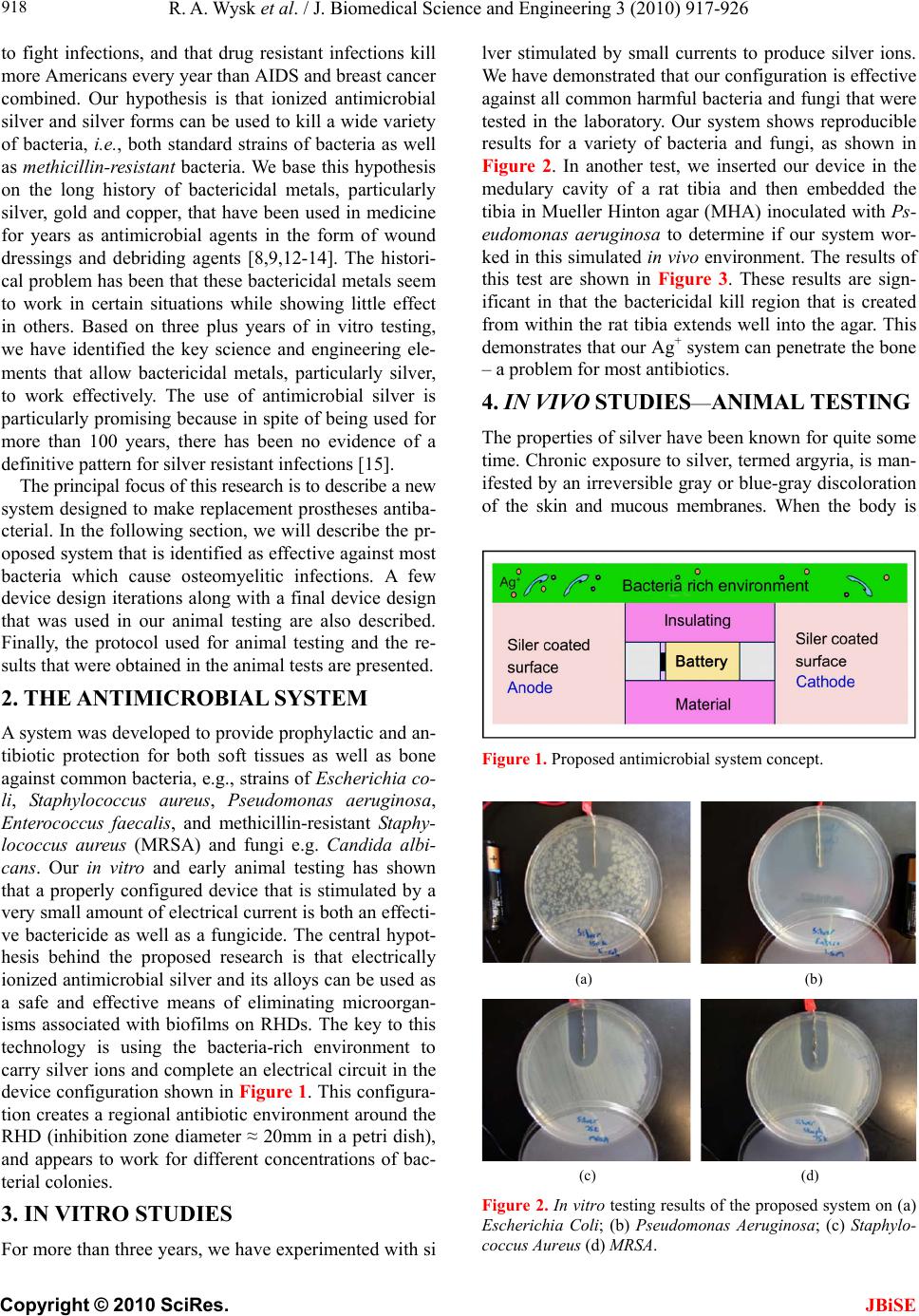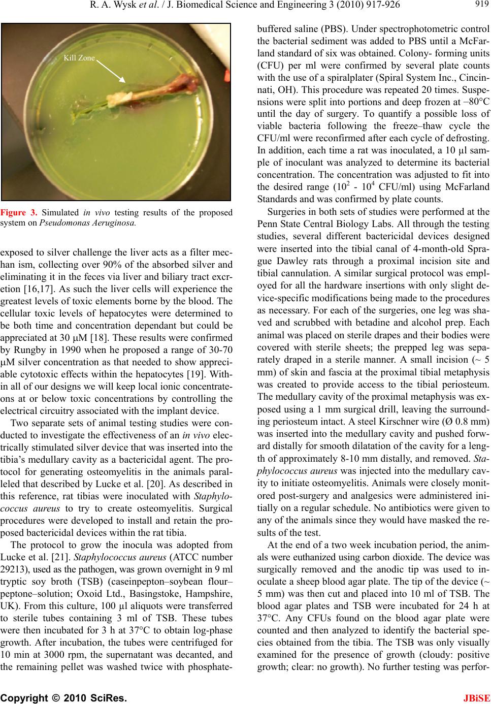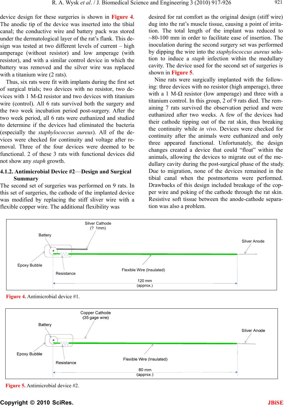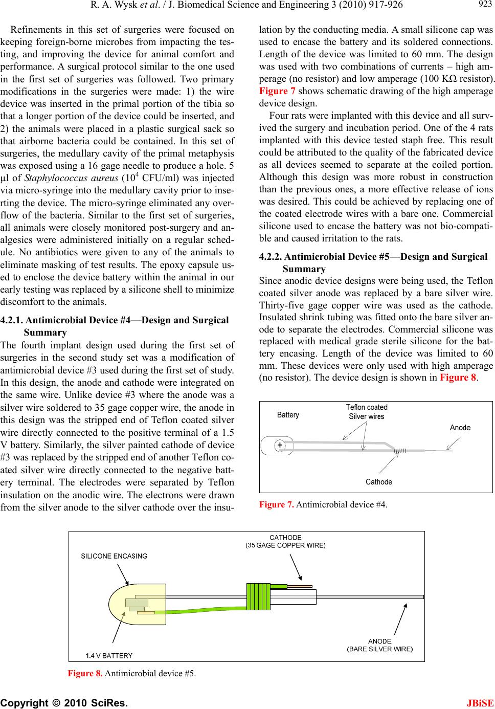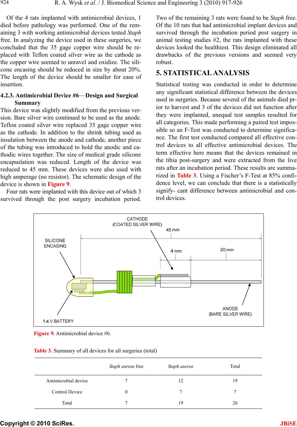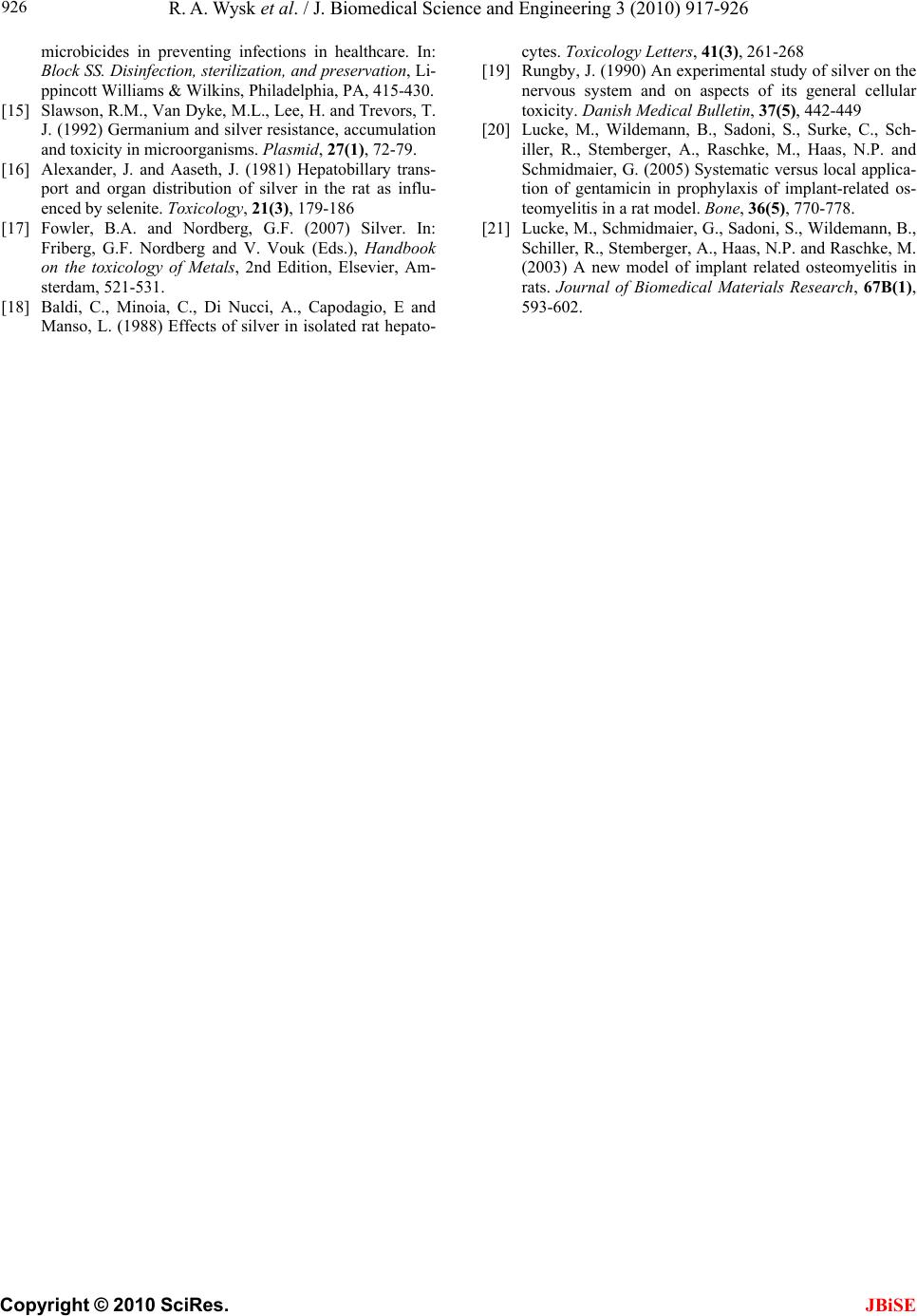
R. A. Wysk et al. / J. Biomedical Science and Engineering 3 (2010) 917-926
Copyright © 2010 SciRes. JBiSE
925
During animal testing studies #1, antimicrobial de-
vices were found to be electrically disconnected proxi-
mal to the tibial cavity insertion point of 2 rats, render-
ing the devices electrically ineffective. If these 2 devices
are considered as control instead of antimicrobial devic-
es, the F-test concludes that there is a statistically sign-
ificant difference between antimicrobial devices and con-
trol devices at 95% confidence level. In addition, each
device design was compared with others to determine
any statistical significance. Strictly statistically speaking,
only antimicrobial device #1 and antimicrobial device #6
showed significant difference from the controls among
all six design iterations. Although 2 out of 3 animals
implanted with working antimicrobial device #1 tested
staph free, the device tip often penetrated the soft tissue
and caused irritation to the animals. On the other hand,
antimicrobial device #6 also resulted in 2 out of 3 ani-
mals being staph free but eliminated the problem associ-
ated with damaging the soft tissue.
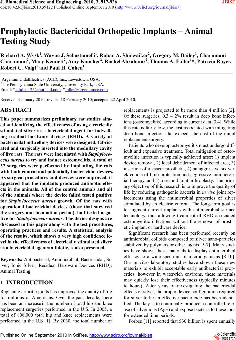
6. DISCUSSION AND CONCLUSIONS
An early conjecture in this research was that a bacteri-
cidal environment would be created if we could get bac-
teria to conduct Ag+. The hope was to create this envi-
ronment within a bone because getting traditional antibi-
otics to penetrate the bone while the antibiotics are still
viable can be very difficult. A basic design concept was
developed and had to go through several iterations gov-
erned by rat comfort after implant as well as bactericidal
performance. Interestingly, the first and the last device
design iterations showed the greatest efficacy.
Based on the surgeries, pathology results and statisti-
cal analysis, the rat osteomyelitic model described in lit-
erature is validated, since all animals without an antimic-
robial device were infected. More importantly, the resu-
lts show that properly configured electrically stimulated
silver is an effective bactericidal agent for indwelling
devices. Of all the surgeries performed using the bacteri-
cidal devices, there is a statistically significant difference
between using no device and an antimicrobial device. As
the antimicrobial devices and surgical procedures were
refined throughout the study, the effectiveness of the
devices was found to be improved. In the last set of sur-
geries, 67% of the harvested animals were free of Sta-
phylococcus aureus even after they were inoculated with
the bacteria and given no antibiotics. The bactericidal
device as configured has a definite ability to reduce/
eliminate bacterial infection. Using such a bactericidal
device in conjunction with a standard treatment of anti-
biotics should have a profound effect on the number of
residual hardware associated bacterial infections.
7. ACKNOWLEDGEMENTS
The technology and designs tested within this study are protected
under U. S. Patent as owned by ArgentumCidalElectrics, Inc.. It is only
with their support that device modifications and manufacturing could
be properly completed and controlled.
REFERENCES
[1] Iorio, R., Robb, W.J., Healy, W.L., Berry, D.J., Hozack,
W.J., Kyle, R.F., Lewallen, D.G., Trousdale, R.T., Jira-
nek, W.A., Stamos, V.P. and Parsley, B.S. (2008) Or-
thopaedic surgeon workforce and volume assessment for
total hip and knee replacement in the united states: Pre-
paring for an epidemic. The Journal of Bone and Joint
Surgery, 90(7), 1598-1605.
[2] Kurtz, S., Ong, K., Lau, E., Mowat F. and Halpern, M.
(2007) Projections of primary and revision hip and knee
arthroplasty in the United States from 2005 to 2030. The
Journal of Bone and Joint Surgery, 89(4), 780-785.
[3] Cumming, D. and Parker, M.J. (2007) Urinary catheteri-
sation and deep wound infection after hip fracture sur-
gery. International Orthopaedics, 31(4), 483-485.
[4] Fitzgerald Jr.R.H. (1992) Total hip arthroplasty sepsis:
Prevention and diagnosis. Orthopedic Clinics of North
America, 23(2), 259-264.
[5] Furst, A. and Schlauder, M.C. (1978) Inactivity of two
noble metals as carcinogens. The Journal of Environ-
mental Pathology, Toxicology and Oncology, 1(1), 51-
57.
[6] Solberg, B.D., Gutow, A.P. and Baumgaertner, M.R.
(1999) Efficacy of gentamycin impregnated resorbable
hydroxyapatite cement in treating osteomyelitis in a rat
model. Journal of Orthopaedic and Trauma, 13(2), 102-
106.
[7] Furno, F., Morley, K.S., Wong, B., Sharp, B.L., Arnold,
P.L., Howdle, S.M., Bayston, R., Brown, P.D., Winship
P.D. and Reid, H.J. (2004) Silver nanoparticles and
polymeric medical devices: A new approach to preven-
tion of infection? The Journal of Antimicrobial Chemo-
therapy, 54(6), 1019-1024.
[8] Klasen, H.J. (2000) Historical review of the use of silver
in the treatment of burns Part 1: Early uses. Burns, 26(2),
117-130.
[9] Klasen, H.J. (2000) Historical review of the use of silver
in the treatment of burns Part 2: Renewed interest for sil-
ver. Burns, 26(2), 131-138.
[10] Samuel, U. and Guggenbichler, J.P. (2004) Prevention of
catheter-related infections: The potential of a new nano-
silver impregnated catheter. The International Journal of
Antimicrobial Agents, 23(Suppl 1), 75-78.
[11] Langreth, R. and Herper, M. (2006) Germ Warfare. Forbes.
http://www.forbes.com/forbes/2006/0619/060.html.
[12] Melaiye, A. and Youngs, W.J. (2005) Silver and its ap-
plication as an antimicrobial agent. Expert Opinion on
Therapeutic Patens, 15(2), 125-130.
[13] Price, W.R. and Wood, M. (1996) Silver nitrate burn
dressing: Treatment of seventy burned persons. American
Journal of Surger , 112(5), 674-680.
[14] Weber, D.J. and Rutala, W.A. (2001) Use of metals as