Open Journal of Ophthalmology, 2012, 2, 116-118
http://dx.doi.org/10.4236/ojoph.2012.24025 Published Online November 2012 (http://www.SciRP.org/journal/ojoph)
Combined Hamartoma of the Optic Disc and Retinal
Pigment Epithelium—3 Years of the Follow-Up with
Semi-Automated Kinetic Perimetry
Katarzyna Nowomiejska1, Tomasz Zarnowski2, Robert Rejdak1,3,4, Anselm Juenemann5
1Department of General Ophthalmology, Medical University, Lublin, Poland; 2Department of Glaucoma Diagnostics and Microsur-
gery, Medical University, Lublin, Poland; 3Department of Pathophysiology of Vision and Neuro-Ophthalmology, University Eye
Hospital, Tuebingen, Germany; 4Department of Experimental Pharmacology , Mossakowski Medical Research Centre, Polish Academy
of Sciences, Warsaw, Poland; 5Department of Ophthalmology, University of Erlangen-Nürnberg, Erlangen, Germany.
Email: katarzynanowomiejska@mailcity.com
Received June 21st, 2012; revised September 18th, 2012; accepted October 7th, 2012
ABSTRACT
Aim: To present a case of hamartoma of the optic disc and Retinal Pigment Epithelium (RPE) and follow up of the visual
function over three-years period. Methods: A seventeen-year-old boy has observed reduced visual acuity in his left eye.
The visual acuity was 0.2 and there was RAPD in the left eye. Fundoscopy revealed an elevation of the optic disc ob-
scuring disc vessels with epiretinal gliosis. Fluorescein angiography demonstrated hyperfluorescent saccular dilatations
with leakage in the late phase. Ocular Coherence Tomography (OCT) showed hyperreflective elevation of the optic disc
and epiretinal membrane. There was a juxtapapillary scotoma in Semi-automated Kinetic Perimetry (SKP). There was
no systemic diseases. Results of blood tests, CT and MRI of CNV were normal. Results: After 3 years period of the
follow-up the visual acuity was 0.1 and there was a progression of the visual field defect to the altitudinal scotoma in
the upper hemisphere. Fluorescein anhiography and OCT revealed the same. Conclusion: Hamartoma of RPE and optic
disc is a rare condition consisting of glial, vascular and RPE cells. It should be differentiated from optic disc oedema
and vascular tumors of the retina. Assessment of the visual function is very important in the longitudinal follow-up.
Keywords: Hamartoma; Perimetry; Optic Disc
1. Case
A 17-year-old male has observed slow, progressive and
painless reduction of the visual acuity of his left eye for
two years. He had no systemic diseases, only meningitis
after virus infection in childhood (8 years old) was re-
ported. He had no significant family history. The best
corrected visual acuity at presentation was 20/20 in the
right eye and 20/80 in the left eye. Intraocular pressure
was 20 mm·Hg in both eyes. Co lor vision and eye move-
ments were normal. There was RAPD in the left eye.
Anterior segment was normal. Fundus was normal in the
right eye. In the left eye fundoscopy revealed elevated
optic disc margins with scattered hiperpigmentstion ob-
scuring major disc vessels (Figure 1). Additionally, there
was an epiretinal gliosis and traction involving the macula.
Flurescein angiography (HRA2, Heidelberg Enginering,
Germany) demonstrated early hyperfluorescence through-
out the lesion and saccular dilatations with leakage in the
late phase (Figure 2), OCT (Cirru s HD-OCT Model 400,
Carl Zeiss, Meditec, Dublin, CA, USA) revealed the dis-
tortion of the retinal architecture, hyperreflective retinal
elevation and epiretinal membrane.
Visual field of the right eye was normal, in the left eye
juxtapapillary scotoma was observed (Figure 3 left).
Perimetry was performed using Semi-automated Kinetic
Perimetry (SKP) implemented in Octopus 101 and Oc-
topus 900 instrument. MRI and CT examinations were
described as normal. In the neurological examination no
pathological signs were observed. The systemic studies
produced no abnormalities.
Three years later the visual acuity was 0.1 in the left
eye, the visual field has progressed to altitudinal scotoma
in the upper hemisphere (Figure 3 right). The fundus and
fluorescein angiography appeared the same. There was
no treatment applied.
2. Discussion
Hamartoma of the optic disc and Retinal Pigment Ep ithe-
lium (RPE) is a rare benign congenital ocular tumor de-
scribed first by Gass [1]. It is single, solitary and unila-
Copyright © 2012 SciRes. OJOph
Combined Hamartoma of the Optic Disc and Retinal Pigment Epithelium—3 Years of the Follow-Up with
Semi-Automated Kinetic Perimetry 117
Figure 1. Fundus photograph of the left eye showing pig-
ment epithelium hamartoma of the optic disc.
Figure 2. Fluorescein angiography (Spectralis HRA2, Hei-
delberg Enginering, Germany) of the left eye showing early
phase (0:21 min.)—at the top and late phase with leakage
(4:51 min.)—below.
Figure 3. Result of semi-automated kinetic perimetry of the
left eye. Three isopters I2e, I4e and III4e. Baseline exami-
nation (Octopus 101)—juxtapapillary scotoma (top). Pe-
rimetry after three years (Octopus 900)—altitudinal scot oma
in the upper hemisphere (bottom).
teral intraocular tumor. Histologically, it is composed of
three cell populations: pigmented, glial and vascular.
Different proportions of these cells are responsible for
various clinical pictures. Usually hamartoma of the retina
and RPE are located close to the optic disc in 76%, in the
macula in 17% and in the periph eral retina in 17%. Only
few cases have been described so far localized within the
optic nerve head [2].
Copyright © 2012 SciRes. OJOph
Combined Hamartoma of the Optic Disc and Retinal Pigment Epithelium—3 Years of the Follow-Up with
Semi-Automated Kinetic Perimetry
Copyright © 2012 SciRes. OJOph
118
The diagnostics is based on fundoscopy and confirmed
by fluorescein angiography and Ocular Coherence To-
mography (OCT). The presenting syndrome is painless
unilateral silent visual loss. The patients are usually as-
ymptomatic until the late childhood. The anterior seg-
ment is not affected. There are anecdotical reports on
systemic associations with neurofibromatosis type II and
tuberous sclerosis. RPE hamartomas usually show no
growth potential. Hamartoma of the retina and RPE can
generate a large papillary and retinal distortion [3], thu s it
is often accompanied with vascular tortuosity and epire-
tinal membrane. Secondary changes as vitreous hemor-
rhage, Choroidal Neovascular Membrane (CNV) or retinal
detachment [4] on some occasions may cause visual loss.
There have been reports describing pars plana vitrectomy
due to epiretinal membrane following hamartoma [5] or
submacular surgery due to CNV [6].
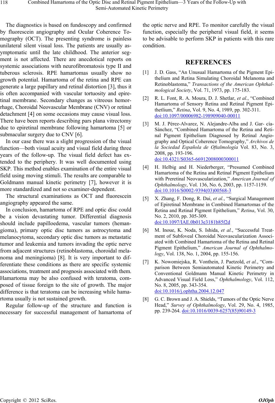
In our case there was a slight progression of the visual
function—both visual acuity and visual field during three
years of the follow-up. The visual field defect has ex-
tended to the periphery. It was well documented using
SKP. This method enables examination of the entire visual
field using moving stimuli. The results are comparable to
Goldmann manual kinetic perimetry [7], however it is
more standardized and not so examiner-dependent.
The structural examinations as OCT and fluorescein
angiography appeared the same.
In conclusion, hamartoma of RPE and optic disc could
be a vision devastating tumor. Differential diagnosis
should include papilloedema, vascular tumors (heman-
gioma), primary optic disc tumors as astrocytoma and
melanocytoma, secondary optic disc tumors as metastatic
tumor and leukemia and tumors invading the optic nerve
from adjacent structures (retinoblastoma, choroidal mela-
noma and meningioma) [8]. It is very important to dif-
ferentiate these conditions as there are specific systemic
associations, treatment and prognosis associated with them.
Hamartoma may be also confused with teratoma, com-
posed of tissue foreign to the site of growth. The major
difference is that teratoma can be increasing while hama-
rtoma usually is not sustained growth.
Regular follow-up of the structure and function is
necessary for successful management of hamartoma of
the optic nerve and RPE. To monitor carefully the visual
function, especially the peripheral visual field, it seems
to be advisable to perform SKP in patients with this rare
condition.
REFERENCES
[1] J. D. Gass, “An Unusual Hamartoma of the Pigment Epi-
thelium and Retina Simulating Choroidal Melanoma and
Retinoblastoma,” Transactions of the American Ophthal-
mological Society, Vol. 71, 1973, pp. 175-183.
[2] R. L. Font, R. A. Moura, D. J. Shetlar, et al., “Combined
Hamartoma of Sensory Retina and Retinal Pigment Epi-
thetlium,” Retina, Vol. 9, No. 4, 1989, pp. 302-311.
doi:10.1097/00006982-198909040-00011
[3] M. J. Pérez-Alvarez, N. Alejandre-Alba and J. Gar- cía-
Sánchez, “Combined Hamartoma of the Retina and Reti-
nal Pigment Epithelium Diagnosed by Retinal Angio-
graphy and Optical Coherence Tomography,” Archivos de
la Sociedad Española de Oftalmología Vol. 83, No. 3,
2008, pp. 193-196.
doi:10.4321/S0365-66912008000300011
[4] H. Helbig and H. Niederberger, “Presumed Combined
Hamartoma of the Retina and Retinal Pigment Epithelium
with Preretinal Neovascularization,” American Journal of
Ophthalmology, Vol. 136, No. 6, 2003, pp. 1157-1159.
doi:10.1016/S0002-9394(03)00568-3
[5] X. Zhang, F. Dong, R. Dai, et al., “Surgical Management
of Epiretinal Membrane in Combined Hamartomas of the
Retina and Retinal Pigment Epithelium,” Retina, Vol. 30,
No. 2, 2010, pp. 305-309.
doi:10.1097/IAE.0b013e3181b85f2d
[6] M. Inoue, K. Noda, S. Ishida, et al., “Successful Treat-
ment of Subfoveal Choroidal Neovascularization Associ-
ated with Combined Hamartoma of the Retina and Retinal
Pigment Epithelium,” American Journal of Ophthalmo-
logy, Vol. 138, No. 1, 2004, pp. 155-156.
[7] K. Nowomiejska, R. Vonthein, J. Paetzold, et al., “Com-
parison Between Semiautomated Kinetic Perimetry and
Conventional Goldmann Manual Kinetic Perimetry in
Advanced Visual Field Loss,” Ophthalmology, Vol. 112,
No. 8, 2005, pp. 343-354.
doi:10.1016/j.ophtha.2004.12.047
[8] G. C. Brown and J. A. Shields, “Tumors of the Optic Nerve
Head,” Survey of Ophthalmology, Vol. 29, No. 4, 1985,
pp. 239-264. doi:10.1016/0039-6257(85)90149-3