Journal of Minerals and Materials Characterization and Engineering, 2012, 11, 982-988
Published Online October 2012 (http://www.SciRP.org/journal/jmmce)
Effect of Magnesium on the Mechanical and Bioactive
Properties of Biphasic Calcium Phosphate
Ponnusamy Kanchana, Chinnathambi Sekar
Department of Bioelectronics and Biosensors, Alagappa University, Karaikudi, India
Email: Sekar2025@gmail.com
Received August 12, 2012; revised September 15, 2012; accepted September 25, 2012
ABSTRACT
Incorporation of trace elements into calcium phosphate structure is of great interest for the development of artificial
bone implants. Biphasic calcium phosphate (BCP) composed of hydroxyapatite (HA) and β-tricalcium phosphate
(β-TCP) have been synthesized in the presence of magnesium (5 M% - 20 M%) by gel method under physiological
conditions. Crystallization of Mg-BCP in the gel medium mimics the Mg intake in the human body. Powder X-ray dif-
fraction and Fourier transform infrared analyses confirmed that the Mg doping leads to the enrichment of β-TCP phase
and suppresses the HA content in BCP. Nanoindentation studies indicate a significant decrease in hardness and elastic
modulus values of BCP due to Mg doping. In vitro bioactivity study has confirmed the formation of apatite layer on the
Mg doped samples making it suitable for bone replacement. The results suggest that the optimum Mg doping promotes
the bioactivity which is perquisite for biomedical applications.
Keywords: Biphasic Calcium Phosphate; Crystal Growth; X-Ray Diffraction; Mechanical Properties
1. Introduction
Biphasic calcium phosphate (BCP) composed of a mix-
ture of hydroxyapatite (HA, Ca10(PO4)6(OH)2) and β-tri-
calcium phosphate (β-TCP, β-Ca3(PO4)2) are interesting
candidates in reconstructive surgery [1-3]. They are par-
ticularly suitable for synthetic bone substitution appli-
cations because the HA provides a permanent scaffold
for new bone formation via osteoconduction and the re-
sorption of the β-TCP oversaturates the local environ-
ment with Ca2+ and ions to accelerate this new
bone formation. The HA/β-TCP ratio dominantly deter-
mines the rate and extent of BCP resorption in vivo, in
that higher β-TCP contents allow faster and more exten-
sive resorption [4]. Magnesium, one of the most impor-
tant divalent ions associated with the biological apatite,
has its own significance in the calcification process and
on bone fragility and has indirect influence on mineral
metabolism. BCP with Mg incorporation has been the
subject of extensive research because of its potential for
developing artificial bone and other biomedical appli-
cations [5]. The amount of HA and β-TCP in BCP can be
tailored by the insertion of Mg. Independently, Mg doped
HA is of great interest for the development of artificial
bones. The presence of Mg2+ within HA lattice sensibly
affects apatite crystallization in solution promoting the
formation of β-TCP and thus forming BCP [6].
3
4
PO
Several methods have been reported to prepare Mg
substituted HA, β-TCP and BCP [6-9]. Ryu et al. [7]
showed that the MgO-doped HA/β-TCP synthesized by
conventional solid state reaction method exhibited high
density and improvements in compressive strength and
fracture toughness. In particular, the compressive strength
shows 1203 MPa, which is three times greater than the
undoped HA/β-TCP composite. Gomes et al. [1] have
reported that the bioactivity of the BCP has improved
due to the presence of Mg atoms in the structure of the
β-TCP phase. Kannan and Ferreira [8] have reported that
the sodium, magnesium and fluorine co-substituted
HA/β-TCP composites have higher thermal stability upto
1400˚C. In general, sol-gel and precipitation methods
need high temperature heating to produce BCP phases,
and the high temperature treatments results in the forma-
tion of minor phases such as CaO and MgO [1].
Earlier we have demonstrated that the strontium (Sr)
doped BCP synthesized by gel method was free from
these impurities [10]. As the gel medium offer the possi-
bility to mimic the growth of various biominerals in en-
vironments similar to natural biomineralization [11,12],
we have carried out the synthesis of BCP in sodium me-
tasilicate gel with various concentration of magnesium.
Effect of Mg addition on the HA/β-TCP ratio, morpho-
logy, bioactive properties and mechanical behavior of
BCP have been investigated.
2. Materials and Method
BCP was prepared by gel method using AR grade re-
Copyright © 2012 SciRes. JMMCE
P. KANCHANA, C. SEKAR 983
agents. Single diffusion gel method was employed to
prepare BCP using sodium metasilicate (SMS,
Na2SiO3·9H2O) gel. The gel was prepared by mixing
SMS solution of specific gravity 1.03 g/cm3 and 0.6 M of
disodium hydrogen phosphate (Na2HPO4) in the ratio 1:1.
The pH of the solution was adjusted to 7.4 using 10%
glacial acetic acid. About 10 ml of this solution was al-
lowed to gel in a test tube over a period of 2 days. After
the gelation, 1 M anhydrous CaCl2 solution was poured
on the gel without disturbing it. Growth experiments
were carried out at room temperature for a period of
three weeks. Finally, samples were harvested by decant-
ing the test tubes and the gel was removed by washing.
The samples were kept in hot air oven (60˚C) for 2 hours.
In another series, supernatant solution was prepared by
mixing 1 M of anhydrous CaCl2 with 0.05, 0.1, 0.15 and
0.2 M of magnesium chloride (MgCl2·2H2O) and the
growth was carried out as described above. BCP crystal-
lites collected from middle layers of each test tube were
subjected to systematic studies to understand the role of
Mg on the growth and mechanical properties. Both the
pristine and Mg doped BCP were sintered at 1000˚C in
ambient atmosphere for 5 hours and the resulting pow-
ders were characterized by commonly used techniques.
Powder X-ray diffraction pattern was recorded on
Bruker AXS D8 advanced diffractometer within the 2θ
range of 10 to 55˚ using CuKα1 as X-ray source (λ =
1.5406 Å). Surface analyses were carried out on BCP
samples using scanning electron microscopy (SEM) JSM
5610 LV JEOL make. Elemental analyses were done us-
ing the OXFORD INCA energy dispersive X-ray spec-
trometer (EDX) attached to SEM. Thermogravimetry of
samples was performed using TA instruments SDT Q600
V8.3 in the temperature range of 30˚C - 1200˚C at the
heating rate of 20˚C/minute in nitrogen atmosphere.
FTIR spectra were recorded on a Perkin Elmer (Spec-
trum RXI) spectrometer in transmission mode in the
wave number range between 400 and 4000 cm1 using
KBr pellets.
To test the in vitro bioactivity, the samples were im-
mersed in simulated body fluid (SBF). The SBF was
prepared according to the procedure described by Ko-
kubo [13]. Samples were pressed into pellets of about 8
mm diameter and 1 mm thickness and the pellet was
immersed in 15 ml of SBF solution in airtight plastic
container. The solution was renewed every 48 h for a
period of three weeks. Then the samples were taken out,
washed with deionized water and dried. The morpho-
logical variation of the pellets surface after soaking in
SBF was studied by SEM.
For characterizing the mechanical response, both pris-
tine and Mg doped BCP were die-pressed and sintered at
600˚C for 5h in ambient atmosphere. Indentation impres-
sions were made by nanobased indentation system
NHTXS/S: 50-0172 with diamond Berkovich indenter.
The force used for all the samples was 100 mN with
loading and unloading rate of 20 mN/min. A minimum of
10 indents were made on each sample. Distance between
two separate indents was more than 20 μm to avoid the
influence of residual stresses from adjacent impressions.
From the load (P)-penetration depth (h) curves, the shape
of the unloading curve can be used as a means of obtain-
ing the elastic properties and hardness of the sample. The
slope S is the initial unloading stiffness, A is the pro-
jected area of contact and Er is the reduced modulus de-
termined by a method that has been developed and de-
scribed by Oliver and Pharr [14].
d2
dπ
π
(or) 2
r
r
P
SE
h
S
E
A
A

The elastic modulus of the sample Es can be calculated
using the relation

22
11
1
s
i
rs i
EE E


where Ei is elastic modulus of the indenter tip, νs and νi
are the Poisson ratio of the sample and indenter tip re-
spectively. The hardness (H) which is defined as the
mean pressure that the material will support under load is
determined by the following relation
max
P
H
A
where Pmax is the maximum applied load.
3. Result and Discussion
3.1. Synthesis of BCP
Biphasic calcium phosphate consisting of HA and β-TCP
was successfully synthesized by gel method. Upon the
addition of CaCl2 solution on the top of the set gel in the
test tubes, Ca2+ diffuses into the gel and reacts with
3
4
PO
producing white precipitate at the gel solution
interface. Just below this, a circular white disc was ob-
served after one day. Subsequently, 12 discs of about 3
mm thickness each have grown in the gel medium over a
period of three weeks. The space between successive
white discs was found to increase towards the bottom of
the test tube from the interface due to the lack of diffu-
sion of “Ca” from top of the gel down to the bottom of
the tube. These white colored circular discs, commonly
known as Liesegang rings, were identified as BCP (Fig-
ure 1(a)).
In Mg added experiments, Liesgang rings have ap-
peared 4 to 5 days after the addition of supernatant solu-
Copyright © 2012 SciRes. JMMCE
P. KANCHANA, C. SEKAR
984
Figure 1. BCP in the form of Liesegang rings (a) undoped
BCP; (b) 0.05 M; (c) 0.1 M; (d) 0.15 M; and (e) 0.2 M Mg
doped BCP.
tion which indicate that the initial nucleation and growth
rate were retarded due to the presence of Mg in the
growth environment. The number of white discs got de-
creased as the concentration of Mg (0.05 - 0.2 M) in-
creased in the supernatant solution (Figures 1(b)-(e)).
We could collect the products grown with 0.05 and 0.1
M Mg only. The white discs formed at higher Mg levels
(0.15 and 0.2 M) were very fragile and got mixed up with
the gel at the time of harvest. It should be noted that the
amount of Ca present in the growth environment re-
mained the same as that of pristine BCP and Mg was
added as additive only. Thus the presences of Mg seem
to inhibit the formation of BCP. This inhibitory effect
may be attributed to the incorporation of smaller Mg2+
(0.66 Ǻ) at larger Ca2+ site (1.00 Ǻ) [15]. The presence of
strontium (Sr2+, 1.13 Å) in the growth environment is
also known to inhibit the formation of BCP [10]. Com-
pared to Sr2+, the Mg2+ has a strong influence on the
growth rate and total yield of BCP. The solubility of the
ionic impurity in the mother solution has been considered
as an indicator of the strength of the interfacial coor-
dination, the highest solubility corresponding to the
weakest interaction [16]. The addition of MgCl2·2H2O
with relatively high solubility affects the solubility of the
growth environment. Thus the Mg addition seems to in-
hibit the less soluble HA growth and lead to the forma-
tion of initial transient phases such as TCP [17]. In both
pure and Mg added growth experiments (0.05, 0.1 M %),
DCPD crystals (CaHPO4·2H2O) have grown in between
the rings. No such crystals were found at higher concen-
tration of Mg. Bigi et al. [18] also found significant inhi-
bition by Mg2+ on DCPD crystals.
3.2. Powder XRD Analyses
The as prepared BCP (undoped) and Mg-BCP (Mg
doped BCP) samples were identified as a mixture of HA
and
-TCP (Figures 2(a) and (b)) in agreement with the
JCPDS standards (09-0169 for β-TCP and 09-0432 for
HA). For Mg-BCP, XRD peaks corresponding to β-TCP
planes (0 2 10, 1 1 0) were found to be highly intensed
and the planes (2 1 1) corresponding to HA was less in-
tensed. Contrary to the BCP, Mg doping enriches β-TCP
content and suppresses the HA formation. A number of
ions are known to inhibit and disrupt nucleation of Ca-P
phases. Salimi et al. [19] showed that the Mg2+ has a
marked inhibiting effect on HA growth. They attributed
the inhibitory effect of Mg2+ to its adsorption at active
growth sites. This kinetically hinders nucleation of HA.
In our earlier work, we found that the Sr2+ doping has
enhanced the formation of HA and reduced the β-TCP
content in the BCP composites which was attributed to
the difference in ionic size of the dopant Sr2+ (1.13 Ǻ) in
place of Ca2+ (1.00 Ǻ).
High temperature annealing (1000˚C) of pristine BCP
resulted in the occurrence of more distinct and sharp
XRD peaks of β-TCP (Figures 3(a) and (b)). Contrary to
this, Mg-BCP samples have mainly β-TCP with traces of
γ-Ca2P2O7 phase. Further, the XRD peaks of the Mg
doped samples have been slightly and continuously
shifted to higher diffraction angles and there was a slight
decrease in their peak intensity (see inset of Figure 3).
Table 1 shows the variation of lattice parameters of the
investigated samples. These results suggest that Mg is
preferentially incorporated into the β-TCP phase, the
replacement of Ca by Mg induces lattice contraction and
the respective displacement toward higher 2θ angles [7].
Gomes et al. [1] reported the occurent of minor impurity
phases CaO, CaCO3 and MgO in the pristine and Mg-
BCP samples sintered at high temperature (500˚C -
Figure 2. XRD patterns of (a) pure BCP and (b) 0.1 M Mg
doped BCP.
Copyright © 2012 SciRes. JMMCE
P. KANCHANA, C. SEKAR 985
Figure 3. XRD patterns of sintered (1000˚C) (a) pure BCP
and (b) 0.1 M Mg doped BCP. Inset figure shows a system-
atic shift towards the higher angle side (2θ values between
30˚ to 35˚).
Table 1. Lattice parameters of pure and Mg doped
-TCP
(1000˚C).
Lattice parameters (Å)
Samples
a c
Volume (Å3)
-TCP 10.4147 37.2776 3501.57
-TCP + 0.1M Mg 10.3408 37.1926 3444.17
1100˚C). In the present case, both the as prepared and
sintered (1000˚C) samples synthesized by gel method did
not indicate the presence of any such impurity phases up
to the resolution limit of the XRD measurement.
3.3. SEM- EDX Analyses
Define abbreviations and acronyms the SEM pictures of
the pure and 0.1 M Mg-BCP samples are shown in Fi-
gures 4(a) and (b). The micrograph of BCP consisted of
fibers with an anisotropic aspect of approximately 6 - 10
m in length and 0.5 - 1.0 m in width (Figure 4(a)).
The Mg-BCP consisted of agglomerates of fibers on the
surface (Figure 4(b)), which confirmed that the presence
of Mg changed the growth pattern. An in vitro bioactivity
test of pure and 0.1 M Mg-BCP samples are shown in
Figures 4(c) and (d). Mg-BCP has revealed the forma-
tion of apatite layer on the surface of the pellets when
compared to that of pure BCP. The newly formed apatite
layer consisted of tiny spherical particles of calcium
phosphates (Figure 4(d)). This is in accordance with the
fact that a bioactive material would form a layer of apa-
tite between bones and implant which helps in bonding
of the implant to bone [13].
EDX measurements were made at different points on
Figure 4. SEM pictures of (a) pure BCP; (b) 0.1 M Mg
doped BCP and SBF soaked samples of (c) pure BCP (d) 0.1
M Mg doped BCP.
the samples and the results show that the BCP is prima-
rily composed of calcium and phosphorus without the
presence of other impurities (Figures 5(a) and (b)). In
BCP, the Ca/P ratio was found to be 1.58 [20]. In
Mg-BCP, (Ca+Mg)/P ratio was estimated at 1.42. The
average atomic percentages of the individual elements
are shown in Table 2. It can be noticed that the addition
of Mg leads to the “Ca” reduction when compared to
BCP sample. The average Mg value in the 0.1 M
Mg-BCP was estimated to be 3.52 atm%.
3.4. Thermogravimetric Analyses
TG curves of BCP and Mg-BCP are shown in Figure 6.
In both the cases, initial weight loss up to 450˚C was
due to the loss of adsorbed water (up to 200˚C) and
lattice water (up to 450˚C) [21]. Above this, weight loss
occurred in several steps between 450˚C and 900˚C
which indicate the transformation of HA into β-TCP
phase in the BCP [22].
The total weight loss up to the measured temperature
of 1200˚C was found to be 10.02% and 12.02% re-
spectively for pristine and Mg doped BCP. In addition, a
slight decrease in the decomposition temperature for Mg
doped sample was observed in the TG curve. These
observations suggest that the Mg doping lead to the re-
duced thermal stability of BCP.
3.5. FTIR Analyses
The FTIR spectra of BCP and Mg-BCP show vibrational
bands characteristic of calcium phosphate compounds
(Figure 7). The band at 1031 cm1 is assigned to the tri-
Copyright © 2012 SciRes. JMMCE
P. KANCHANA, C. SEKAR
986
Table 2. EDX data of pure and Mg doped BCP.
Element Pure BCP (atm. %) Mg doped BCP (atm. %)
Ca 61.25 55.18
P 38.75 41.30
Mg - 3.52
Ca/P = 1.58 (Ca+Mg)/P = 1.42
Figure 5. EDX spectra of (a) BCP and (b) 0.1M Mg doped
BCP.
Figure 6. TG curves of (a) BCP and (b) 0.1 M Mg doped
BCP.
ply degenerate ν3 asymmetric P-O stretching mode. The
peak at 961 cm1 is attributed to ν1, the non-degenerate
P-O symmetric stretching mode. The peaks at 601 and
562 cm1 correspond to the triply degenerate ν4, O-P-O
bending mode and the band at 460 cm1 is attributed to
the doubly degenerate ν2, O-P-O bending mode. The
bands at 1640 and 3441 cm1 are assigned to the ad-
sorbed water [23]. The low intensity hydroxyl group at
3553 cm1 indicates the presence of HA/TCP [24]. The
low intensity OH group at 3553 cm1 of the BCP was
not visible in the case of Mg-BCP sample. Moreover,
broadening of phosphate band at 1000 - 1100 cm1 and
the weak bands at 869, 1371 cm1 due to the P-O(H)
stretching in groups have appeared in the Mg-
2
4
HPO
Wave number (cm–1)
Figure 7. FTIR spectra of (a) BCP and (b) 0.1 M Mg doped
BCP.
BCP only [25,26]. The peak broadening of phosphate
group and new peaks have confirmed the formation of
tricalcium phosphate due to Mg doping.
3.6. Nanoindentation Studies
Mechanical behavior of the BCP samples was investi-
gated in terms of hardness and elastic modulus obtained
by nanoindentation. Figure 8 shows the typical load-
penetration depth (p-h) curves obtained for BCP and
Mg-BCP samples. For comparative analysis, the p-h
curve of Sr doped BCP is also incorporated [10]. In all
the three cases, the p-h curves are associated with the
displacement bursts or pop-ins as indicated by arrow
marks in Figure 8. Similar pop-ins observed in the p-h
curves were reported to be associated with pile-ups
around the indent mark for HA and β-TCP single crys-
tals [27]. The mean value of elastic modulus (E) and
hardness values (H) of BCP, Sr and Mg doped BCP cal-
culated from the p-h curves are shown in Table 3. It can
be noticed that the E and H values for the Mg doped BCP
is lower when compared to that of pure and Sr doped
samples. The observed decrease in hardness values could
be attributed to the enrichment of β-TCP phase and the
difference in sizes between Ca (1.00 Å) and the dopants
Sr (1.13 Å) and Mg (0.66 Ǻ). However, all the p-h curves
show an elastic recovery after the removal of the applied
load.
4. Conclusion
The Mg doped biphasic calcium phosphate was success-
fully synthesized by the simple gel method. The as pre-
pared and sintered (1000˚C) samples did not indicate the
presence of impurities like CaO, CaCO3, MgO and ni-
trates. The presence of Mg2+ ions in the growth environ-
Copyright © 2012 SciRes. JMMCE
P. KANCHANA, C. SEKAR 987
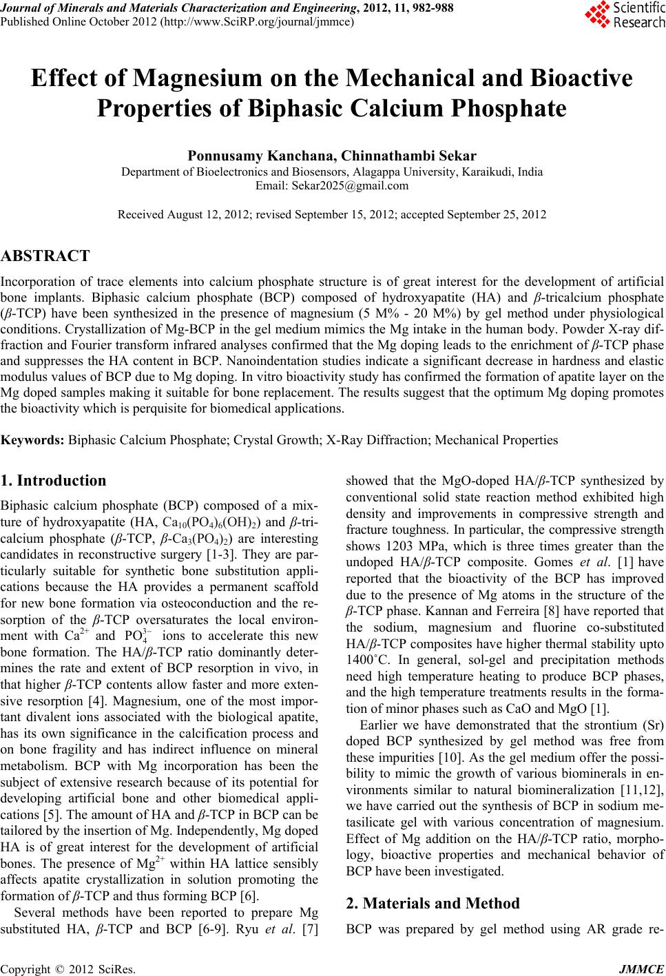
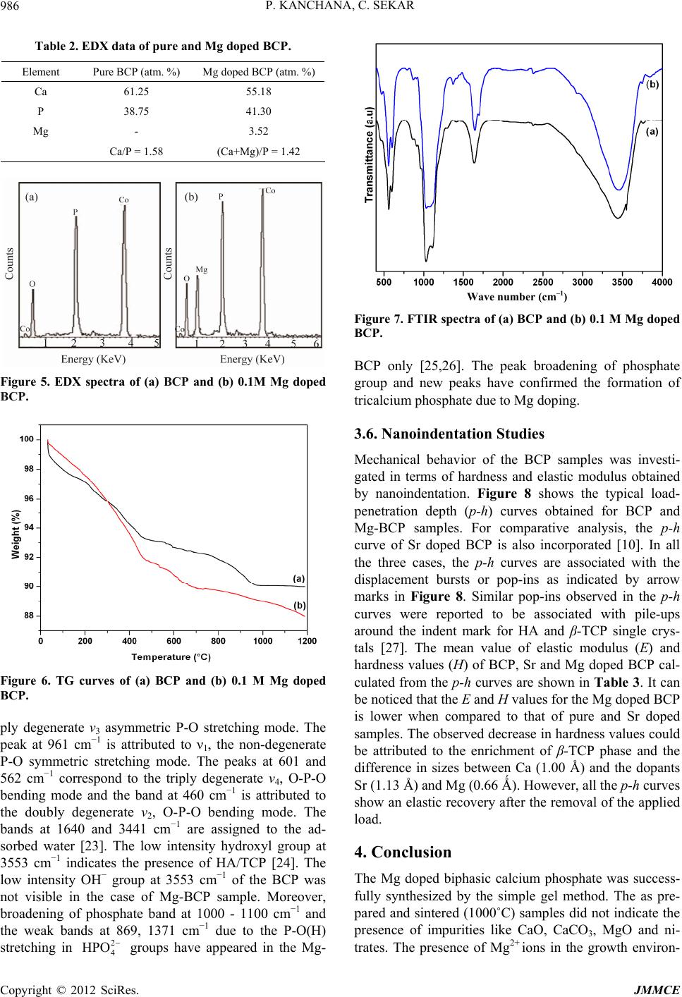
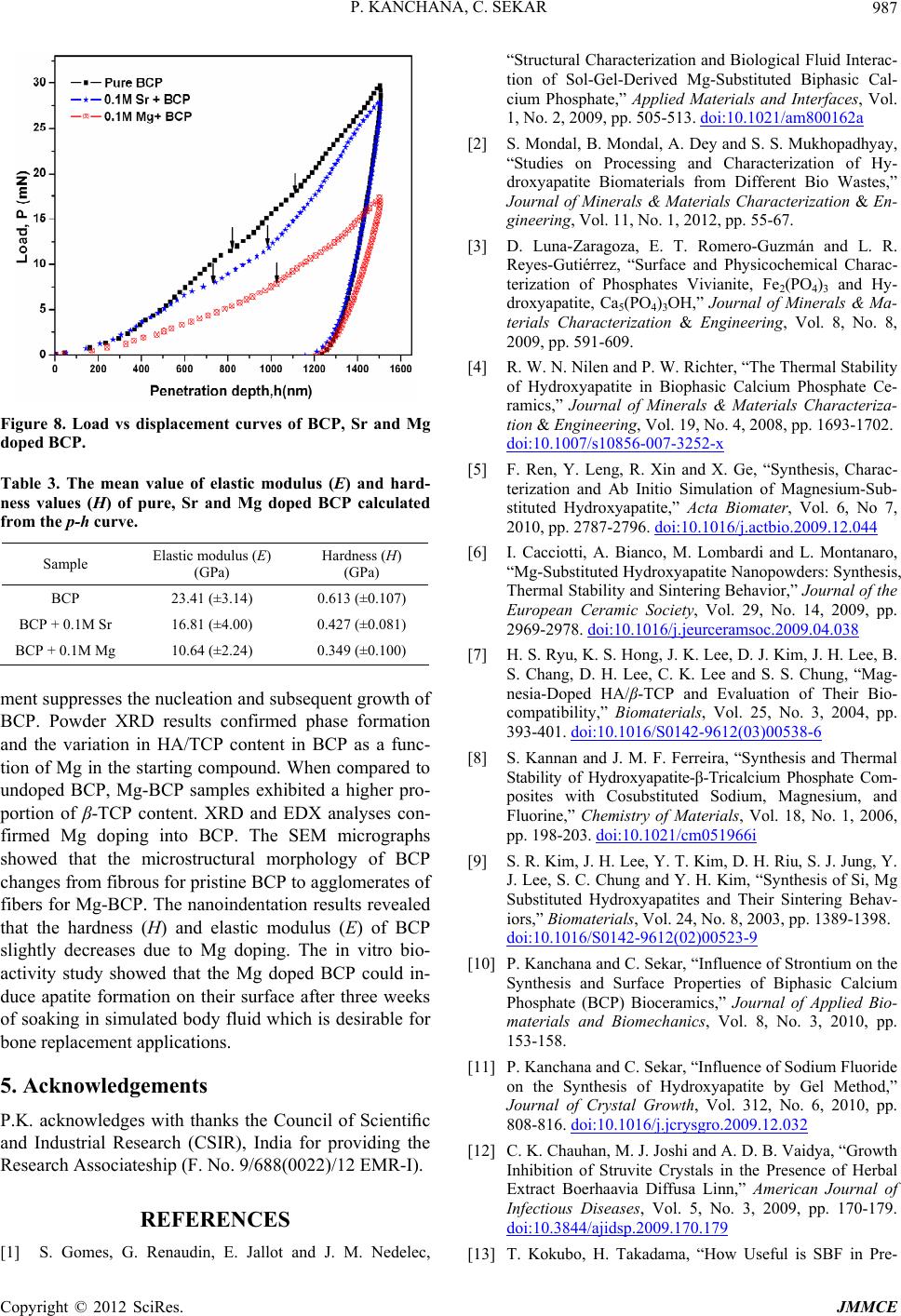
Figure 8. Load vs displacement curves of BCP, Sr and Mg
doped BCP.
Table 3. The mean value of elastic modulus (E) and hard-
ness values (H) of pure, Sr and Mg doped BCP calculated
from the p-h curve.
Sample Elastic modulus (E)
(GPa)
Hardness (H)
(GPa)
BCP 23.41 (±3.14) 0.613 (±0.107)
BCP + 0.1M Sr 16.81 (±4.00) 0.427 (±0.081)
BCP + 0.1M Mg 10.64 (±2.24) 0.349 (±0.100)
ment suppresses the nucleation and subsequent growth of
BCP. Powder XRD results confirmed phase formation
and the variation in HA/TCP content in BCP as a func-
tion of Mg in the starting compound. When compared to
undoped BCP, Mg-BCP samples exhibited a higher pro-
portion of β-TCP content. XRD and EDX analyses con-
firmed Mg doping into BCP. The SEM micrographs
showed that the microstructural morphology of BCP
changes from fibrous for pristine BCP to agglomerates of
fibers for Mg-BCP. The nanoindentation results revealed
that the hardness (H) and elastic modulus (E) of BCP
slightly decreases due to Mg doping. The in vitro bio-
activity study showed that the Mg doped BCP could in-
duce apatite formation on their surface after three weeks
of soaking in simulated body fluid which is desirable for
bone replacement applications.
5. Acknowledgements
P.K. acknowledges with thanks the Council of Scientic
and Industrial Research (CSIR), India for providing the
Research Associateship (F. No. 9/688(0022)/12 EMR-I).
REFERENCES
[1] S. Gomes, G. Renaudin, E. Jallot and J. M. Nedelec,
“Structural Characterization and Biological Fluid Interac-
tion of Sol-Gel-Derived Mg-Substituted Biphasic Cal-
cium Phosphate,” Applied Materials and Interfaces, Vol.
1, No. 2, 2009, pp. 505-513. doi:10.1021/am800162a
[2] S. Mondal, B. Mondal, A. Dey and S. S. Mukhopadhyay,
“Studies on Processing and Characterization of Hy-
droxyapatite Biomaterials from Different Bio Wastes,”
Journal of Minerals & Materials Characterization & En-
gineering, Vol. 11, No. 1, 2012, pp. 55-67.
[3] D. Luna-Zaragoza, E. T. Romero-Guzmán and L. R.
Reyes-Gutiérrez, “Surface and Physicochemical Charac-
terization of Phosphates Vivianite, Fe2(PO4)3 and Hy-
droxyapatite, Ca5(PO4)3OH,” Journal of Minerals & Ma-
terials Characterization & Engineering, Vol. 8, No. 8,
2009, pp. 591-609.
[4] R. W. N. Nilen and P. W. Richter, “The Thermal Stability
of Hydroxyapatite in Biophasic Calcium Phosphate Ce-
ramics,” Journal of Minerals & Materials Characteriza-
tion & Engineering, Vol. 19, No. 4, 2008, pp. 1693-1702.
doi:10.1007/s10856-007-3252-x
[5] F. Ren, Y. Leng, R. Xin and X. Ge, “Synthesis, Charac-
terization and Ab Initio Simulation of Magnesium-Sub-
stituted Hydroxyapatite,” Acta Biomater, Vol. 6, No 7,
2010, pp. 2787-2796. doi:10.1016/j.actbio.2009.12.044
[6] I. Cacciotti, A. Bianco, M. Lombardi and L. Montanaro,
“Mg-Substituted Hydroxyapatite Nanopowders: Synthesis,
Thermal Stability and Sintering Behavior,” Journal of the
European Ceramic Society, Vol. 29, No. 14, 2009, pp.
2969-2978. doi:10.1016/j.jeurceramsoc.2009.04.038
[7] H. S. Ryu, K. S. Hong, J. K. Lee, D. J. Kim, J. H. Lee, B.
S. Chang, D. H. Lee, C. K. Lee and S. S. Chung, “Mag-
nesia-Doped HA/β-TCP and Evaluation of Their Bio-
compatibility,” Biomaterials, Vol. 25, No. 3, 2004, pp.
393-401. doi:10.1016/S0142-9612(03)00538-6
[8] S. Kannan and J. M. F. Ferreira, “Synthesis and Thermal
Stability of Hydroxyapatite-β-Tricalcium Phosphate Com-
posites with Cosubstituted Sodium, Magnesium, and
Fluorine,” Chemistry of Materials, Vol. 18, No. 1, 2006,
pp. 198-203. doi:10.1021/cm051966i
[9] S. R. Kim, J. H. Lee, Y. T. Kim, D. H. Riu, S. J. Jung, Y.
J. Lee, S. C. Chung and Y. H. Kim, “Synthesis of Si, Mg
Substituted Hydroxyapatites and Their Sintering Behav-
iors,” Biomaterials, Vol. 24, No. 8, 2003, pp. 1389-1398.
doi:10.1016/S0142-9612(02)00523-9
[10] P. Kanchana and C. Sekar, “Influence of Strontium on the
Synthesis and Surface Properties of Biphasic Calcium
Phosphate (BCP) Bioceramics,” Journal of Applied Bio-
materials and Biomechanics, Vol. 8, No. 3, 2010, pp.
153-158.
[11] P. Kanchana and C. Sekar, “Influence of Sodium Fluoride
on the Synthesis of Hydroxyapatite by Gel Method,”
Journal of Crystal Growth, Vol. 312, No. 6, 2010, pp.
808-816. doi:10.1016/j.jcrysgro.2009.12.032
[12] C. K. Chauhan, M. J. Joshi and A. D. B. Vaidya, “Growth
Inhibition of Struvite Crystals in the Presence of Herbal
Extract Boerhaavia Diffusa Linn,” American Journal of
Infectious Diseases, Vol. 5, No. 3, 2009, pp. 170-179.
doi:10.3844/ajidsp.2009.170.179
[13] T. Kokubo, H. Takadama, “How Useful is SBF in Pre-
Copyright © 2012 SciRes. JMMCE
P. KANCHANA, C. SEKAR
Copyright © 2012 SciRes. JMMCE
988
dicting in Vivo Bone Bioactivity?” Biomaterials, Vol. 27,
No. 15, 2006, pp. 2907-2915.
doi:10.1016/j.biomaterials.2006.01.017
[14] W. C. Oliver and G. M. Pharr, “An Improved Technique
for Determining Hardness and Elastic Modulus Using
Load and Displacement Sensing Indentation Experi-
ments,” Journal of Materials Research, Vol. 7, No. 6,
1992, pp. 1564-1583. doi:10.1557/JMR.1992.1564
[15] J. F. Ferguson and P. L. McCarty, “Effects of Carbonate
and Magnesium on Calcium Phosphate Precipitation,”
Environmental Science and Technology, Vol. 5, No. 6,
1971, pp. 534-540. doi:10.1021/es60053a005
[16] S. Veintemillas-Verdaguer, “Chemical Aspects of the
Effects of Impurities in Crystal Growth,” Progress in
crystal growth and characterization of materials, Vol. 32,
No. 1-2, 1996, pp. 75-109.
doi:10.1016/0960-8974(96)00005-8
[17] E. Boanini, M.Gazzano and A. Bigi, “Ionic Substitutions
in Calcium Phosphates Synthesized at Low Tempera-
ture,” Acta Biomaterialia, Vol. 6, No. 6, 2010, pp. 1882-
1894. doi:10.1016/j.actbio.2009.12.041
[18] A. Bigi, M. Gazzano, A. Ripamonti and N. Roveri, “Ef-
fect of Foreign Ions on the Conversion of Brushite and
Octacalcium Phosphate into Hydroxyapatite,” Journal of
inorganic biochemistry, Vol. 32, No. 4, 1988, pp. 251-257.
doi:10.1016/0162-0134(88)85004-9
[19] M. H. Salami, J. C. Heughebaert and G. H. Nancollas,
“Crystal Growth of Calcium Phosphates in the Presence
of Magnesium Ions.” Langmuir, Vol. 1, No. 1, 1985, pp.
119-122. doi:10.1021/la00061a019
[20] E. C. Victoria and F. D. Gnanam, “Synthesis and Charac-
terization of Biphasic Calcium Phosphate,” Trends in
Biomaterials & Artificial Organs, Vol. 16, No. 1, 2002,
pp. 12-14.
[21] W. L. Suchanek, K. Byrappa, P. Shuk, R. E. Riman, V. F.
Janas and K. S. Ten Huisen, “Mechanochemical-Hydro-
thermal Synthesis of Calcium Phosphate Powders with
Coupled Magnesium and Carbonate Substitution,” Jour-
nal of solid state chemistry, Vol. 177, No. 3, 2004, pp.
793-799. doi:10.1016/j.jssc.2003.09.012
[22] P. N. Kumta, C. Sfeir, D. H. Lee, D. Olton and D. Choi,
“Nanostructured Calcium Phosphates for Biomedical Ap-
plications: Novel Synthesis and Characterization,” Acta
Biomaterialia, Vol.1, No. 1, 2005,pp. 65-83.
doi:10.1016/j.actbio.2004.09.008
[23] I. Sopyan and A. N. Natasha, “Preparation of Nanostruc-
tured Manganese-Doped Biphasic Calcium Phosphate
Powders via Sol-Gel Method,” Ionics, Vol. 15, No. 62009,
pp. 735-741. doi:10.1007/s11581-009-0330-8
[24] I. Manjubala and M. Sivakumar, “In-Situ Synthesis of
Biphasic Calcium Phosphate Using Microwave Irradia-
tion,” Materials Chemistry and Physics, Vol. 71, No. 3,
2001, pp. 272-278. doi:10.1016/S0254-0584(01)00293-0
[25] J. Pena and M. Vallet-Regi, “Hydroxyapatite, Tricalcium
Phosphate and Biphasic Materials Prepared by a Liquid
Mix Technique,” Journal of the European Ceramic Soci-
ety, Vol. 23, No. 10, 2003, pp. 1687-1696.
doi:10.1016/S0955-2219(02)00369-2
[26] K. P. Sanosh, M. C. Chu, A. Balakrishnan, T. N. Kim, S.
J. Cho, “Sol-Gel Synthesis of Pure Nano Sized β-Tricalcium
Phosphate Crystalline Powders,” Current Applied Physics,
Vol. 10, No. 1, 2010, pp. 68-71.
doi:10.1016/j.cap.2009.04.014
[27] B. Viswanath, R. Raghavan, U. Ramamurthy and N. Rav-
ishankar, “Mechanical Properties and Anisotropy in Hy-
droxyapatite Single Crystal,” Scripta Materialia, Vol. 57,
No. 4, pp. 361-364. doi:10.1016/j.scriptamat.2007.04.027