Journal of Modern Physics, 2012, 3, 1663-1669
http://dx.doi.org/10.4236/jmp.2012.330203 Published Online October 2012 (http://www.SciRP.org/journal/jmp)
Diagnostic Study of Nickel Plasma Produced by
Fundamental (1064 nm) and Second Harmonics
(532 nm) of an Nd: YAG Laser
M. Hanif1, M. Salik2,3, M. A. Baig2
1MCS, National University of Sciences & Technology, Rawalpindi, Pakistan
2National Center for Physics, Quaid-i-Azam University Campus, Islamabad, Pakistan
3School of Science, Jiaaotong University, Beijing, China
Email: drhanif-mcs@nust.edu.pk
Received August 23, 2012; revised September 23, 2012; accepted September 30, 2012
ABSTRACT
In the present work, we have studied the spatial evolution of the nickel alloy plasma produced by the fundamental (1064
nm) and second (532 nm) harmonics of a Q-switched Nd: YAG laser by placing the target material in air at atmospheric
pressure. The four Ni I lines at 335.10 nm, 394.61 nm, 481.19 nm and 515.57 nm are used for the determination of
electron temperature (Te) using Boltzmann plot method. The electron temperature is calculated as a function of distance
from the target surface for both modes of Nd: YAG laser. In case of fundamental (1064 nm) mode of laser, the tem-
perature varies from 13700 - 10270 K as the distance is varied from 0 to 2 mm. Whereas, in the case of second (532 nm)
mode of laser it varies from 13270 - 9660 K for the same distance variation. The electron temperature has also been
determined by varying the energy of the laser from 90 to 116 mJ, for the fundamental (1064 nm) harmonic and from 58
to 79 mJ for the second (532 nm) harmonics of the laser. The temperature increases from 14192 to 15765 K in the first
case and from 13,170 to 14,800 K for the second case. We have also studied the spatial behavior of the electron number
density in the plasma plume. The electron number density (Ne) in the case of fundamental (1064 nm) harmonic of the
laser having pulse energy 125 mJ varies from 2.81 × 10¹⁶ to 9.81 × 1015 cm3 at distances of 0 mm to 2.0 mm, whereas,
in the case of second (532 nm) harmonic, with pulse energy 75 mJ it varies from 3.67 × 10¹⁶ to 1.48 × 1016 cm3 for the
same distance variation by taking Ni I line at 227.20 nm in both the cases.
Keywords: Laser Plasma; Laser Induced Breakdown Spectroscopy (LIBS); Electron Temperature and Electron
Number Density
1. Introduction
In the present study, Laser induced breakdown spectros-
copy (LIBS) has been employed, which is an analytical
promising detection technique for solid, liquid and gase-
ous samples and is based on optical detection of certain
atomic and molecular species by monitoring their emis-
sion signals from the laser induced plasma. This tech-
nique is very simple as compared to many other types of
elemental analysis methods because of its straightfor-
ward experimental set-up. In it, one requires a pulsed
laser for generating micro plasma on the target surface
and the elemental analysis is accomplished by studying
the emission of the plasma plume. The nature and dy-
namic of the laser induced plasma depends on different
parameters such as, laser wavelength, spot size, pulse
width and ambient environment etc. By using this tech-
nique, experiments can be performed either in air or in
the presence of some ambient gas. During ablation proc-
ess, laser energy is used in dissipation into the sample
through heat conduction, melting and vaporization of the
target material to generate plasma plume [1-5]. The ele-
ment of nickel being good metal and vast applications in
engineering remained under research since long. More-
over, after the invention of LASER, many researchers
studied it by focusing on various aspects of interest [6-
13].
In the present work, we have used LIBS technique to
study the spatial evolution of the nickel plasma generated
by the fundamental (1064 nm) and second (532 nm)
harmonics of a Q-switched Nd: YAG laser. The experi-
mentally observed line profiles of neutral nickel (Ni I)
have been used to extract the electron temperature using
the Boltzmann plot method. Whereas, the electron num-
ber density has been determined from the Stark broaden-
ing. Beside we have studied the variation of electron
temperature and electron number density as a function of
C
opyright © 2012 SciRes. JMP
M. HANIF ET AL.
1664
laser energy.
2. Experimental Details
The schematic diagram of experimental system as shown
in Figure 1 is same as described in our previous work
[14-16]. Briefly we used a Q-switched Nd: YAG
(Quantel Brilliant) pulsed laser having pulse duration of
5 ns and 10 Hz repetition rate which is capable of deliv-
ering 400 mJ at 1064 nm, and 200 mJ at 532 nm. The
laser pulse energy was varied by the flash lamp Q-switch
delay through the laser controller, and the pulse energy
was measured by a Joule meter (Nova-Quantel 01507).
The laser beam was focused on the target using convex
lens of 20 cm focal length. The sample was mounted on a
three dimensional sample stage, which was rotated to
avoid the non-uniform pitting of the target. The distance
between the focusing lens and the sample was kept less
than the focal length of the lens to prevent any break-
down of the ambient air in front of the target. The spectra
were obtained by averaging 10 data of single shot under
identical experimental conditions. The radiation emitted
by the plasma were collected by a fiber optics (high-OH,
core diameter: 600 m) having a collimating lens (0˚ -
45˚ field of view) placed at right angle to the direction of
the laser beam. The optical fiber was connected with the
LIBS-2000 detection system (Ocean Optics Inc.), to
measure the plasma emission. The emission signal was
corrected by subtracting the dark signal of the detector
through the LIBS software. The LIBS-2000 detection
system is equipped with five spectrometers each having
slit width of 5 m, covering the range between 220 - 720
nm. Each spectrometer has 2048 element linear CCD
array and an optical resolution of 0.05 nm by scanning a
narrow bandwidth dye laser. In the experiments, the time
delay between the laser pulses and the start of the data
acquisition is about 3.5 µs, whereas the system inte-
Figure 1. Schematic diagram of experimental se t up.
gration time is 2.1 ms. In order to record the emission
spectrum, the LIBS-2000 detection system was synchro-
nized with the Q-switch of the Nd: YAG laser. The flash
lamp out of the Nd: YAG laser triggered detection sys-
tem through a four-channel digital delay/Pulse generator
(SRS DG 535). The LIBS-2000 detection system trig-
gered the Q-switch of the Nd: YAG laser.
3. Results and Discussion
3.1. Optical Emission Spectra
In the present work, we have produced nickel alloy
plasma using fundamental (1064 nm) and second (532
nm) harmonics of a Q-switched Nd: YAG laser. In the
first set of experiments, the fundamental (1064 nm) laser
having 400 mJ pulse energy and 5 ns pulse width was
focused on the target placed in the air at atmospheric
pressure. The emission spectra of the plasma produced at
the surface of the target is recorded at different distances
along the direction of expansion of the plume. The
ground state configuration of nickel is 3d8(3F) 4s2 which
yields several levels.
In the Figure 2, we show the emission spectrum of Ni
alloy plasma covering the spectral region from 320 to
400 nm. The lines at 324.84 , 335.10 and 394.61 nm be-
long to neutral nickel and are identified as 3d9(2D)4 s
3d8(3F)4s4p(3P˚), 3d8(3F)4s203d8(3F)4s4p(3P˚) and 3d8
(3F)4s2 3d8(3F)4s4p(3P˚) respectively.
In Figure 3, we show the emission spectrum of Ni al-
loy plasma covering the spectral region from 450 to 485
nm. All observed transition lines in this region also be-
long to neutral nickel and iron. The dominating lines at
468.62, 472.92, and 481.19 nm belong to neutral nickel
(Ni I) and are identified as 3d8(3F) 4s4p(3P˚)
3d84s(4F)5s, 3d8(3F)4s4p(3P˚) 3d84s(2F)5s and
3d9(2D)4p 3d8(1S)4s2 respectively.
All the observed lines in the investigated spectral re-
Figure 2. The emission spectrum of Ni alloy plasma gener-
ated by fundamental (1064 nm) harmonic of the laser cov-
ering the region from 320 to 400 nm.
Copyright © 2012 SciRes. JMP
M. HANIF ET AL.
Copyright © 2012 SciRes. JMP
1665


Figure 3. The emission spectrum of Ni alloy plasma gener-
ated the by fundamental (1064 nm) harmonic of the laser
covering the region from 450 to 485 nm.
gion along-with their assignments are listed in Table 1
(given at the end) based on the data given in the NBS
Tables [17,18].
3.2. Determination of Electron Temperature
Plasma temperature is one of the most important proper-
ties of any excitation source, and its determination is
important to understand the dissociation, ionization and
excitation processes taking place in the plasma [19].
When the laser light interacts with the target surface,
outer most electrons of the atoms get excited, and when
the energy is greater than the binding energy of the target
material, bond breaking occurs and evaporation of target
material starts. The appearance of the plasma in front of
the target changes the character of thermal and mechani-
cal influence of laser radiation on the target. As the ioni-
zation potential of atoms is very large as compare to the
laser energy, hence ionization is due to multi-photon io-
nization. The electron temperature is determined using
the Boltzmann plot method from the relative intensities
of the observed line, which are normally proportional to
the population of the pertinent upper levels. The follow-
ing relation has been used to extract the plasma tempera-
ture [20]:
ln ln
ki kik
ki k
NT
I
E
A
gUTkT








(1)
where, Iki is the integrated line intensity of the transition
involving an upper level (k) and a lower level (i), λki is
the transition wavelength, Aki is the transition probability,
gk is the statistical weight of level (k), N(T) is the total
number density, U(T) is the partition function, Ek is the
energy of the upper level, k is the Boltzmann constant
and T is the electron temperature. A plot of ln (λI/gA)
versus the term energy Ek gives a straight line with a
slope equal to (1/kT). Thus the electron temperature can
be determined without the knowledge of the total number
density or the partition function. Errors are bound to be
present in the determination of the electron temperature
by this method therefore; the electron temperature is de-
termined with 10% uncertainty, coming mainly from
the transition probabilities and the measurement of the
integrated intensities of the spectral lines. The line iden-
tifications and different spectroscopic parameters such as
wavelength (λ), statistical weight (g), transition probabil-
ity (A) and term energy (E) listed in the Table 1.
The four neutral nickel (Ni I) lines at 335.10, 394.61,
481.19 and 515.57 nm are used for the determination of
electron temperature using Boltzmann plot method as
shown in Figure 4. The electron temperature has been
calculated as a function of distance from the target sur-
face for both modes of the laser as shown in the Figure 5.
In the case of fundamental (1064 nm) laser, the tempera-
ture varies from 13700 to 10270 K as the distance is var-
ied from 0.05 to 2 mm. Whereas, it varies from 13270 to
9660 K in the case of second (532 nm) harmonic of the
laser over the same variation of the distance.
3.3. Determination of Electron Number Density
During the evolution of laser induced plasma (LIP), ex-
citation and ionization of the evaporated material occur.
It is then important to determine the thermodynamic pa-
rameters of LIP such as electron number density and
Table 1. Spectroscopic parameters of the Ni I lines.
Statistical weightEnergy (cm1)
Sr Wavelength
(nm) Transitions
gk gi
Transition
probability Aki (s1) Ek E
i
1 227.20 3d9 (2D)4s3D3 3d8(3P)4s4p(3P˚)5D˚3 9 7 2.3 × 10⁶ 44206.099 204.787
2 335.10 3d8 (3F)4s23F4 3d8(3F)4s4p(3P˚)5F˚3 7 9 2.8 × 10⁸ 29832.779 0
3 394.61
3d8 (3F)4s2 3F4 3d8(3F)4s4p(3P°)5D˚3 7 7 2.1 × 10² 26665.887 1332.164
4 481.19 3d9 (2D)4p3P01 3d8(1S)4s21S0 1 3 9.5 × 10⁶ 50276.321 29500.674
5 515.57 3d9 (2D)4p1D˚2 3d9(2D3/2)4d2(5/2)3 7 5 2.9 × 10⁷ 50832.001 31441.635
M. HANIF ET AL.
1666
Figure 4. Boltzmann plot for the Ni I spectral lines at 0.05
mm from the target surface using fundamental (1064 nm)
harmonic of the laser.
Figure 5. Variation of the electron temperature, along the
direction of propagation of the plasma plume, using the
fundamental (1064 nm) and second (532 nm) harmonics of
the Nd: YAG laser.
electron temperature. One of the most reliable techniques
to determine the electron number density is from the
measured Stark broadened line profile of an isolated line
of either neutral atom or single charge ion. The electron
number density (Ne), related to the full width at half
maximum (FWHM) of the Stark broadening lines is
given by the following relation [3,5,20]:
14
12 16 16
3
23.5 1
4
10 10
ee
D
NN
AN

 
 
 
 
13
16
10
e
N






(2)
where,
is the electron impact width parameter, A is the
ion broadening parameter, Ne is the electron number den-
sity and ND is the number of particles in the Debye
sphere. The first term in Equation (2) refers to the broad-
ening due to the electron contribution, whereas, the sec-
ond term is attributed to the ion broadening. Since the
contribution of the ionic broadening is normally very
small, therefore, it can be neglected. The electron number
densities have been determined from the line profiles of
the isolated nickel neutral line at 227.20 nm using the
relation (3) by neglecting the contribution of the ion im-
pact broadening and Doppler broadening in a relation
(2):
12 16
210
e
N




(3)
The value of
corresponding to different electron
temperatures is obtained from the reference data [13].
In the Figure 6, we show the line profile of the neutral
nickel line at 515.57 nm recorded from the plasma gen-
erated by the second harmonic (532 nm) of the laser. The
laser energy was varied from 52 to 75 mJ for the various
corresponding values of Q switch delay from 10 to 80 µs.
The width of the line profile increases as the laser energy
is increased and its value is maximum at 40 µs delay.
In the Figure 7, we show the Stark broadened profile
of neutral nickel line at 335.10 nm recorded from the
plasma using the first harmonic (1064 nm) of the laser.
The full line represents the Lorentizian fit to the experi-
mental data points. The full width half maxima (FWHM)
of the spectra are used to estimate the electron number
density. The spatial behavior of the electron number den-
sity in the plume is determined using the above relation
3.
The electron number density (Ne) in the case of fun-
damental harmonic (1064 nm) of the laser having pulse
energy 125 mJ varies from 2.81 × 10¹⁶ to 9.81 × 1015 cm3
Figure 6. (Color line) Variation in the signal intensity and
width of the neutral nickel line at 515.57 nm using second
harmonic (532 nm) of the Nd: YAG laser.
Copyright © 2012 SciRes. JMP
M. HANIF ET AL. 1667
Figure 7. Stark broadening profile of Ni I line at 335.10 nm.
The dots represent the experimental profile and the solid
line is Lorentizion fit.
at distances of 0.05 to 2.0 mm as shown in the Figure 8.
In the case of second harmonic (532 nm) of the laser
with pulse energy 75 mJ it varies from 3.67 × 10¹⁶ to
1.48 × 1016 cm3 at a distance of 0.05 to 2.0 mm from the
target surface by taking neutral nickel line at 227.20 nm
in both the cases. It is evident that the electron number
density is higher for second harmonic (532 nm) as com-
pared to first harmonic (1064 nm) of Nd: YAG laser,
which demonstrates that the mass ablation rate is maxi-
mum for the shorter wavelength laser. It is observed that
electron number density close to the target surface (0.05
mm) is maximum and decreases as the distance from the
target is increased. The electron temperature and electron
number density are both maximum close to the target
surface (0.05 mm), since the region close to the surface
continuously absorbs the laser radiation during the laser
pulse. When the plasma expands it thermalizes by trans-
ferring the energy to its surroundings. Moreover, it is
transparent to the laser pulse; therefore, the electron
temperature and the electron number density decrease
along the direction of expansion of the plume. The elec-
tron temperature and electron number density are differ-
ent for the two modes of the Nd: YAG laser, because of
the difference in the energy per photon in each mode.
3.4. Variation of Plasma Parameters
In the second set of experiments, we have determined the
electron temperature (Te) and electron number density
(Ne) for different values of the laser energy by using both
modes of the Nd: YAG laser at 1064 and 532 nm wave-
lengths. We have observed that the intensities and widths
of the spectral lines increase with the increase in the laser
energy. The electron temperature has also been deter-
mined by varying the energy of the laser from 90 to 116
mJ, for the fundamental (1064 nm) harmonic and from
Figure 8. (Color line) Variation of the electron number den-
sity with the distance using the fundamental (1064 nm) and
second (532 nm) harmonics of the Nd: YAG laser.
58 to 79 mJ for the second harmonic (532 nm) of the
laser. The temperature increases from 14192 to 15765 K
in the first case and from 13170 to 14800 K for the sec-
ond case as shown in the Figu re s 9 (a) and (b).
The electron temperature near the target surface is
found to be higher and it increases with the wavelength,
which is likely to be resulted from higher laser plasma
energy transfer. Since the region near the surface of the
target material constantly absorbs radiation during the
time interval of the laser pulse, causing a higher tempe-
rature near the target surface. Decrease in electron tem-
perature is due to the fact that the thermal energy is rap-
idly converted into kinetic energy when the plasma is
attaining maximum expansion velocities, causing the
temperature to drop for the expanding plasma.
In the Figures 10(a) and (b), we show the variation in
the electron number density as a function of the laser
energy. In case of fundamental harmonic (1064 nm) of
the laser, with the variation of laser energy from 110 to
122 mJ, the corresponding electron number densities
varies from 1.83 × 10¹⁵ to 1.54 × 1016 cm3. Whereas, in
case of second harmonic (532 nm) of the laser, with the
variation of laser energy from 49 to 70 mJ, the corre-
sponding electron number densities varies from 9.6 ×
1015 to 1.2 × 1016 cm3. The observed increase in Ne and
Te by the increase of the laser energy is due to the ab-
sorption and/or reflection of the laser photon by the
plasma, which depends upon the plasma frequency. In
our experiment, for both modes of the laser, the corre-
sponding frequencies are 2.8 × 10¹⁴ and 5.6 × 10¹⁴ Hz
respectively, whereas the plasma frequency is p= 8.9 ×
103 Ne. The electron number density is Ne 1016 cm3,
therefore,
p = 3.6 × 10¹² Hz which is less then the laser
frequency (1014 Hz), which shows that, the energy loss
due to the reflection of the laser from the plasma is in-
significant. The use of the emission spectroscopy for the
measurement of the temperature and electron number
density requires optically thin spectral lines. The self ab-
Copyright © 2012 SciRes. JMP
M. HANIF ET AL.
1668
(a)
(b)
Figure 9. (a) Variation of the electron temperature (Te) with
the laser energy using fundamental (1064 nm) harmonic of
the Nd: YAG laser; (b) Variation of the electron tempera-
ture (Te) with the laser energy using second harmonic (532
nm) of the Nd: YAG laser.
sorption depends on the oscillator strength, level energies
degeneracy, broadening parameters and also on the plas-
ma parameters. The Ni-Fe alloy plasma is observed to be
optically thin as in case of self absorption a strong line
appears to have a dip at the central frequency (self ab-
sorption).
3.5. Validity of Local Thermodynamic
Equilibrium Condition
The use of the emission spectroscopy for the determina-
tion of the electron temperature and electron number
density requires optically thin spectral lines. The self
absorption depends on the oscillator strength, level ener-
gies degeneracy, broadening parameters and also on the
plasma parameters. The Ni alloy plasma is observed to
be optically thin as in case of self absorption a strong line
appears to have a dip at the central frequency (self ab-
sorption). In the present work we did not find any dip at
(a)
(b)
Figure 10. (a) Variation of the electron numbe r density with
the laser energy using the fundamental harmonic (1064 nm)
of the Nd: YAG laser; (b) Variation of the electron number
density with the laser energy using the second (532 nm)
harmonic of the Nd: YAG laser.
the central frequency of the observed emission lines.
The condition that the atomic states should be populated
and depopulated predominantly by electron collisions,
rather than by radiation, requires an electron density
which is sufficient to ensure the high collision rate. The
corresponding lower limit of the electron density is given
by Mc Whirter criterion, which is the condition for at-
taining the minimum number density to check the valid-
ity of the local thermodynamic equilibrium [3,21,22]:

3
1212
1.610 T
e
NE  (4)
where, Ne (cm3) is the electron number density, T (K) is
the electron temperature and
E (eV) is the difference in
the energies between the upper and lower states of all the
investigated transitions.
4. Conclusion
We have used a Q-switched Nd: YAG laser at its funda-
mental harmonic (1064 nm) and second harmonic (532
Copyright © 2012 SciRes. JMP
M. HANIF ET AL.
Copyright © 2012 SciRes. JMP
1669
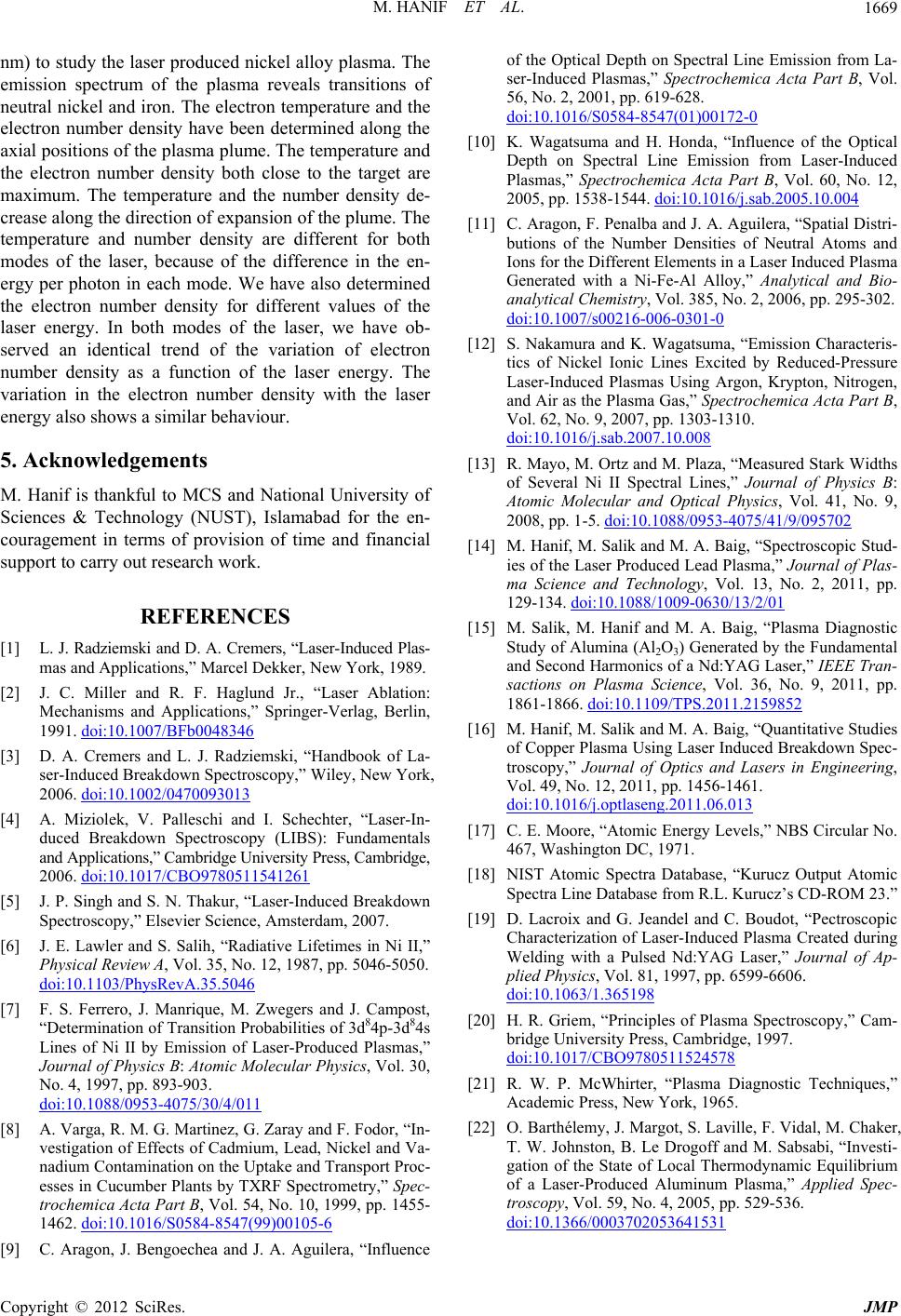
nm) to study the laser produced nickel alloy plasma. The
emission spectrum of the plasma reveals transitions of
neutral nickel and iron. The electron temperature and the
electron number density have been determined along the
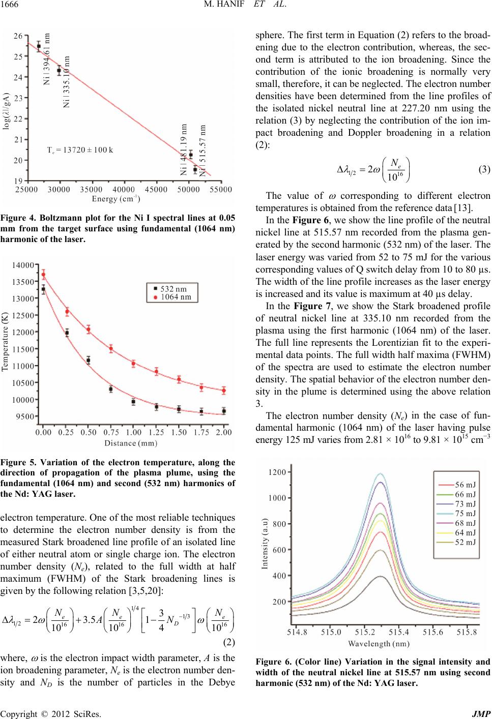
axial positions of the plasma plume. The temperature and
the electron number density both close to the target are
maximum. The temperature and the number density de-
crease along the direction of expansion of the plume. The
temperature and number density are different for both
modes of the laser, because of the difference in the en-
ergy per photon in each mode. We have also determined
the electron number density for different values of the
laser energy. In both modes of the laser, we have ob-
served an identical trend of the variation of electron
number density as a function of the laser energy. The
variation in the electron number density with the laser
energy also shows a similar behaviour.
5. Acknowledgements
M. Hanif is thankful to MCS and National University of
Sciences & Technology (NUST), Islamabad for the en-
couragement in terms of provision of time and financial
support to carry out research work.
REFERENCES
[1] L. J. Radziemski and D. A. Cremers, “Laser-Induced Plas-
mas and Applications,” Marcel Dekker, New York, 1989.
[2] J. C. Miller and R. F. Haglund Jr., “Laser Ablation:
Mechanisms and Applications,” Springer-Verlag, Berlin,
1991. doi:10.1007/BFb0048346
[3] D. A. Cremers and L. J. Radziemski, “Handbook of La-
ser-Induced Breakdown Spectroscopy,” Wiley, New York,
2006. doi:10.1002/0470093013
[4] A. Miziolek, V. Palleschi and I. Schechter, “Laser-In-
duced Breakdown Spectroscopy (LIBS): Fundamentals
and Applications,” Cambridge University Press, Cambridge,
2006. doi:10.1017/CBO9780511541261
[5] J. P. Singh and S. N. Thakur, “Laser-Induced Breakdown
Spectroscopy,” Elsevier Science, Amsterdam, 2007.
[6] J. E. Lawler and S. Salih, “Radiative Lifetimes in Ni II,”
Physical Review A, Vol. 35, No. 12, 1987, pp. 5046-5050.
doi:10.1103/PhysRevA.35.5046
[7] F. S. Ferrero, J. Manrique, M. Zwegers and J. Campost,
“Determination of Transition Probabilities of 3d84p-3d84s
Lines of Ni II by Emission of Laser-Produced Plasmas,”
Journal of Physics B: Atomic Molecular Physics, Vol. 30,
No. 4, 1997, pp. 893-903.
doi:10.1088/0953-4075/30/4/011
[8] A. Varga, R. M. G. Martinez, G. Zaray and F. Fodor, “In-
vestigation of Effects of Cadmium, Lead, Nickel and Va-
nadium Contamination on the Uptake and Transport Proc-
esses in Cucumber Plants by TXRF Spectrometry,” Spec-
trochemica Acta Part B, Vol. 54, No. 10, 1999, pp. 1455-
1462. doi:10.1016/S0584-8547(99)00105-6
[9] C. Aragon, J. Bengoechea and J. A. Aguilera, “Influence
of the Optical Depth on Spectral Line Emission from La-
ser-Induced Plasmas,” Spectrochemica Acta Part B, Vol.
56, No. 2, 2001, pp. 619-628.
doi:10.1016/S0584-8547(01)00172-0
[10] K. Wagatsuma and H. Honda, “Influence of the Optical
Depth on Spectral Line Emission from Laser-Induced
Plasmas,” Spectrochemica Acta Part B, Vol. 60, No. 12,
2005, pp. 1538-1544. doi:10.1016/j.sab.2005.10.004
[11] C. Aragon, F. Penalba and J. A. Aguilera, “Spatial Distri-
butions of the Number Densities of Neutral Atoms and
Ions for the Different Elements in a Laser Induced Plasma
Generated with a Ni-Fe-Al Alloy,” Analytical and Bio-
analytical Chemistry, Vol. 385, No. 2, 2006, pp. 295-302.
doi:10.1007/s00216-006-0301-0
[12] S. Nakamura and K. Wagatsuma, “Emission Characteris-
tics of Nickel Ionic Lines Excited by Reduced-Pressure
Laser-Induced Plasmas Using Argon, Krypton, Nitrogen,
and Air as the Plasma Gas,” Spectrochemica Acta Part B,
Vol. 62, No. 9, 2007, pp. 1303-1310.
doi:10.1016/j.sab.2007.10.008
[13] R. Mayo, M. Ortz and M. Plaza, “Measured Stark Widths
of Several Ni II Spectral Lines,” Journal of Physics B:
Atomic Molecular and Optical Physics, Vol. 41, No. 9,
2008, pp. 1-5. doi:10.1088/0953-4075/41/9/095702
[14] M. Hanif, M. Salik and M. A. Baig, “Spectroscopic Stud-
ies of the Laser Produced Lead Plasma,” Journal of Plas-
ma Science and Technology, Vol. 13, No. 2, 2011, pp.
129-134. doi:10.1088/1009-0630/13/2/01
[15] M. Salik, M. Hanif and M. A. Baig, “Plasma Diagnostic
Study of Alumina (Al2O3) Generated by the Fundamental
and Second Harmonics of a Nd:YAG Laser,” IEEE Tran-
sactions on Plasma Science, Vol. 36, No. 9, 2011, pp.
1861-1866. doi:10.1109/TPS.2011.2159852
[16] M. Hanif, M. Salik and M. A. Baig, “Quantitative Studies
of Copper Plasma Using Laser Induced Breakdown Spec-
troscopy,” Journal of Optics and Lasers in Engineering,
Vol. 49, No. 12, 2011, pp. 1456-1461.
doi:10.1016/j.optlaseng.2011.06.013
[17] C. E. Moore, “Atomic Energy Levels,” NBS Circular No.
467, Washington DC, 1971.
[18] NIST Atomic Spectra Database, “Kurucz Output Atomic
Spectra Line Database from R.L. Kurucz’s CD-ROM 23.”
[19] D. Lacroix and G. Jeandel and C. Boudot, “Pectroscopic
Characterization of Laser-Induced Plasma Created during
Welding with a Pulsed Nd:YAG Laser,” Journal of Ap-
plied Physics, Vol. 81, 1997, pp. 6599-6606.
doi:10.1063/1.365198
[20] H. R. Griem, “Principles of Plasma Spectroscopy,” Cam-
bridge University Press, Cambridge, 1997.
doi:10.1017/CBO9780511524578
[21] R. W. P. McWhirter, “Plasma Diagnostic Techniques,”
Academic Press, New York, 1965.
[22] O. Barthélemy, J. Margot, S. Laville, F. Vidal, M. Chaker,
T. W. Johnston, B. Le Drogoff and M. Sabsabi, “Investi-
gation of the State of Local Thermodynamic Equilibrium
of a Laser-Produced Aluminum Plasma,” Applied Spec-
troscopy, Vol. 59, No. 4, 2005, pp. 529-536.
doi:10.1366/0003702053641531