Modern Plastic Surgery, 2012, 2, 83-86
http://dx.doi.org/10.4236/mps.2012.24020 Published Online October 2012 (http://www.SciRP.org/journal/mps) 83
Assessing Injection Techniques in the Treatment of
Trigger Finger
John R. Fowler1, Lauren Ogrich2, Perry Evangelista3, Alyssa A. Schaffer1
1Temple University Hospital, Department of Orthopaedics, Philadelphia, USA; 2Temple University School of Medicine, Philadelphia,
USA; 3University of Pennyslvania School of Medicine, Philadelphia, USA.
Email: john.fowler@tuhs.temple.edu
Received June 7th, 2012; revised July 8th, 2012; accepted August 6th, 2012
ABSTRACT
Background: Trigger finger is characterized by the inability to smoothly flex and extend the digit. Corticosteroids are
an accepted non-surgical treatment option and can be delivered via two techniques. While the palmar approach is more
commonly used, some have suggested that the mid-axial approach may be less painful for patients and yield higher in-
trasheath injection rates. The purpose of this study is to compare the accuracy of the palmar and midaxial approaches
for delivery of corticosteroids into the flexor tendon sheath using radio-opaque dye in a cadaver model. Methods: A
total of 50 injections were performed, 25 via midaxial technique and 25 via palmar technique. A one inch, 25-gauge
needle was used to inject 1 mL of Isovue contrast dye into the flexor tendon sheath under live fluoroscopy. The fluoro-
scopic images were examined after injection to determine intrasheath versus extrasheath delivery of the dye, with visu-
alization of contrast filling the sheath defining a successful injection. Results: The midaxial approach had a success rate
of 52% compared to the conventional palmar approach success rate of 36%, p = 0.5. The ring finger is the most com-
mon location of trigger finger and the rates of success were equal between groups for this digit (80%). Conclusions:
Based on our findings, there is no statistical difference in the accuracy of intrasheath injection between the midaxial
technique and palmar technique. The midaxial technique can be considered as an alternative to the palmar technique for
trigger finger injection.
Keywords: Trigger Finger; Injection; Corticosteroids; Palmar and Midaxial Approaches
1. Introduction
Trigger finger, also known as stenosing tenosynovitis or
stenosing tendovaginitis, is a common condition affect-
ing nearly 100,000 Americans annually [1]. It is charac-
terized by the inability to smoothly flex and extend the
digit [1]. The patho physiology of stenosing tenosynovitis
is related to a disproportion between the volume of the
flexor sheath and its contents, the flexor tendons [1].
Non-surgical management consists of activity modifica-
tion, non-steroidal anti-inflammatory medications, and
corticosteroid injection [1]. The reported effectiveness of
corticosteroid injections has ranged from 25% - 77% [2-
6]. A recent systematic review examining the effective-
ness of corticosteroid injections found corticosteroid in-
jections to be effective in only 57% of patients [7].
Although several studies have found extra-sheath cor-
ticosteroid injections to have some benefit in the treat-
ment of trigger finger [3,8], the gold standard remains
delivery of the corticosteroid into the flexor tendon
sheath [1,9]. Two major techniques exist to deliver the
corticosteroid into the flexor tendon sheath: the palmar
approach and the midaxial approach [1,2,9]. While the
palmar approach is more commonly used, some have
suggested that the mid-axial approach may be less pain-
ful for patients and yield higher intrasheath injection
rates [1,2,9]. Studies examining the accuracy of intra-
sheath injection by conventional techniques found deliv-
ery into the tendon sheath occurs in only 37% - 50% of
patients [8,10]. If intrasheath delivery of therapeutic me-
dications is an important factor in the treatment of trigger
finger, then determining the optimal injection technique
is critical.
The purpose of this study is to compare the accuracy
of the palmar and midaxial approaches for delivery of
corticosteroids into the flexor tendon sheath using radio-
opaque dye in a cadaver model.
2. Methods
The left hands of ten embalmed cadavers were used in
this study. The cadaver hands were randomly assigned to
receive injection of corticosteroid into the flexor tendon
sheath by either the midaxial or palmar approach. For the
Copyright © 2012 SciRes. MPS
Assessing Injection Techniques in the Treatment of Trigger Finger
84
palmar injection technique, the needle was introduced at
the level of the A1 pulley and was advanced until resis-
tance was felt. The needle was withdrawn slightly to en-
sure it was no within the tendon (see Figure 1) and then
the contrast was injected. The midaxial technique was
performed as described by Carlson [7]. From the radial
border of the finger, the needle was inserted into the
midlateral area of the proximal phalanx (see Figure 2).
The needle was inserted until resistance was felt an d th en
finger was then flexed and extended to confirm that the
needle was not in the tendon itself. The contrast dye was
then injected.
A one inch, 25-gauge needle was used to inject 1 mL
of Isovue contrast dye into the flexor tendon sheath under
live fluoroscopy. Isovue dye was provided by the De-
partment of Radiology. The fluoroscopic images were
examined after injection to determine intrasheath versus
extrasheath delivery of the dye. A positive outcome was
defined as contrast medium tracking proximally and dis-
tally through the flexor tendon sheath on fluoroscopic
Figure 1. Photograph demonstrating palmar injection tech-
nique.
Figure 2. Photograph demonstrating the mid-axial injection
technique.
images (see Figure 3). A negative outcome was defined
as an injection that did not fill th e tendon sheath on fluo-
roscopic images (see Figure 4).
Each finger was injected on each hand for a total of 50
fingers, 25 midaxial and 25 palmar. All injections were
performed by a senior orthopaedic resident. The finger,
technique, and result were recorded for each hand and
images saved for review by the senior author. Statistical
analysis was performed using Graphpad (La Jolla, CA)
Software. Fisher’s exact test was used to determine sta-
tistical significance. A power analysis was performed
prior to the initiation of the study, which found a sample
size of 25 injections would be needed in each group to
detect a difference of 30% between groups, assuming an
alpha value of 0.05 and a beta value of 0.8.
Institutional Review Board ex emption was obtained as
this was a cadaver study withou t live subjects.
3. Results
The midaxial approach had a success rate of 52% com-
pared to the conventional palmar approach success rate
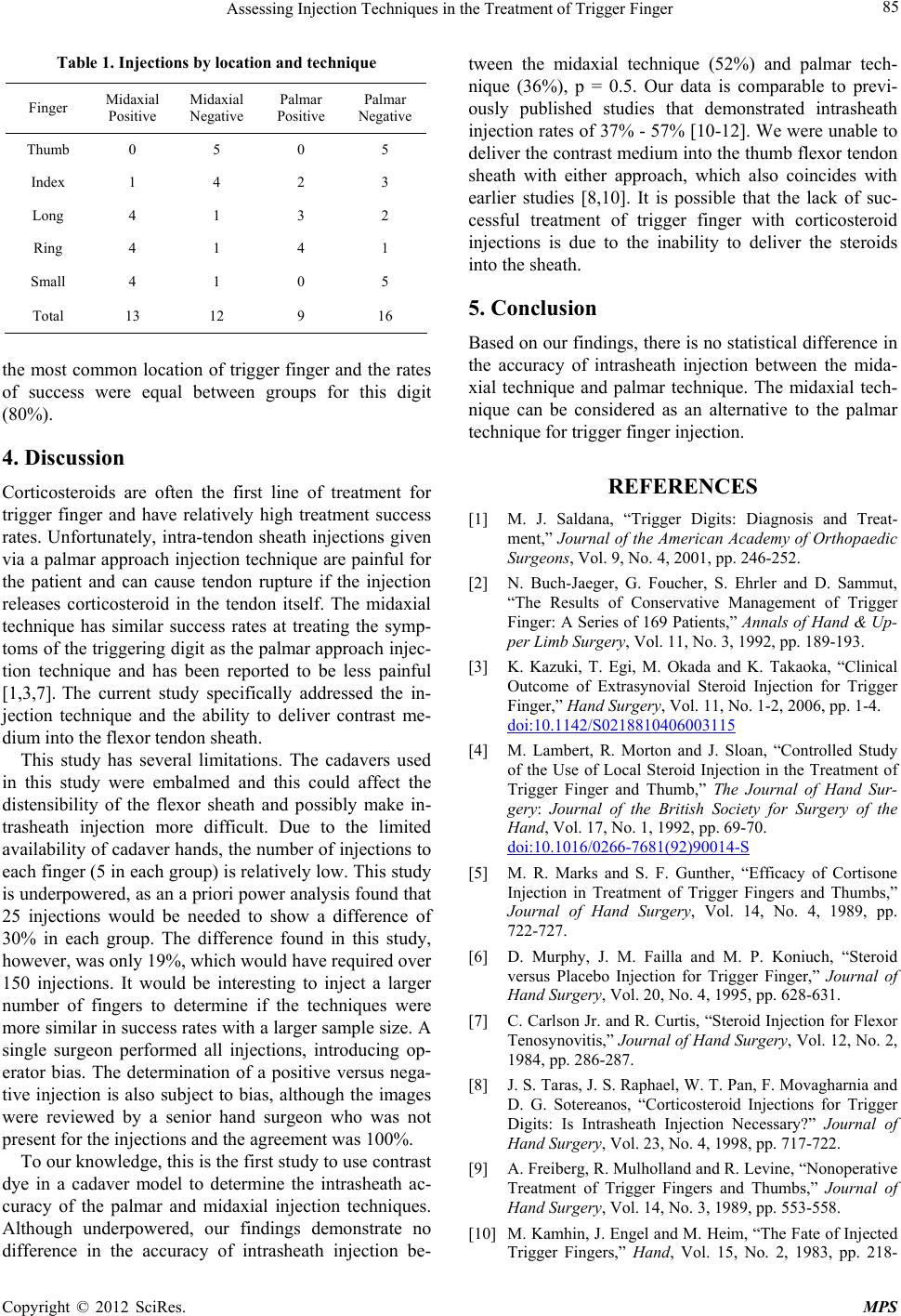
of 36%, p = 0.5. See Table 1. Injection of the thumb was
difficult regardless of the approach, yielding an overall
success rate of 0%. If this data is removed, the success
rates for the other fingers improves to 65% for the mi-
daxial and 45% for the palmar approach, p = 0.4. The
palmar approach was also found to have a 0% success
rate in the small finger, whereas the midxial approach
had a success rate of 80% in this digit. The rin g finger is
Figure 3. Positive intra-sheath injection, showing contrast
tracking throughout the sheath.
Figure 4. Negative intra-sheath injection, showing contrast
tracking outside the sheath.
Copyright © 2012 SciRes. MPS
Assessing Injection Techniques in the Treatment of Trigger Finger 85
Table 1. Injections by loca tion and technique
Finger Midaxial
Positive Midaxial
Negative Palmar
Positive Palmar
Negative
Thumb 0 5 0 5
Index 1 4 2 3
Long 4 1 3 2
Ring 4 1 4 1
Small 4 1 0 5
Total 13 12 9 16
the most common location of trigger finger and the rates
of success were equal between groups for this digit
(80%).
4. Discussion
Corticosteroids are often the first line of treatment for
trigger finger and have relatively high treatment success
rates. Unfortunately, intra-tendon sheath injections given
via a palmar approach injection technique are painful for
the patient and can cause tendon rupture if the injection
releases corticosteroid in the tendon itself. The midaxial
technique has similar success rates at treating the symp-
toms of the triggering d igit as the palmar approach injec-
tion technique and has been reported to be less painful
[1,3,7]. The current study specifically addressed the in-
jection technique and the ability to deliver contrast me-
dium into the flexor tendo n sheath.
This study has several limitations. The cadavers used
in this study were embalmed and this could affect the
distensibility of the flexor sheath and possibly make in-
trasheath injection more difficult. Due to the limited
availability of cadaver hand s, the number of injections to
each finger (5 in each group) is relatively low. This study
is underpowered, as an a priori power analysis found that
25 injections would be needed to show a difference of
30% in each group. The difference found in this study,
however, was only 19%, which would have required over
150 injections. It would be interesting to inject a larger
number of fingers to determine if the techniques were
more similar in success rates with a larger sample size. A
single surgeon performed all injections, introducing op-
erator bias. The determination of a positive versus nega-
tive injection is also subject to bias, although the images
were reviewed by a senior hand surgeon who was not
present for the injections and the agreement was 100%.
To our knowledge, this is the first study to use contrast
dye in a cadaver model to determine the intrasheath ac-
curacy of the palmar and midaxial injection techniques.
Although underpowered, our findings demonstrate no
difference in the accuracy of intrasheath injection be-
tween the midaxial technique (52%) and palmar tech-
nique (36%), p = 0.5. Our data is comparable to previ-
ously published studies that demonstrated intrasheath
injection rates of 37% - 57% [10-12]. We were unable to
deliver the contrast medium into the thumb flexor tendon
sheath with either approach, which also coincides with
earlier studies [8,10]. It is possible that the lack of suc-
cessful treatment of trigger finger with corticosteroid
injections is due to the inability to deliver the steroids
into the sheath.
5. Conclusion
Based on our findings, there is no statistical difference in
the accuracy of intrasheath injection between the mida-
xial technique and palmar technique. The midaxial tech-
nique can be considered as an alternative to the palmar
technique for trigger finger injection.
REFERENCES
[1] M. J. Saldana, “Trigger Digits: Diagnosis and Treat-
ment,” Journal of the American Academy of Orthopaedic
Surgeons, Vol. 9, No. 4, 2001, pp. 246-252.
[2] N. Buch-Jaeger, G. Foucher, S. Ehrler and D. Sammut,
“The Results of Conservative Management of Trigger
Finger: A Series of 169 Patients,” Annals of Hand & Up-
per Limb Surgery, Vol. 11, No. 3, 1992, pp. 189-193.
[3] K. Kazuki, T. Egi, M. Okada and K. Takaoka, “Clinical
Outcome of Extrasynovial Steroid Injection for Trigger
Finger,” Hand Surgery, Vol. 11, No. 1-2, 2006, pp. 1-4.
doi:10.1142/S0218810406003115
[4] M. Lambert, R. Morton and J. Sloan, “Controlled Study
of the Use of Local Steroid Injection in the Treatment of
Trigger Finger and Thumb,” The Journal of Hand Sur-
gery: Journal of the British Society for Surgery of the
Hand, Vol. 17, No. 1, 1992, pp. 69-70.
doi:10.1016/0266-7681(92)90014-S
[5] M. R. Marks and S. F. Gunther, “Efficacy of Cortisone
Injection in Treatment of Trigger Fingers and Thumbs,”
Journal of Hand Surgery, Vol. 14, No. 4, 1989, pp.
722-727.
[6] D. Murphy, J. M. Failla and M. P. Koniuch, “Steroid
versus Placebo Injection for Trigger Finger,” Journal of
Hand Surgery, Vol. 20, No. 4, 1995, pp. 628-631.
[7] C. Carlson Jr. and R. Curtis, “Steroid Injection for Flexor
Tenosynovitis,” Journal of Hand Surgery, Vol. 12, No. 2,
1984, pp. 286-287.
[8] J. S. Taras, J. S. Rapha el, W. T. Pan, F. Movagharnia and
D. G. Sotereanos, “Corticosteroid Injections for Trigger
Digits: Is Intrasheath Injection Necessary?” Journal of
Hand Surgery, Vol. 23, No. 4, 1998, pp. 717-722.
[9] A. Freiberg, R. Mulholland and R. Levine, “Nonoperative
Treatment of Trigger Fingers and Thumbs,” Journal of
Hand Surgery, Vol. 14, No. 3, 1989, pp. 553-558.
[10] M. Kamhin, J. Engel and M. Heim, “The Fate of Injected
Trigger Fingers,” Hand, Vol. 15, No. 2, 1983, pp. 218-
Copyright © 2012 SciRes. MPS
Assessing Injection Techniques in the Treatment of Trigger Finger
Copyright © 2012 SciRes. MPS
86
220. doi:10.1016/S0072-968X(83)80018-7
[11] S. B. Fleisch, K. P. Spindler and D. H. Lee, “Corticoste r-
oid Injections in the Treatment of Trigger Finger: A Level
I and II Systematic Review,” Journal of the American
Academy of Orthopaedic Surgeons, Vol. 15, No. 3, 2007,
pp. 166-171.
[12] D. Ring, S. Lozano-Calderón, R. Shin, P. Bastian, C.
Mudgal and J. Jupiter, “A Prospective Randomized Con-
trolled Trial of Injection of Dexamethasone versus Tri-
amcinolone for Idiopathic Trigger Finger,” Journal of
Hand Surgery, Vol. 33, No. 4, 2008, pp. 516-522.