Pharmacology & Pharmacy, 2012, 3, 404-408
http://dx.doi.org/10.4236/pp.2012.34054 Published Online October 2012 (http://www.SciRP.org/journal/pp) 1
Relationship of Uric Acid with Superoxide Dismutase (Sod)
in Induced Hyperuricemic Rat Model
Shiza Batool1, Iftikhar Ahmed2*, Muhammad Sarwar3, Hafeez ul Hassan4
1Biochemistry Department, Aziz Fatima Medical and Dental College, Faisalabad, Pakistan; 2Biochemistry Department, Baqai Medi-
cal University, Karachi, Pakistan; 3Chemical Pathology Department, Al Jouf University, Al-Jawf, KSA; 4Physiology Department,
Baqai Medical Unive rsity, Karachi, Pa ki st an.
Email: *dr_iftikharahmed@yahoo.com, *driftikharahmed68@gmail.com
Received July 16th, 2012; revised August 18th, 2012; accepted September 12th, 2012
ABSTRACT
Increase uric acid levels have been found in oxidative stress. Urate radicals do not react with oxygen to form another
peroxy radical, thus increasing the efficacy of uric acid as an antioxidant. Therefore, this study is designed to measure
the level of uric acids and find out the relationship of uric acid with superoxide dismutase in induced hyperuricemic
model. Forty male albino rats with an average weight of 180 ± 2 g were selected. The rats were grouped. The animals
were fed on standard diet and given tap water ad libitum until treatment. Albino rats were divided into four groups.
Group A (10)—con trol giv en only stand ard diet, gr oup B (10) fed on 60% fructose w ith standard diet, group C (10) fed
on fructose, standard diet and intraperitonially oxonic acid 250 mg/kg and group D (10) only on injection intraperotoni-
ally oxonic acid 250 mg/kg. At the end of study 10 mL of blood was drawn from heart of rats. Then blood was esti-
mated for superoxide dismutase and uric acids done by kit methods randox-manual/Rx monza UA230/UA 233. Results:
In Group C superoxide dismutase was found to be 32% (244 ± 2.23 mg/dL) more than control. In the same group the
uric acid concentration was highly significantly correlated with control. Conclusion: The uric acid concentration in-
creases when we take fructose up to 60% in our diet. It also increases superoxide dismutase concentration. More than
this value may have inverse effect on the uric acid level and its role as an antioxidant may become inversed.
Keywords: Uric Acid; Superoxide Dismutase; Albino Rats; Fructose Induced Hyperuricemia
1. Introduction
Free radical can be defined as any molecular species ca-
pable of independent existence that contains an unpaired
electron in an atomic orbital [1]. They are capable of tr ig-
gering chain reactions which can damage the different
cell constituents.
The most important free radicals in many disease
states are oxygen derivatives, particularly superoxide and
the hydroxyl radical. Superoxide is formed from several
molecules by oxidation including adrenaline, flavine nu-
cleotides, thiol compounds and glucose. Superoxide is
also produce during important biological reactions in-
cluding electron transport chain in mitochondria [2].
In order to check free radicals formation to avoid oxi-
dative stress, body has different anti-oxidant defense
systems. An antioxidant can be defined as: “any sub-
stance that when present in low concentrations compared
to that of an oxidisable substrate significantly delays or
inhibits the oxidatio n of that substrate. The physiolog ical
role of antioxidants, as this definition suggests, is to pre-
vent damage to cellular components arising as a conse-
quence of chemical reactions involving free radicals [3].
Superoxide dismutase is believed to serve as first line of
defense against toxicity of superoxide radicals. Also
takes part in cell signaling regarding to reactive oxygen
species levels [4]. Supero xide is one of the main reactive
oxygen species in the cell and as such, SOD serves a key
antioxidant role. The physiological importance of SODs
is illustrated by the severe pathologies evident in mice
genetically engineered to lack these enzymes. Mice lack-
ing SOD2 die several days after birth, amid massive oxi-
dative stress [5] Mice lacking SOD1 develop a wide range
of pathologies, includ ing hepatocellular carcinoma [6] an
acceleration of age-related muscle mass loss [7], an ear-
lier incidence of cataracts and a reduced lifespan. Mice
lacking SOD3 do not show any obvious defects and ex-
hibit a normal lifespan, though they are more sensitive to
hyperoxic injury [8].
Uric acid now is not considered as merely a metabolic
waste. It ha s been proposed that increase in life span ob-
served in human evolution to some extent might be due
to protective action of uric acid [9]. Increase uric acid
*Corresponding a uthor.
Copyright © 2012 SciRes. PP
Relationship of Uric Acid with Superoxide Dismutase (Sod) in Induced Hyperuricemic Rat Model 405
levels have been found in oxidative stress and ischemia
which might be compensatory mechanism of protection
against free radicals [10]. Urate radicals do not react with
oxygen to form another peroxy radical which is seen with
the ascorbic acid, thus increasing the efficacy of uric acid
as an antioxidant [11]. Uric acid cause inactivation of
Nitric oxide and peroxynitrite radicals [12,13]. Another
important function of urate is found in its ability to form
chelating agents with transition metals ions like iron and
copper thus scavenging them. We carried out a study to
assess the relationship of uric acid with superoxide dis-
mutase in an induced hyperuricemic rats.
2. Methodology
Locally bred fort y (40) male Albino ra ts with an averag e
weight of 180 ± 20 g were purchased. The rats were
grouped and housed in environmentally controlled room
(ambient temperature 24˚C ± 2˚C and relative humidity
of 55% ± 5%) in the animal house and acclimatized for
07 days. The animals were fed standard diet and given
tap water ad libitum until treatment. The protocols for
experimentation was approved and performed in strict
accordance with the Guide for the care and use of labo-
ratory animals (Institute of Laboratory Animal Resources
on Life Sciences, US National Research Council, 1996)
and the Institutional Animal Ethical Committee (IAEC)
of Baqai Medical University, Karachi. Pakistan. The
cage size was 8" × 18" × 10" to keep a group of 5 ani-
mals in the cage to prevent from cannibalism.
Sodium Tungstate 10%, 2/3N sulphuric acid, 10% so-
dium bicarbonate, LiCO3, 40% Formaline, Acetic acid,
Fructose, Oxonic acid. Spectrosol grade reagents and
acids from B.D.H., Poole, UK, were employed. All puri-
fied enzymes, coenzymes, substrates, standards and buff-
ers will be purchased from Sigma Chemicals Company,
USA. All other chemicals were of analytical grade and
will be procured from SRL and Qualigens, USA.
All animals housed in standard conditions were ini-
tially fed standard diet and allowed adaptation of one
(1) week. Albino rats were divided in four (4) groups: A,
B, C & D.
Group A: Ten (10) male albino rats as Control were
kept as control and were fed standard diet and water ad
libitum for 10 weeks. Group B: Ten (10) male albino rats
[F] were fed 60% fructose mixed in standard diet and
water ad libitum for 10 weeks. Group C: Ten (10) male
albino rats [FO] were fed 60% fructose mixed in standard
diet and water ad libitum for 10 weeks. They were also
injected intraperitonealy oxonic acid 250 mg/kg every
third day for 10 weeks. Group D: Ten (10) male albino
rats [O] were injected intraperitonealy oxonic acid 250
mg/kg every third day for 10 weeks. They were fed
standard diet and water ad libitum for 10 weeks. Body
weights were measured at the commencement and at the
end of study. The amount of diet was measured before
giving and then subtracted from the amount of food left
over daily. At the end of study, rats were dissected in a
nearby room separate from experiment area. Approxi-
mately 10 mls of blood was drawn from heart using dis-
posable syringe. 8 mls of blood was transferred in he pa rin -
ized tube, mixed and centrifuged to separate plasma and
divided in two epindorf cups for estimation of uric acid
and SOD done by kit methods by randox-manual /Rx
monza UA230/UA 233.
3. Results
3.1. Graph 1
It demonstrates the comparison of mean plasma uric acid
levels of Control with rest of the groups. Mean plasma
level of uric acid of Control is found to be 1.97 mg/dL
(±0.09). Group “F” (fructose) showed mean plasma uric
acid of 3.15 mg/dL (±0.17). This reflects that uric acid
was raised to 37% in rats which were exposed to diet
comprising 60% Fructose than control. On comparing
both groups i.e., Control with Group “F” (highly signifi-
cant statistical correlation (P < 0.001) was observed.
The mean plasma uric acid levels of Group “O”
SERUM URIC ACID
GROUPS CONTROL GROUP = F GROUP = O GROUP = F + O
M.V. 1.97 3.15 3.63 4.43
S.D 0.3 0.55 0.63 0.43.
Graph 1. Comparison of serum uric acid levels of all study groups.
Copyright © 2012 SciRes. PP
Relationship of Uric Acid with Superoxide Dismutase (Sod) in Induced Hyperuricemic Rat Model
406
(oxonic acid) was 3.63 mg/dL (±0.22) which is 45%
higher than Control. The probability calculated was high l y
significant (P < 0.0 01 ) w hen both groups were eval uat ed .
While comparing Group “F + O” (Fructose + Oxonic
acid) with Control, highly sign ificant correlation was ob-
served (P < 0.001). It was due to high mean plasma se-
rum uric acid level of Group “F + O” which was 4.41
mg/dL (±0.14). The combinatio n of fructose with uricase
inhibitor, Oxonic acid raises uric acid to 55% from con-
trol and this level is highest of all these groups.
3.2. Graph 2
It shows the comparison of mean plasma SOD levels of
Control with rest of the groups. Mean plasma level of
SOD of Control was found to be 165.15 mg/dL (±2.65).
Group “F” (Fructose) showed mean plasma SOD level of
176.65 mg/dL (±2.60) reflecting SOD levels were raised
to 6.5% in rats which were exposed to diet comprising
60% Fructose. On comparing both groups i.e., Control
with Group “F” significant statistical correlation (P <
0.01) was observed. The mean plasma SOD levels of
Group “O” (oxonic acid) has been measured to 181.2
mg/dL (±3.52) which is again 8% more than Control.
Therefore, significant (P < 0.01) correlation was ob-
served on comparison of both groups.
While comparing Group “F + O” (Fructose + Oxonic
acid) with Control, highly significant correlation was
observed (P < 0.001). It was due to high mean plasma
SOD levels of Group “F + O” which was 244 mg/dL
(±2.23) which is high est of all these groups. In this gro up
SOD was 32% more than control which might be due to
antioxidant action of uric acid .
4. Discussion
One of the important features of this study was the
method by which hyperuricemia have been induced in
animal model. The group B was given fructose, group D
was treated with “oxonic acid” and group C was offered
both fructose and oxonic acid (G = Fructose + Oxonic
acid). The principle hyperuricemic factor in this study
was fructose as it is extensively used in beverages and
food. Its a rather controversial factor as number of stud-
ies both animals and human, are in the favour that fruc-
tose can induce hyperuricemia [14] but many studies
have opposed this hypothesis [15] and even mixed re-
sponse has been shown [16]. Present investigation has
tried to verify this theory. Very few studies have used
this combined model of fructose plus oxonic acid. In
order to make conditions similar to human, uricase in-
hibitor oxonic acid was incorporated to ab olish the effect
of this enzyme in rats. Also these different regimens
were used to establish the extent of hyperuricemia caused
by fructose.
Superoxide dismutase levels were found to be raised in
all three groups in comparison to control. The highest
level was obser ved in gro up C of 244 mg/dL as sh own in
graph 1. They were 32% more than the control. This was
in agreement with H. Ulrich Hink and Nalini Santanam
et al. The possible explanation can be drawn from num-
ber of studies showing that superoxide dismutase during
catalyzing dismutation of 2 to H2O2 can form copper
bound hydroxyl radical from hydrogen peroxide H2O2
[17]. Hydroxyl radical when gets bounded to SOD, then
it can attack adjacent histidine residue which is attached
to copper resulting in inactivation of both SOD1 and
SOD3 [18]. This might be prevented when small anions
or reductants including Uric acid are co incubated [19] as
shown in graph 1 the comparison of mean plasma SOD
levels of Control with rest of the groups. Mean plasma
level of SOD of Control was found to be 165.15 mg/dL
(±2.65). Group B (Fructose) showed mean plasma SOD
level of 176.65 mg/dL (±2.60) reflecting SOD levels
were raised to 6.5% in rats which were exposed to diet
comprising 60% Fructose. On comparing both groups, i.e.
Control with Group B significant statistical correlation (P <
0.01) was observed.
O
SUPEROXIDE DISMUTASE
GROUPS CONTROL GROUP = F GROUP = O GROUP = F + O
M.V. 165.15 176.65 181.25 244
S.D 8.38 8.23 9.96 6.69
Graph 2. Comparison of serum SOD levels of all study groups.
Copyright © 2012 SciRes. PP
Relationship of Uric Acid with Superoxide Dismutase (Sod) in Induced Hyperuricemic Rat Model 407
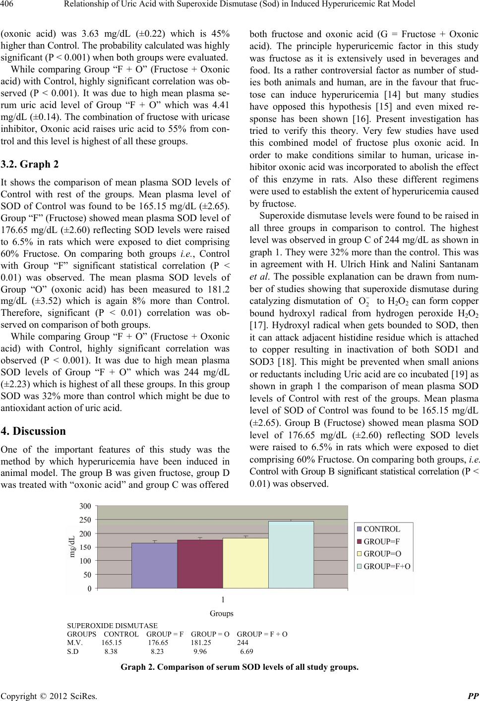
The mean plasma SOD levels of Group D (oxonic acid)
has been measured to 181.2 mg/dL (±3.52) which is
again 8% more than Control. Therefore, significant (P <
0.01) correlation was observed on comparison of both
groups.
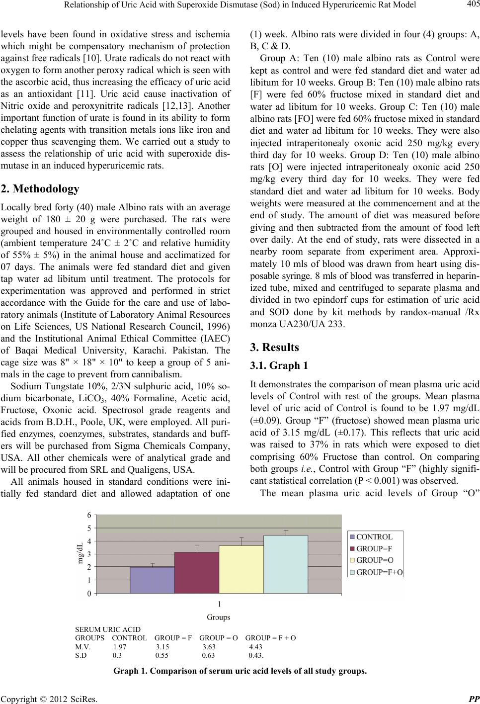
While comparing Group C (Fructose + Oxonic acid)
with Control, highly significant co rrelation was observed
(P < 0.001). It was due to high mean plasma SOD levels
of Group “F + O” which was 244 mg/dL (±2.23) which
is highest of all these groups. In this group SOD was
32% more than control which might be due to antioxi-
dant action of uric acid.
It has been suggested that in group C there was signi-
ficant increase in the concen tration of uric acid. This was
in accordance with some studies done on human that
high dietary intake of fructose contributes significantly to
hyperuricemia [20]. In a large study in the United States,
consumption of four or more sugar-sweetened soft drinks
per day gave an odds ratio of 1.82 for hyperuricemia [21].
Increased pr oduction of uric acid is th e result of interfer-
ence, by a product of fructose metabolism, in purine me-
tabolism. This interference has a dual action, both in-
creasing the conversion of ATP to inosine and increasing
the synthesis of purine [22] http://en.wikipedia.org/wiki/
Hyperuricemia-cite_note-pmid8213607-14. Fructose also
inhibits the excretion of uric acid, apparently by compet-
ing with uric acid for access to the transport protein
SLC2A9 [23]. The effect of fructose in reducing excre-
tion of uric acid is increased in people with a hereditary
(genetic) predisposition toward hyperuricemia and/or
gout [22] .
Starvation causes the body to metabolize its own
(purine-rich) tissues for energy. Thus, like a high purine
diet, starvation increases the amount of purine converted
to uric acid. A very low calorie diet without carbohydrate
can induce extreme hyperuricemia; including some car-
bohydrate (and redu cing the protein) reduces th e level of
hyperuricemia [24]. Starvation also impairs the ability of
the kidney to excrete uric acid, due to competition for
transport between uric acid and ketones [25]. Many
studies are controversial to our results all were done on
human some supported that high doses of fructose (200
g/day for 2 weeks) raise the blood pressure and cause the
features of metabolic syndrome. Some suggested that
lowering of the uric acid level prevents the increase in
mean arterial blood pressure. Excessive intake of fruc-
tose may have a role in the current epidemics of obesity
and diabetes [26]. Some authors also suggested that in-
creased dietary fructose was not associated with increase
uric acid level [27].
5. Conclusion
The uric acid concentration does increase when we take
fructose up to 60% in our diet, e.g., beverages soft drinks.
It also increases superoxide dismutase concentration. It
has been concluded that more than this value of fructose
may have inverse effect on the uric acid level and its role
as an antioxidant may become inversed. Therefore, it is
suggested from our study that further work need to be
done on the effect of fructose on uric acid levels in hu-
man.
REFERENCES
[1] B. Halliwell and J. M. C. Gutteridge, “Role of Free Radi-
cals and Catalytic Metal Ions in Human Disease: An O ve r-
view,” Methods in Enzymology, Vol. 186, 1990, pp. 1-85.
[2] L. B. Becker, T. L. Vanden Hoek, Z. H. Shao, C. Q. Li
and P. T. Schumacker, “Generation of Superoxide in Car-
diomyocytes during Ischemia before Reperfusion,” Ameri-
can Journal of Physiology, Vol. 277, No. 6, 1999, pp.
H2240-H2246.
[3] I. S. Young and J. V. Woodside, “Antioxidants in Health
and Disease,” Journal of Clinical Pathology, Vol. 54, No.
3, 2001, pp. 176-186.
[4] A. J. Augustin, “Was It oxidativer Stress?” Klinische Mo-
natsblätter für Augenheilkunde, Vol. 227, No. 2, 2010, pp.
90-98. doi:10.1055/s-0029-1245125
[5] Y. Li, T. T. Huang, E. J. Carlson, S. Melov, P. C. Ursell,
J. L. Olson, L. J. Noble, M. P. Yoshimura, C. Berger, P.
H. Chan, D. C. Wallace and C. J. Epstein, “Dilated Card io-
myopathy and Neonatal Lethality in Mutant Mice Lack-
ing Manganese Superoxide Dismutase,” Nature Genetics,
Vol. 11, No. 4, 1995, pp. 376-377.
doi:10.1038/ng1295-376
[6] S. Elchuri, T. D. Oberley, W. Qi, R. S. Eisenstein, L. J.
Roberts, H. Van Remmen, C. J. Epstein and T.-T. Huang,
“CuZnSOD Deficiency Leads to Persistent and Wide-
spread Oxidative Damage and Hepatocarcinogenesis Lat er
in Life,” Oncogene, Vol. 24, No. 3, 2005, pp. 367- 380.
doi:10.1038/sj.onc.1208207
[7] F. L. Muller, W. Song, Y. Liu, A. Chaudhuri, S. Pieke-
Dahl, R. Strong, T.-T. Huang, C. J. Epstein, L. J. Roberts,
M. Csete, J. A. Faulkner and H. Van Remmen, “Absence
of CuZn Superoxide Dismutase Leads to Elevated Oxida-
tive Stress and Acceleration of Age-Dependent Skeletal
Muscle Atrophy,” Free Radical Biology & Medicine, Vol.
40, No. 11, 2006, pp. 1993-1004.
doi:10.1016/j.freeradbiomed.2006.01.036
[8] M. L. Sentman, M. Granström, H. Jakobson, A. Reaume,
S. Basu and S. L. Marklund, “Phenotypes of Mice Lack-
ing Extracellular Superoxide Dismutase and Copper- and
Zinc-Containing Superoxide Dismutase,” Journal of Bio-
logical Chemistry, Vol. 281, No. 11, 2006, pp. 6904-6909.
doi:10.1074/jbc.M510764200
[9] B. N. Ames, R. Cathcart, E. Schwiers and P. Hochstein,
“Uric Acid Provides an Antioxidant Defense in Humans
against Oxidant- and Radical-Caused Aging and Cancer:
A Hypothesis,” Proceedings of the National Academy of
Sciences of USA, Vol. 78, No. 11, 1981, pp. 6858-6862.
doi:10.1073/pnas.78.11.6858
[10] F. J. Nieto, C. Iribarren, M. D. Gross, G. W. Comstock, R.
Copyright © 2012 SciRes. PP
Relationship of Uric Acid with Superoxide Dismutase (Sod) in Induced Hyperuricemic Rat Model
408
G. Cutler, “Uric Acid and Serum Antioxidant Capacity: A
Reaction to Atherosclerosis,” Atherosclerosis, Vol. 148,
No. 1, 2000, pp. 131-139.
doi:10.1016/S0021-9150(99)00214-2
[11] W. S. Waring, “Uric Acid: An Important Antioxidant in
Acute Ischaemic St roke,” QJM: An International Journal
of Medicine, Vol. 95, No. 10, 2002, pp. 691-693.
doi:10.1093/qjmed/95.10.691
[12] M. Whiteman, U. Kestsakul and B. Halliwell, “A Reas-
sessment of the Peroxynitrite Scavenging Activity of Uric
Acid,” Annals of the New York Academy of Sciences, Vol.
962, 2002, pp. 242-259.
[13] G. Toncev, B. Milicic, S. Toncev an d G. Samardzic, “High-
Dose Methylprednisolone Therapy in Multiple Sclerosis
Increases Serum Uric Acid Levels,” Clinical Chemistry
and Laboratory Medicine, Vol. 40, No. 5, 2002, pp. 505-
508. doi:10.1515/CCLM.2002.088
[14] R. J. Johnson, S. E. Perez-Pozo, Y. Y. Sautin, J. Manitius,
L. G. Sanchez-Lozada, D. I. Feig, M. Shafiu, M. Segal, R.
J. Glassock, M. Shimada, C. Roncal and T. Nakagawa,
“Hypothesis: Could Excessive Fructose Intake and Uric
Acid Cause Type 2 Diabetes,” Endocrine Reviews, Vol.
30, No. 1, 2009, pp. 96-116. doi:10.1210/er.2008-0033
[15] P. A. Crapo and O. G. Kolterman, “The Me tabol ic E ffect s
of 2-Week Fructose Feeding in Normal Subjects,” Ameri-
can Journal of Clinical Nutrition, Vol. 39, No. 4, 1984,
pp. 525-534.
[16] S. Z. Sun, B. D Flickinger, P. S. Williamson-Hughes and
M. W. Empie, “Lack of Association between Dietary Fruc-
tose and Hyperuricemia Risk in Adults,” Nutrition &
Metabolism, Vol. 7, No. 1, 2010, pp. 12-16.
doi:10.1186/1743-7075-7-16
[17] H. U. Hink, N. Santanam, S. Dikalov, L. McCann, A. D.
Nguyen, S. Parthasarathy, D. G. Harrison and T. Fukai,
“Peroxidase Properties of Extracellular Superoxide Dis-
mutase. Role of Uric Acid in Modulating in Vivo Activ-
ity,” Arteriosclerosis, Thrombos is, and Vascular Biology,
Vol. 22, No. 9, 2002, pp. 1506-1508.
doi:10.1161/01.ATV.0000027524.86752.02
[18] S. L. Jewett, A. M. Rocklin, M. Ghanevati, J. M. Abel
and J. A. Marach, “A New Look at a Time-Worn System:
Oxidation of CuZn-SOD by H2O2,” Free Radical Biology
& Medicine, Vol. 26, No. 7-8, 1999, pp. 905-918.
doi:10.1016/S0891-5849(98)00274-3
[19] S. P. A. Goss, R. J. Singh and B. Kalyanaraman, “Bicar-
bonate Enhances the Peroxidase Activity of Cu,Zn-Su-
peroxide Dismutase. Role of Carbonate Anion Radical,”
The Journal of Biological Chemistry, Vol. 274, No. 40,
1999, pp. 28233-28239. doi:10.1074/jbc.274.40.28233
[20] A. Miller and K. Adeli, “Dietary Fructose and the Meta-
bolic Syndrome,” Current Opinion in Gastroenterology,
Vol. 24, No. 2, 2008, pp. 204-209.
[21] J. W. Choi and E. S. Ford, “Sugar Sweete ned Soft Drinks,
Diet Soft Drinks, and Serum Uric Acid Level: The Third
National Health and Nutrition Examination Survey,” Ar-
thritis & Rheumatism, Vol. 59, No. 1, 2008, pp. 109-116.
doi:10.1002/art.23245
[22] P. A. Mayes, “Intermediary Metabolism of Fructose,” Ame-
rican Journal of Clinical Nutrition, Vol. 58, Suppl. 5, 1993,
pp. 754S-765S.
[23] V. Vitart and I. Rudan, “SLC2A9 Is a Newly Identified
Urate Transporter Influencing Serum Urate Concentration,
Urate Excretion and Gout,” Nature Genetics, Vol. 40, No.
4, 2008, pp. 437-442. doi:10.1038/ng.106
[24] A. N. Howard, “Historical Development, Efficacy and
Safety of Very Low Calori e Diets,” International Journal
of Obesity, Vol. 5, No. 3, 1981, pp. 195-208.
[25] W. Kirch and C. Von Gicycki, “Renal Function in Thera-
peutic Starvation,” Wiener klinische Wochenschrift, Vol.
92, No. 8, 1980, pp. 263-266.
[26] S. E. Perez Pozo, J. Schold and T. Nakagawa, “Excessive
Fructose Intake Induces the Features of Metabolic Syn-
drome in Healthy Adult Men, Role of Uric Acid in Hy-
pertensive Response,” International Journal of Obesity,
Vol. 34, No. 3, 2010, pp. 454-461.
doi:10.1038/ijo.2009.259
Copyright © 2012 SciRes. PP