 Advances in Infectious Diseases, 2012, 2, 53-61 http://dx.doi.org/10.4236/aid.2012.23008 Published Online September 2012 (http://www.SciRP.org/journal/aid) 53 A wzt Mutant Burkholderia mallei Is Attenuated and Partially Protects CD1 Mice against Glanders Aloka B. Bandara Center for Molecular Medicine and Infectious Diseases, Virginia-Maryland Regional College of Veterinary Medicine, Virginia Poly- technic Institute and State University, Blacksburg, USA. Email: abandara@vt.edu Received April 23rd, 2012; revised May 25th, 2012; accepted June 27th, 2012 ABSTRACT Burkholderia mallei is the etiologic agent of glanders in solipeds and humans. Lipopolysaccharide (LPS) is a major component of cell envelop of this pathogen. O-antigen, the most external component of LPS, is a virulence factor and a protective antigen in many pathogenic bacteria. Two putative proteins named Wzm (integral membrane protein) and Wzt (hydrophilic ATP-binding protein) are believed to make up an ABC-2 transporter of B. mallei that facilitates transport of components of O-antigen from cytosol to outer-membrane. We studied the importance of wzt (encoding Wzt) to growth, LPS O-antigen profile, and pathogenicity of B. mallei. A wzt mutant strain was generated by deleting a portion of the wzt in B. mallei wild type strain ATCC 23344 by gene replacement. Compared to the wild type strain, the wzt mutant displayed slower growth in v it r o and less lethality in CD1 mice when inoculated intraperitoneally. The 50% lethal doses (LD50) of the wild type and the wzt mutant strains were 5.9 × 10⁵ and 9.1 × 10⁵ cfu, respectively. CD1 mice inoculated with a non-lethal dose of the wzt mutant produced specific serum immunoglobulins IgG1 and IgG2a and were partially protected against challenge with 11.2 times LD50 of the wild type strain. These findings suggest that the wzt is required for optimal in vitro growth and pathogenesis of B. mallei, and a wzt mutant protects CD1 mice against glanders. Keywords: Burkholderia mallei; ABC-2 Transporter; wzm Integral Membrane Protein; wzm Hydrophilic ATP-Binding Protein; Glanders; CD1 Mice; Pathogenicity; Protection 1. Introduction Burkholderia mallei, the causative agent of glanders, is a Gram-negative, aerobic bacillus. This bacterium is pri- marily responsible for disease in horses, mules, donkeys and occasionally humans [1-3]. Relatively little is known about the mechanisms of B. mallei pathogenesis [4,5]. In gram-negative bacteria, lipopolysaccharides (LPS), com- monly referred to as endotoxins, are a major component of cell envelopes [6,7]. Bacterial outermembranes pro- vide the “barrier function” largely due to the presence of LPS [8]. Bacterial strains expressing a smooth phenotype synthesize LPS molecules that are composed of three covalently linked domains: an O-polysaccharide antigen (O-antigen), a core region, and a lipid A moiety [9]. The O-antigen is the most external component of LPS, and it consists of a polymer of oligosaccharide repeating units. Chemical composition of O-antigens varies among dif- ferent bacterial species, as a result of the genetic varia- tion in the genes involved in O-antigen biosynthesis, designated the wb cluster. The genetics of O-antigen biosynthesis have been intensively studied in the En- terobacteriaceae, and it has been shown that the wb clusters usually contain genes involved in biosynthesis of activated sugars, glycosyl transferases, O-antigen poly- merases, and O-antigen export [10,11]. Burtnick et al. [12] identified the gene cluster responsible for O-antigen bio- synthesis in B. mallei ATCC 23344, and determined the physical structure of the B. mallei ATCC 23344 O-anti- gen. In bacteria, LPS O-antigen is exported to the cell sur- face using three distinctive pathways, as reviewed by Samuel and Reeves [13]. One of these pathways called ATP-binding cassette (ABC) transporter-dependent path- way, which has been found in E. coli O8 and O9 and Klebsiella pneumoniae O1 and O12 [14-17] is comprised of an integral membrane protein, Wzm, and a hydrophilic protein containing an ATP-binding motif, Wzt. The me- chanism for the biosynthesis of LPS O-antigen in B. mal- lei is largely unknown. The genome of B. mallei strain ATCC 23344 carries a wzm gene encoding a putative Wzm integral membrane protein of an ABC-2 transporter complex, and a wzt gene encoding a putative Wzt hydro- philic protein of this complex [12]. In this communica- tion, we report the influence of wzt on in vitro growth and in vivo pathogenicity of B. mallei, and the protective Copyright © 2012 SciRes. AID  A wzt Mutant Burkholderia mallei Is Attenuated and Partially Protects CD1 Mice against Glanders 54 efficacy of a wzt mutant as a vaccine candidate against glanders. 2. Materials and Methods 2.1. DNA and Protein Sequence Analyses The nucleotide sequences of the B. mallei wzm and wzt genes encoding respectively the putative Wzm and the putative Wzt proteins were analyzed with DNASTAR software (DNASTAR, Inc., Madison, WI). The presence of any signal sequence of Wzm and Wzt proteins was predicted by using the SignalP 3.0 server (http://www. cbs.dtu.dk/) [18]. The destination of the Wzm and Wzt proteins upon translation and processing was predicted using the Subloc v1.0 server (http://www.bioinfo.tsinghua. edu.cn/). The identity of B. mallei Wzm and Wzt to pro- teins of the EMBL/GenBank/DDBJ databases was ana- lyzed using the BLAST software [19]. 2.2. Bacterial Strains, Plasmids, and Reagents B. mallei strains ATCC 23344 and 23344∆sacB [20]. were obtained from our culture collection. B. mallei strains were grown in trypticase soy broth or trypticase soy agar (Difco Laboratories, Sparks, MD) supplemented with 4% glycerol (TSB-G and TSA-G, respectively) at 37˚C in the presence of 5% CO2 as previously described [21]. Es- cherichia coli XL1Blue was used for general cloning, and E. coli S17-1 [22] was used as a mobilizing strain for constructing mutants. The suicide vector pGRV2 [23] that carries the counter-selectable marker sacB was em- ployed in generating the mutant B. mallei strains. Bacte- ria containing plasmids were grown in the presence of polymyxin at 15 g/ml. Genomic DNA from B. mallei strain ATCC 23344 and plasmid DNA from recombinant E. coli strains were harvested by use of kits obtained from Qiagen (Qiagen Inc., Valencia, CA). Restriction di- gests, Klenow reactions, and ligations of DNA were per- formed using standard procedures [24]. All experiments with live B. mallei were performed in a Biosafety Level 3 facility in the Infectious Disease Unit of the Virginia- Maryland Regional College of Veterinary Medicine per CDC-approved standard operating procedures. 2.3. Construction of a ∆wzt Mutant Strain of B. mallei PCR primers were designed to amplify the 5’ and 3’ ends of the wzt gene (BMA1985) on chromosome I of strain ATCC 23344 (NC_006348). The primer sequences (5’ to 3’) were as follows: wzt-1, CGGCCATGGATGTCCT CTGAATTGCCG; wzt-2, CGGGGATCCCCGCACG- TACATGCCGCTCG; wzt-3 CGGGGATCCGCTGCG CAACGACGTGGAGT; and wzt-4 CGGCCATGGTT CAGATTTCATTGCGCC. The 5’ end of the primers wzt-1 and wzt-4 carried NcoI sites (in bold case), whereas the 5’ ends of primers wzt-2 and wzt-3 carried BamHI (in bold case). The PCR amplifications were performed us- ing nearly 100 ng of ATCC 23344 genomic DNA and 20 pmoles oligodeoxyribonucleotide primers. The fragment-1 was amplified using primers wzt-1 and wzt-2, and the fragment-2 was amplified using wzt-3 and wzt-4. Each fragment was restricted digested with NcoI and BamHI. The digested two fragments were then cloned into the NcoI digested plasmid pGRV2 [23] to produce pABwzt. The plasmid pABwzt was harvested from XL1Blue, and introduced into competent E. coli S17-1 cells by electroporation to produce S17-1 (pABwzt). The plasmid was then delivered to B. mallei 23344∆sacB [20] via conjugation with S17-1 (pABwzt) by using a membrane filter mating technique, as described elsewhere [20]. Strain 23344∆sacB was chosen as the platform strain, as its resistance to sucrose was useful as a non-antibiotic marker in selecting the recombinant stains [20]. Poly- myxin was used to counterselect E. coli. One of the 23344∆sacB: pABwzt colonies was used to inoculate TSB-G. Ten-fold dilutions of the overnight culture were spread onto TSA-G + 5% sucrose. Six sucrose-resistant colonies were screened by PCR for the deletion in the wzt gene (data not shown). One of the colonies carrying the deletion was chosen for further work and designated 23344∆sacB∆wzt. 2.4. RNA Isolation and Reverse Transcription-PCR (RT-PCR) Extraction of RNA, treatment with DNase, and RT-PCR were perfomed as described elsewhere [20]. The primers wbiA-Forward (5’ TAGATTCCATACGAGTAGTC 3’) and wbiA-Reverse (5’ ATGTGGCGCCTGACGCTCAA 3’) were used for PCR amplifycation of wbiA, whereas, primers wzt-1 and wzt-4 (see section 2.3) were used for amplification of wzt. 2.5. Extraction and Analysis of LPS and Other Cellular Components Single colonies of the wild type, the sacB mutant, and the wzt mutant were patched on TSA-G, and incubated at 37˚C for 96 h in 5% CO2. The cells were harvested and treated with 0.5% phenol for 72 h at 4˚C. The lysate was used for extraction of LPS using a modified hot aque- ous-phenol procedure [25,26]. Following extraction, the resulting phenol and aqueous phases were combined and dialyzed in distilled water to remove phenol. The dialys- ates were then clarified by centrifugation and concen- trated by lyophilization. The crude preparations were solubilized in 10 mM Tris-HCl (pH 7.5), 1 mM MgCl2, 1 mM CaCl2, 50 μg·ml–1 RNase A and 50 μg·ml–1 DNase I, Copyright © 2012 SciRes. AID  A wzt Mutant Burkholderia mallei Is Attenuated and Partially Protects CD1 Mice against Glanders 55 and incubated for 3 h with shaking at 37˚C. Proteinase K was then added to a final concentration of 50 μg·ml–1 and the digests were incubated for an additional 3 h at 60˚C. The enzymatic digests were clarified by centrifugation, and LPS was isolated from the supernatants as precipi- tated gels following three rounds of ultracentrifugation at 100,000 g and 4˚C. After the final spin, the gel-like pel- lets were resuspended in pyrogen-free water and lyophi- lized. The purified LPS were electrophorased using 16% Tricine gels (Invitrogen) as described elsewhere [27], and the products were stained with silver using the procedure of Tsai and Frasch [28]. Western blotting was performed using standard pro- cedures [24]. Briefly, proteins separated by SDS-PAGE were transferred to a nitrocellulose membrane by using a Trans-blot semidry system (Bio-Rad Laboratories, Her- cules, CA). The membranes were blocked with a solution of 1.5% non-fat milk powder plus 1.5% bovine serum albumin. For analysis of O-antigen profile, the mem- branes were incubated with mouse monoclonal antibody 3D11 that is specific to B. pseudomallei LPS O-antigen (Research Diagnostics, Inc.) overnight and subsequently developed with goat anti-mouse IgG (whole molecule) conjugated with horseradish peroxidase (Sigma Chemical Co). 2.6. Thin-Section Immune-Electron Microscopy Bacteria were grown for 3 days on TSA-G and gently suspended in PBS to 109 colony forming units (CFU) ml–1. Mouse monoclonal antibody 3D11 was diluted 1:40 in PBS and incubated with the cells for 1 h at 37˚C. The bacteria were washed with PBS, suspended in 0.5 ml of a 1:20 dilution of protein A-20 nm gold particles (Poly- sciences, Warrington, PA), incubated at 37˚C for 1 hour, and washed in PBS at 2000 × g for 15 min. The final pellet was suspended in 0.1 M phosphate buffer (pH 7.3), mixed with molten agar, solidified, and cut into small blocks. The cubes were fixed in 2.5% gluteraldehyde/0.1 M L-lysine for 25 min, then 2.5% gluteraldehyde for 90 minutes at room temperature, and stored in 0.1 M phos- phate buffer at 4˚C. Samples were dehydrated with a series of ethanol washes at 30, 50, 70, and 80% ethanol in 0.1 M phosphate buffer (pH 7.4) for 15 minutes each at room temperature. Samples were dehydrated once more with 2 parts LR White, 1 part 80% ethanol for 15 minutes at RT, infiltrated with LR white for 39 hours, and polymerized at 60˚C for 20 hours. Thin sections on copper grids were stained with 1.7% lead citrate and 2% uranyl acetate, and viewed with a JEOL 100 CX-II trans- mission electron microscope (Zeiss 10C; Carl Zeiss Inc., New York, NY) with ×25,000 magnification. 2.7. Serum Bactericidal Assay The bactericidal activity of 20% guinea pig serum (PCS; which contains no antibody) for B. mallei was determined as previously described [29] Control tubes contained heat- inactivated serum. 2.8. Pathogenicity of B. mallei Strains in Mice The cultures of the wild type, the sa c B mutant, and the wzt mutant strains were grown in TSB-G for 24 h at 37˚C with shaking (200 rpm). The cells were harvested by centrifugation at 2000 × g for 20 min, washed with PBS, and resuspended in 10 ml of PBS. The dilutions of cul- tures were plated on TSA-G plates to determine the cfu/ml. Seven-week-old female CD1 mice (Charles River Lab- oratories, Wilmington, MA) were allowed 1 week of ac- climatization. Groups of five mice each were intraperi- toneally injected with saline or three different doses (4.4 × 10⁵, 6.6 × 10⁵, or 8.8 × 10⁵ cfu/mouse) of each strain: wild type, sacB mutant, and wzt mutant. Survival of ani- mals for 36 days post-inoculation was monitored, and ab- normal animal behaviors of surviving animals (any hud- dling or fur ruffling) were recorded. The 50% lethal dose (LD50) of the treatment groups was calculated using the Probit.exe program of STAT 2050 server of the University of Guelph, Canada (http://www.uoguelph.ca/~jhubert/stat 2050/software/software_2050.html). The number of ani- mals dying up to 6 d post-inoculation was used in LD50 calculations. The animals that received wild type or sacB strains and survived were sacrificed by exposing to CO2 on day 36 post-inoculation. Their spleens and livers were homogenized and cultured to determine the presence of the inoculated strain [21]. 2.9. Immune and Protective Responses in CD1 Mice Inoculated with the B. mallei wzt Mutant Blood samples were collected by retro-orbital bleeding from mice injected/inoculated with saline or the wzt mu- tant, on day 30 post-inoculation/injection. Sera were col- lected by centrifugation at 3,000 g for 5 min, and serum IgG1 and IgG2a levels were determined by enzyme-linked immunosorbent assay as described elsewhere [20]. The heat-killed wild type strain ATCC 23344 suspended in 0.06 M sodium carbonate buffer (pH 9.6) was used as the antigen to coat polystyrene plates. At day 36 post-inoculation, those mice that were in- jected with saline (5 mice) or the wzt mutant (8 mice) and survived were challenged intraperitoneally with 6.6 × 10⁶ cfu/mouse (11.2 times the LD50) of wild type strain ATCC 23344. Survival of the mice for 15 days post- challenge was monitored, and abnormal animal behave- iors were recorded. On day 15 post-challenge, the sur- viving animals were killed by CO2 asphyxiation. Their spleens and livers were homogenized and cultured to determine the presence of B. mallei. Copyright © 2012 SciRes. AID  A wzt Mutant Burkholderia mallei Is Attenuated and Partially Protects CD1 Mice against Glanders 56 3. Results 3.1. Organization of LPS O-Antigen Biosynthetic Gene Cluster, and Nucleotide and Protein Sequences of wzm and wzt The goals of this study were to elucidate the influence of the wzt gene encoding the putative hydrophilic ATP- binding protein of the polysaccharide ABC transporter system (also called Wzt) on LPS O-antigen biosynthesis and pathogenicity of B. mallei, and evaluate the protect- tive efficacy of a wzt mutant against glanders infection in mice. The gene cluster believed to encode O-antigen biosynthesis is comprised of at least 15 individual genes, and is located in Chromosome I of B. mallei strain ATCC 23344 (GenBank accession AY028370 and NC006348) [12]. The wzm gene (BMA1986) encoding the putative per- mease protein of the polysaccharide ABC transporter system (also called Wzm) is 833-bp long. The DNA se- quence analyses predicted that Wzm is a non-secretory protein without a clear N-terminal signal sequence (Sig- nal peptide probability: 0.043). The predicted subcellular localization of Wzm is cytoplasmic (Reliability Index: RI = 1; Expected Accuracy = 63%). The wzt gene (BMA1985) encoding the putative ATP-binding protein of the poly- saccharide ABC transporter system (also called Wzt) is 1397-bp long. The ATG starting codon of wzt is located just 2-bp downstream the stop codon of wzm. The DNA sequence analyses predicted that Wzt is also a non-sec- retory protein without a clear N-terminal signal sequence (Signal peptide probability: 0.00). The predicted subcel- lular localization of Wzt is cytoplasmic (Reliability Index: RI = 6; Expected Accuracy = 98%). 3.2. Genomic and Transcriptomic Characterization of the wzt Mutant A wzt mutant strain of B. mallei was constructed by dis- rupting the wzt gene of the sacB mutant 23344∆sacB [20], and designated as 23344∆sac B ∆wzt. The sucrose resis- tance of strain 23344∆sacB was used as the non-antibiotic marker in selecting the wzt mutant. A PCR assay with the primer pair wzt-1/wzt-4 (see Materials and Methods) produced a predicted 1.4-kb amplicon from wild type B. mallei, and an approximately 0.9-kb amplicon from the wzt mutant strain 23344∆sacB∆wzt (data not shown), in- dicating that due to homologous recombination event, a 484-bp region was deleted from the wzt gene. Reverse-transcription (RT)-PCR with the primer pair wzt-1/wzt-4 produced a 1.4-kb product from the wild type and the sacB mutant strains (lanes 1 and 2 of Figure 1), but no product from the wzt mutant (lane 3 of Figure 1). These results suggest that both the wild type and the sacB mutant expressed a full-length wzt mRNA, whereas, the wzt mutant failed to express a wzt mRNA as a result of the deletion event in the wzt gene. In order to charac- terize any polar effect induced by this deletion event, the expression of mRNA of wbiA was analyzed by RT-PCR using the primer pair wbiA-Forward/wbiA-Reverse (see Materials and Methods). The wbiA gene was chosen for this assay since it is located immediately downstream of wzt in LPS O-antigen cluster. Just like the wild type and the sacB mutant strains, the wzt mutant produced an ap- proximately 0.8-kb amplicon (lanes 4, 5, and 6 of Figure 1), suggesting that expression of wbiA mRNA was not affected due to the deletion in wzt. 3.3. Growth Characteristics of B. mallei Strains The growth rates of B. mallei strains in trypticase soy broth supplemented with 4% glycerol (TSB-G) was meas- ured. During log phase, the growth of the wzt mutant was slower (approximately 4.5 h doubling time) than that of the wild type or the sacB mutant (approximately 2 h doubling time). 3.4. LPS Profiles of B. Mallei Strains LPS extracts were electrophoresed in 16% Tricine gels, and stained with silver to analyze any visible differences among strains with regard to expression of cellular com- ponents at translational level (Figure 2). No major dif- ferences were visible between strains with regard to products in the range of 20 to 200-kDa. When the LPS extracts were analyzed by western immunoblot proce- dure using the mouse monoclonal antibody 3D11 that is specific to B. pseudomallei LPS O-antigen, no differ- ences were seen between the wild type and the wzt mu- tant (data not shown). The results suggest that the mu- tation in wzt did not affect O-antigen biosynthesis. 3.5. Transport of O-Antigen to the Cell Surface of B. mallei In order to find the effect of mutation in wzt on O-antigen export, the presence of this carbohydrate on cell surface of strains was determined by immune-electron micros- copy. The gold particles were found attached to the cell surface of both the wild type and the wzt mutant (data not shown). The results suggest that the O-antigen was transported to the cell surface of the wzt mutant similar to that of the wild type. 3.6. Resistance of B. mallei Strains to Serum Bactericidal Killing The wild type and the wzt mutant strains were completely resistant to the bactericidal action of guinea pig sera at 20% (v/v) (data not shown). The serum sensitive Fran- cisella tularensis mutant strain WptIG191V [30] that was used as the control was completely killed by 20% guinea pig sera. The results suggest that disruption of wzt did not Copyright © 2012 SciRes. AID 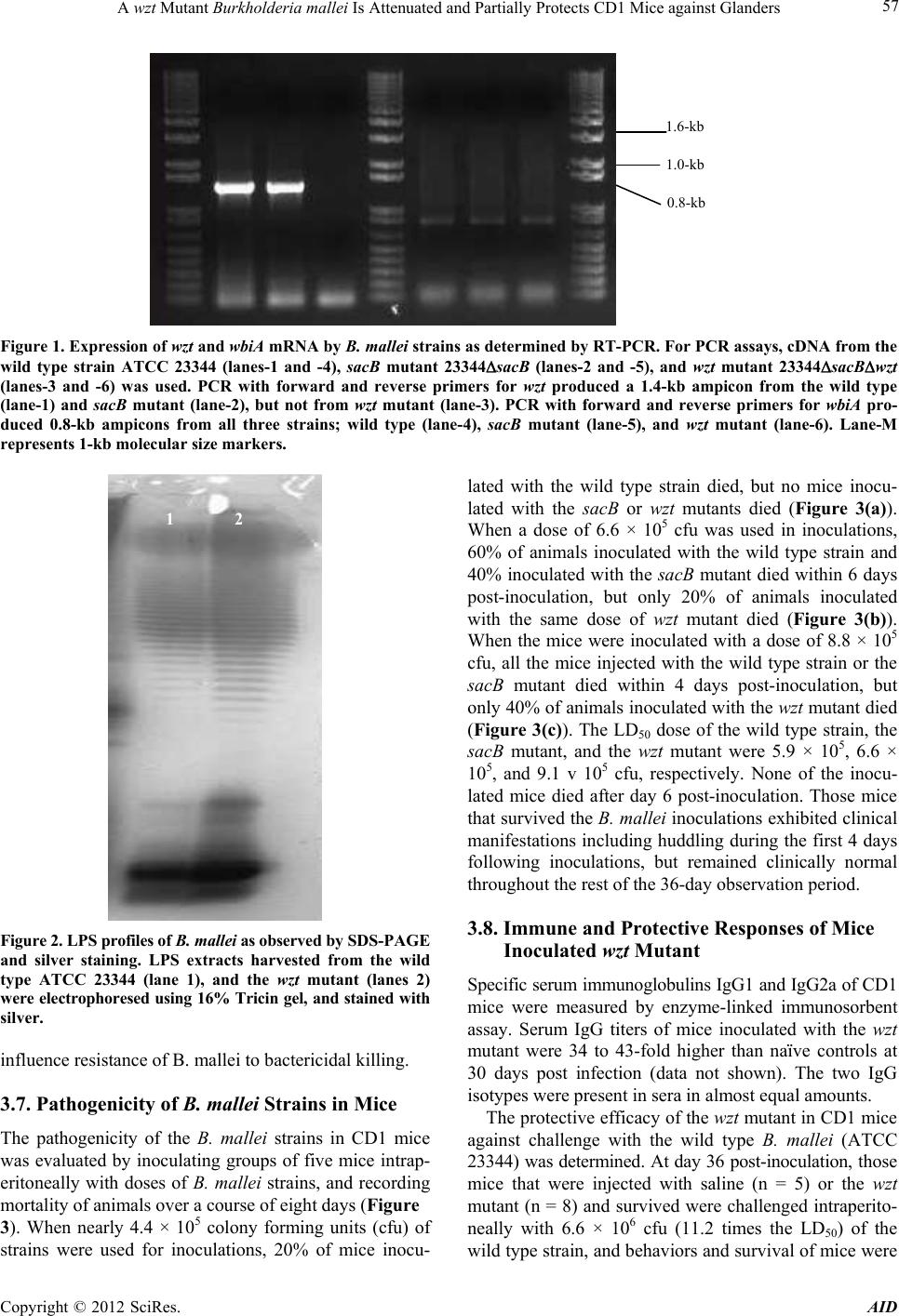 A wzt Mutant Burkholderia mallei Is Attenuated and Partially Protects CD1 Mice against Glanders Copyright © 2012 SciRes. AID 57 1.0-kb 1.6-kb 0.8-kb Figure 1. Expression of wzt and wbiA mRNA by B. mallei strains as determined by RT-PCR. For PCR assays, cDNA from the wild type strain ATCC 23344 (lanes-1 and -4), sacB mutant 23344sacB (lanes-2 and -5), and wzt mutant 23344sacBwzt (lanes-3 and -6) was used. PCR with forward and reverse primers for wzt produced a 1.4-kb ampicon from the wild type (lane-1) and sacB mutant (lane-2), but not from wzt mutant (lane-3). PCR with forward and reverse primers for wbiA pro- duced 0.8-kb ampicons from all three strains; wild type (lane-4), sacB mutant (lane-5), and wzt mutant (lane-6). Lane-M represents 1-kb molecular size markers. lated with the wild type strain died, but no mice inocu- lated with the sacB or wzt mutants died (Figure 3(a)). When a dose of 6.6 × 10⁵ cfu was used in inoculations, 60% of animals inoculated with the wild type strain and 40% inoculated with the sacB mutant died within 6 days post-inoculation, but only 20% of animals inoculated with the same dose of wzt mutant died (Figure 3(b)). When the mice were inoculated with a dose of 8.8 × 10⁵ cfu, all the mice injected with the wild type strain or the sacB mutant died within 4 days post-inoculation, but only 40% of animals inoculated with the wzt mu t a n t died (Figure 3(c)). The LD50 dose of the wild type strain, the sacB mutant, and the wzt mutant were 5.9 × 10⁵, 6.6 × 10⁵, and 9.1 v 105 cfu, respectively. None of the inocu- lated mice died after day 6 post-inoculation. Those mice that survived the B. mallei inoculations exhibited clinical manifestations including huddling during the first 4 days following inoculations, but remained clinically normal throughout the rest of the 36-day observation period. 1 2 3.8. Immune and Protective Responses of Mice Inoculated wzt Mutant Figure 2. LPS profiles of B. mallei as observed by SDS-PAGE and silver staining. LPS extracts harvested from the wild type ATCC 23344 (lane 1), and the wzt mutant (lanes 2) were electrophoresed using 16% Tricin gel, and stained with silver. Specific serum immunoglobulins IgG1 and IgG2a of CD1 mice were measured by enzyme-linked immunosorbent assay. Serum IgG titers of mice inoculated with the wzt mutant were 34 to 43-fold higher than naïve controls at 30 days post infection (data not shown). The two IgG isotypes were present in sera in almost equal amounts. influence resistance of B. mallei to bactericidal killing. 3.7. Pathogenicity of B. mallei Strains in Mice The protective efficacy of the wzt mutant in CD1 mice against challenge with the wild type B. mallei (ATCC 23344) was determined. At day 36 post-inoculation, those mice that were injected with saline (n = 5) or the wzt mutant (n = 8) and survived were challenged intraperito- neally with 6.6 × 10⁶ cfu (11.2 times the LD50) of the wild type strain, and behaviors and survival of mice were The pathogenicity of the B. mallei strains in CD1 mice was evaluated by inoculating groups of five mice intrap- eritoneally with doses of B. mallei strains, and recording mortality of animals over a course of eight days (Figure 3). When nearly 4.4 × 10⁵ colony forming units (cfu) of strains were used for inoculations, 20% of mice inocu- 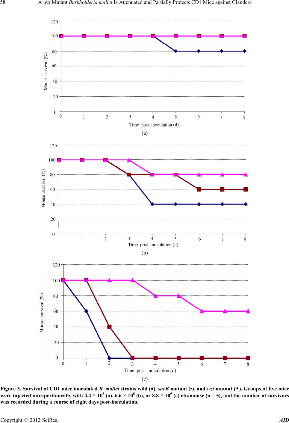 A wzt Mutant Burkholderia mallei Is Attenuated and Partially Protects CD1 Mice against Glanders 58 (a) (b) (c) Figure 3. Survival of CD1 mice inoculated B. mallei strains wild (♦), sacB mutant (▪), and wzt mutant (▲). Groups of five mice were injected intraperitoneally with 4.4 × 10⁵ (a), 6.6 × 10⁵ (b), or 8.8 × 10⁵ (c) cfu/mouse (n = 5), and the number of survivors was recorded during a course of eight days post-inoculation. Copyright © 2012 SciRes. AID 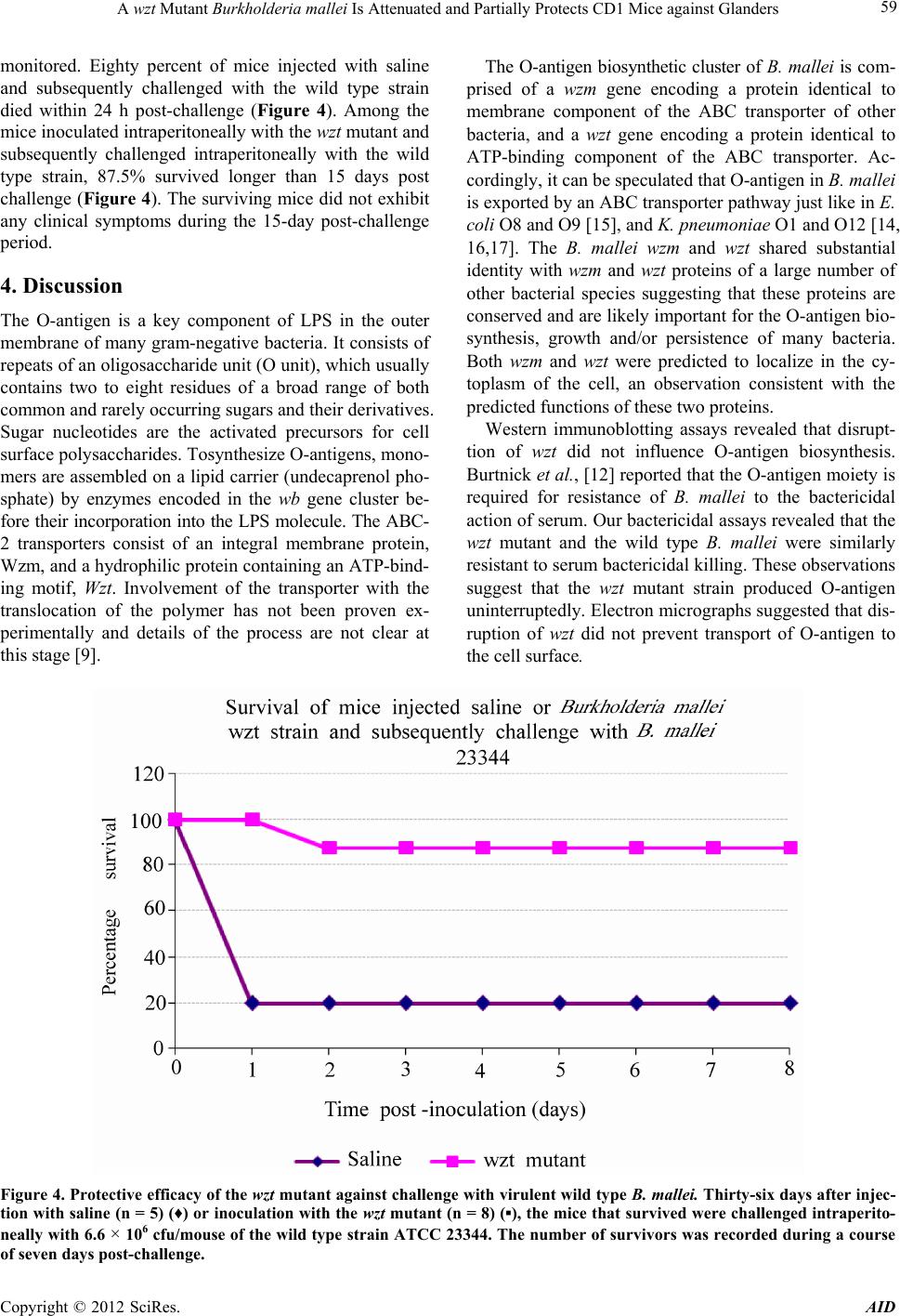 A wzt Mutant Burkholderia mallei Is Attenuated and Partially Protects CD1 Mice against Glanders 59 monitored. Eighty percent of mice injected with saline and subsequently challenged with the wild type strain died within 24 h post-challenge (Figure 4). Among the mice inoculated intraperitoneally with the wzt mutant and subsequently challenged intraperitoneally with the wild type strain, 87.5% survived longer than 15 days post challenge (Figure 4). The surviving mice did not exhibit any clinical symptoms during the 15-day post-challenge period. 4. Discussion The O-antigen is a key component of LPS in the outer membrane of many gram-negative bacteria. It consists of repeats of an oligosaccharide unit (O unit), which usually contains two to eight residues of a broad range of both common and rarely occurring sugars and their derivatives. Sugar nucleotides are the activated precursors for cell surface polysaccharides. Tosynthesize O-antigens, mono- mers are assembled on a lipid carrier (undecaprenol pho- sphate) by enzymes encoded in the wb gene cluster be- fore their incorporation into the LPS molecule. The ABC- 2 transporters consist of an integral membrane protein, Wzm, and a hydrophilic protein containing an ATP-bind- ing motif, Wzt. Involvement of the transporter with the translocation of the polymer has not been proven ex- perimentally and details of the process are not clear at this stage [9]. The O-antigen biosynthetic cluster of B. mallei is com- prised of a wzm gene encoding a protein identical to membrane component of the ABC transporter of other bacteria, and a wzt gene encoding a protein identical to ATP-binding component of the ABC transporter. Ac- cordingly, it can be speculated that O-antigen in B. mallei is exported by an ABC transporter pathway just like in E. coli O8 and O9 [15], and K. pneumoniae O1 and O12 [14, 16,17]. The B. mallei wzm and wzt shared substantial identity with wzm and wzt proteins of a large number of other bacterial species suggesting that these proteins are conserved and are likely important for the O-antigen bio- synthesis, growth and/or persistence of many bacteria. Both wzm and wzt were predicted to localize in the cy- toplasm of the cell, an observation consistent with the predicted functions of these two proteins. Western immunoblotting assays revealed that disrupt- tion of wzt did not influence O-antigen biosynthesis. Burtnick et al., [12] reported that the O-antigen moiety is required for resistance of B. mallei to the bactericidal action of serum. Our bactericidal assays revealed that the wzt mutant and the wild type B. mallei were similarly resistant to serum bactericidal killing. These observations suggest that the wzt mutant strain produced O-antigen uninterruptedly. Electron micrographs suggested that dis- ruption of wzt did not prevent transport of O-antigen to the cell surface. Figure 4. Protective efficacy of the wzt mutant against challenge with virulent wild type B. mallei. Thirty-six days after injec- tion with saline (n = 5) (♦) or inoculation with the wzt mutant (n = 8) (▪), the mice that survived were challenged intraperito- neally with 6.6 × 10⁶ cfu/mouse of the wild type strain ATCC 23344. The number of survivors was recorded during a course of seven days post-challenge. Copyright © 2012 SciRes. AID 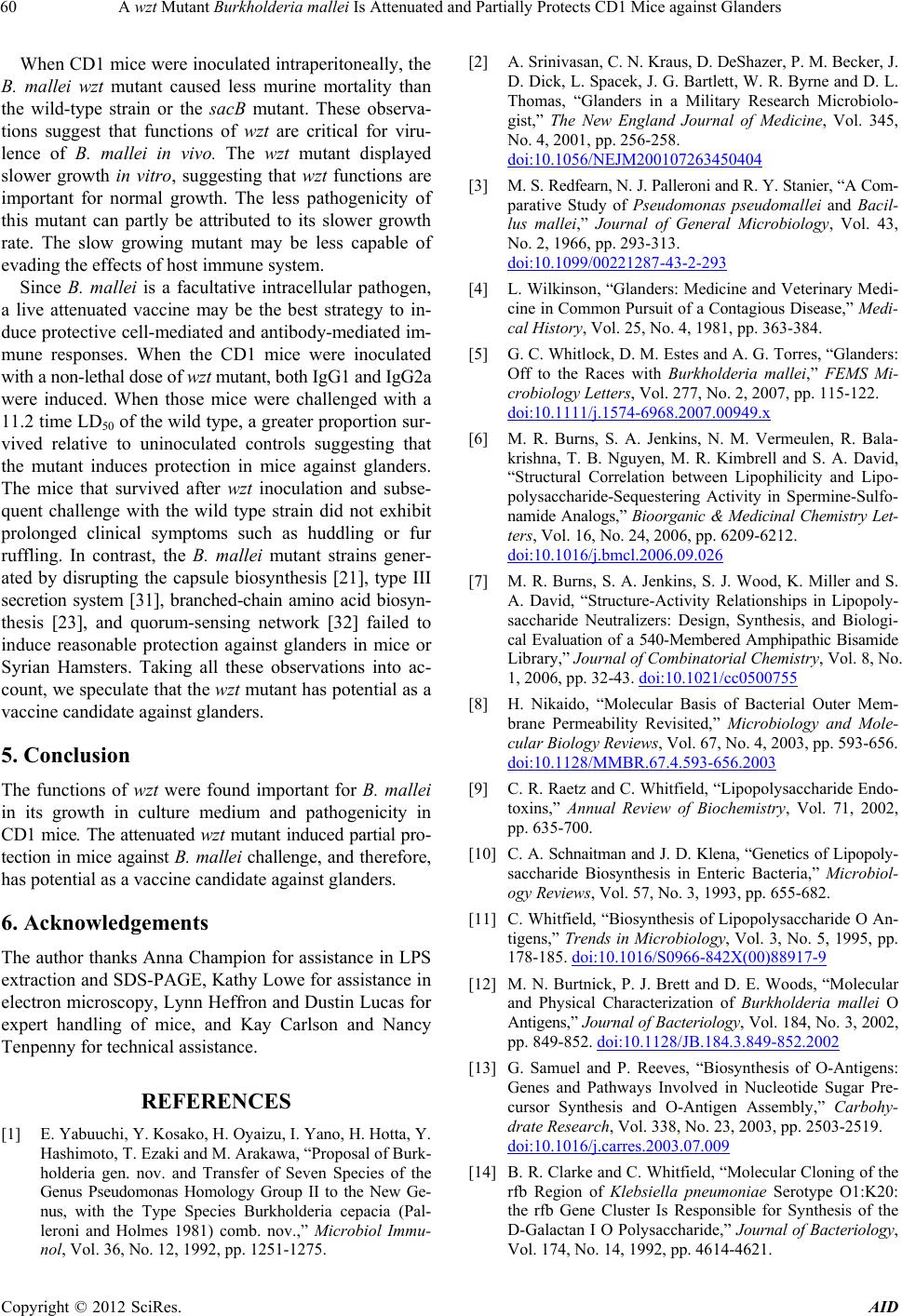 A wzt Mutant Burkholderia mallei Is Attenuated and Partially Protects CD1 Mice against Glanders 60 When CD1 mice were inoculated intraperitoneally, the B. mallei wzt mutant caused less murine mortality than the wild-type strain or the sacB mutant. These observa- tions suggest that functions of wzt are critical for viru- lence of B. mallei in vivo. The wzt mutant displayed slower growth in vitro, suggesting that wzt functions are important for normal growth. The less pathogenicity of this mutant can partly be attributed to its slower growth rate. The slow growing mutant may be less capable of evading the effects of host immune system. Since B. mallei is a facultative intracellular pathogen, a live attenuated vaccine may be the best strategy to in- duce protective cell-mediated and antibody-mediated im- mune responses. When the CD1 mice were inoculated with a non-lethal dose of wzt mutant, both IgG1 and IgG2a were induced. When those mice were challenged with a 11.2 time LD50 of the wild type, a greater proportion sur- vived relative to uninoculated controls suggesting that the mutant induces protection in mice against glanders. The mice that survived after wzt inoculation and subse- quent challenge with the wild type strain did not exhibit prolonged clinical symptoms such as huddling or fur ruffling. In contrast, the B. mallei mutant strains gener- ated by disrupting the capsule biosynthesis [21], type III secretion system [31], branched-chain amino acid biosyn- thesis [23], and quorum-sensing network [32] failed to induce reasonable protection against glanders in mice or Syrian Hamsters. Taking all these observations into ac- count, we speculate that the wzt mutant has potential as a vaccine candidate against glanders. 5. Conclusion The functions of wzt were found important for B. mallei in its growth in culture medium and pathogenicity in CD1 mice. The attenuated wzt mutant induced partial pro- tection in mice against B. mallei challenge, and therefore, has potential as a vaccine candidate against glanders. 6. Acknowledgements The author thanks Anna Champion for assistance in LPS extraction and SDS-PAGE, Kathy Lowe for assistance in electron microscopy, Lynn Heffron and Dustin Lucas for expert handling of mice, and Kay Carlson and Nancy Tenpenny for technical assistance. REFERENCES [1] E. Yabuuchi, Y. Kosako, H. Oyaizu, I. Yano, H. Hotta, Y. Hashimoto, T. Ezaki and M. Arakawa, “Proposal of Burk- holderia gen. nov. and Transfer of Seven Species of the Genus Pseudomonas Homology Group II to the New Ge- nus, with the Type Species Burkholderia cepacia (Pal- leroni and Holmes 1981) comb. nov.,” Microbiol Immu- nol, Vol. 36, No. 12, 1992, pp. 1251-1275. [2] A. Srinivasan, C. N. Kraus, D. DeShazer, P. M. Becker, J. D. Dick, L. Spacek, J. G. Bartlett, W. R. Byrne and D. L. Thomas, “Glanders in a Military Research Microbiolo- gist,” The New England Journal of Medicine, Vol. 345, No. 4, 2001, pp. 256-258. doi:10.1056/NEJM200107263450404 [3] M. S. Redfearn, N. J. Palleroni and R. Y. Stanier, “A Com- parative Study of Pseudomonas pseudomallei and Bacil- lus mallei,” Journal of General Microbiology, Vol. 43, No. 2, 1966, pp. 293-313. doi:10.1099/00221287-43-2-293 [4] L. Wilkinson, “Glanders: Medicine and Veterinary Medi- cine in Common Pursuit of a Contagious Disease,” Medi- cal History, Vol. 25, No. 4, 1981, pp. 363-384. [5] G. C. Whitlock, D. M. Estes and A. G. Torres, “Glanders: Off to the Races with Burkholderia mallei,” FEMS Mi- crobiology Letters, Vol. 277, No. 2, 2007, pp. 115-122. doi:10.1111/j.1574-6968.2007.00949.x [6] M. R. Burns, S. A. Jenkins, N. M. Vermeulen, R. Bala- krishna, T. B. Nguyen, M. R. Kimbrell and S. A. David, “Structural Correlation between Lipophilicity and Lipo- polysaccharide-Sequestering Activity in Spermine-Sulfo- namide Analogs,” Bioorganic & Medicinal Chemistry Let- ters, Vol. 16, No. 24, 2006, pp. 6209-6212. doi:10.1016/j.bmcl.2006.09.026 [7] M. R. Burns, S. A. Jenkins, S. J. Wood, K. Miller and S. A. David, “Structure-Activity Relationships in Lipopoly- saccharide Neutralizers: Design, Synthesis, and Biologi- cal Evaluation of a 540-Membered Amphipathic Bisamide Library,” Journal of Combinatorial Chemistry, Vol. 8, No. 1, 2006, pp. 32-43. doi:10.1021/cc0500755 [8] H. Nikaido, “Molecular Basis of Bacterial Outer Mem- brane Permeability Revisited,” Microbiology and Mole- cular Biology Reviews, Vol. 67, No. 4, 2003, pp. 593-656. doi:10.1128/MMBR.67.4.593-656.2003 [9] C. R. Raetz and C. Whitfield, “Lipopolysaccharide Endo- toxins,” Annual Review of Biochemistry, Vol. 71, 2002, pp. 635-700. [10] C. A. Schnaitman and J. D. Klena, “Genetics of Lipopoly- saccharide Biosynthesis in Enteric Bacteria,” Microbiol- ogy Reviews, Vol. 57, No. 3, 1993, pp. 655-682. [11] C. Whitfield, “Biosynthesis of Lipopolysaccharide O An- tigens,” Trends in Microbiology, Vol. 3, No. 5, 1995, pp. 178-185. doi:10.1016/S0966-842X(00)88917-9 [12] M. N. Burtnick, P. J. Brett and D. E. Woods, “Molecular and Physical Characterization of Burkholderia mallei O Antigens,” Jo urnal of Bacteriology, Vol. 184, No. 3, 2002, pp. 849-852. doi:10.1128/JB.184.3.849-852.2002 [13] G. Samuel and P. Reeves, “Biosynthesis of O-Antigens: Genes and Pathways Involved in Nucleotide Sugar Pre- cursor Synthesis and O-Antigen Assembly,” Carbohy- drate Research, Vol. 338, No. 23, 2003, pp. 2503-2519. doi:10.1016/j.carres.2003.07.009 [14] B. R. Clarke and C. Whitfield, “Molecular Cloning of the rfb Region of Klebsiella pneumoniae Serotype O1:K20: the rfb Gene Cluster Is Responsible for Synthesis of the D-Galactan I O Polysaccharide,” Journal of Bacteriology, Vol. 174, No. 14, 1992, pp. 4614-4621. Copyright © 2012 SciRes. AID  A wzt Mutant Burkholderia mallei Is Attenuated and Partially Protects CD1 Mice against Glanders 61 [15] H. C. Flemming and K. Jann, “Biosynthesis of the O9 An- tigen of Escherichia coli. Growth of the Polysaccharide Chain,” European Journal of Biochemistry, Vol. 83, No. 1, 1978, pp. 47-52. doi:10.1111/j.1432-1033.1978.tb12066.x [16] L. Izquierdo, N. Coderch, N. Pique, E. Bedini, M. M. Cor- saro, S. Merino, S. Fresno, J. M. Tomas and M. Regue, “The Klebsiella pneumoniae wabG Gene: Role in Bio- synthesis of the Core Lipopolysaccharide and Virulence,” Journal of Bacteriology, Vol. 185, No. 24, 2003, pp. 7213- 7221. doi:10.1128/JB.185.24.7213-7221.2003 [17] L. Izquierdo, S. Merino, M. Regue, F. Rodriguez and J. M. Tomas, “Synthesis of a Klebsiella pneumoniae O-An- tigen Heteropolysaccharide (O12) Requires an ABC 2 Transporter,” Journal of Bacteriology, Vol. 185, No. 5, 2003, pp. 1634-1641. doi:10.1128/JB.185.5.1634-1641.2003 [18] J. D. Bendtsen, H. Nielsen, G. von Heijne and S. Brunak, “Improved Prediction of Signal Peptides: SignalP 3.0,” Journal of Molecular Biology, Vol. 340, No. 4, 2004, pp. 783-795. doi:10.1016/j.jmb.2004.05.028 [19] S. F. Altschul, W. Gish, W. Miller, E. W. Myers and D. J. Lipman, “Basic Local Alignment Search Tool,” Journal of Molecular Biology, Vol. 215, No. 3, 1990, pp. 403-410. [20] A. B. Bandara, D. DeShazer, T. J. Inzana, N. Srirangana- than, G. G. Schurig and S. M. Boyle, “A Disruption of ctpA Encoding carboxy-Terminal Protease Attenuates Burk- holderia mallei and Induces Partial Protection in CD1 Mice,” Microbial Pathogenesis, Vol. 45, No. 3, 2008, pp. 207-216. doi:10.1016/j.micpath.2008.05.005 [21] D. DeShazer, D. M. Waag, D. L. Fritz and D. E. Woods, “Identification of a Burkholderia mallei Polysaccharide Gene Cluster by Subtractive Hybridization and Demon- stration That the Encoded Capsule Is An Essential Viru- lence Determinant,” Microbial Pathogenesis, Vol. 30, No. 5, 2001, pp. 253-269. doi:10.1006/mpat.2000.0430 [22] R. Simon, U. Priefer and A. Puhler, “A Broad Host Range Mobilization System for in Vivo Genetic Engineering: Transposon Mutagenesis in Gram Negative Bacteria,” Bio/Technology, Vol. 1, No. 9, 1983, pp. 784-791. [23] R. L. Ulrich, K. Amemiya, D. M. Waag, C. J. Roy and D. DeShazer, “Aerogenic Vaccination with a Burkholderia mallei Auxotroph Protects against Aerosol-Initiated Glan- ders in Mice,” Vaccine, Vol. 23, No. 16, 2005, pp. 1986- 1992. doi:10.1016/j.vaccine.2004.10.017 [24] J. F. Sambrook and E. F. Maniatis, “Molecular Cloning: A Laboratory Manual,” 2nd Edition, Cold Spring Harbor Laboratory Press, Cold Spring Harbor, 1989. [25] P. J. Brett, M. N. Burtnick, D. S. Snyder, J. G. Shannon, P. Azadi and F. C. Gherardini, “Burkholderia mallei Ex- presses a Unique Lipopolysaccharide Mixture That Is a Potent Activator of Human Toll-Like Receptor 4 Com- plexes,” Molecular Microbiology, Vol. 63, No. 2, 2007, pp. 379-390. doi:10.1111/j.1365-2958.2006.05519.x [26] M. B. Perry, L. L. MacLean, T. Schollaardt, L. E. Bryan and M. Ho, “Structural Characterization of the Lipopoly- saccharide O Antigens of Burkholderia pseudomallei,” Infection and Immunity, Vol. 63, No. 9, 1995, pp. 3348- 3352. [27] U. K. Laemmli, “Cleavage of Structural Proteins during the Assembly of the Head of Bacteriophage T4,” Nature, Vol. 227, No. 5259, 1970, pp. 680-685. doi:10.1038/227680a0 [28] C. M. Tsai, R. Boykins and C. E. Frasch, “Heterogeneity and Variation among Neisseria meningitidis Lipopolysac- charides,” Journal of Bacteriology, Vol. 155, No. 2, 1983, pp. 498-504. [29] T. J. Inzana and P. Anderson, “Serum Factor-Dependent Resistance of Haemophilus influenzae Type b to Antibody to Lipopolysaccharide,” The Journal of Infectious Dis- eases, Vol. 151, No. 5, 1985, pp. 869-877. [30] J. Li, C. Ryder, M. Mandal, F. Ahmed, P. Azadi, D. S. Snyder, R. D. Pechous, T. Zahrt and T. J. Inzana, “Atten- uation and Protective Efficacy of an O-antigen-Deficient Mutant of Francisella tularensis LVS,” Microbiology, Vol. 153, No. Pt 9, 2007, pp. 3141-3153. doi:10.1099/mic.0.2007/006460-0 [31] R. L. Ulrich and D. DeShazer, “Type III Secretion: A Vir- ulence Factor Delivery System Essential for the Patho- genicity of Burkholderia mallei,” Infection and Immunity, Vol. 72, No. 2, 2004, pp. 1150-1154. doi:10.1128/IAI.72.2.1150-1154.2004 [32] R. L. Ulrich, D. Deshazer, H. B. Hines and J. A. Jeddeloh, “Quorum Sensing: A Transcriptional Regulatory System Involved in the Pathogenicity of Burkholderia mallei,” In- fection and Immunity, Vol. 72, No. 11, 2004, pp. 6589- 6596. Copyright © 2012 SciRes. AID
|