Advances in Microbiology, 2012, 2, 303-309
http://dx.doi.org/10.4236/aim.2012.23036 Published Online September 2012 (http://www.SciRP.org/journal/aim)
Biodegradation of Melamine and Cyanuric Acid by a
Newly-Isolated Microbacterium Strain
Naofumi Shiomi*, Maiko Ako
School of Human Sciences, Kobe College, Nishinomiya, Japan
Email: *n-shiomi@mail.kobe-c.ac.jp
Received April 25, 2012; revised May 28, 2012; accepted June 11, 2012
ABSTRACT
We searched for a superior melamine-degrading bacterium for the bioremediation of melamine. Cyanuric acid, which is
a by-product produced during the biodegradation of melamine, shows strong nephrotoxicity. Therefore, the mela-
mine-degrading bacterium is also required to show a high ability to degrade cyanuric acid. We selected a mela-
mine-degrading strain (MEL1) among ten cyanuric acid-degrading bacteria isolated from the soil. The species of MEL1
strain was Microbacterium esteramaticum or was extremely similar to that species, and the enzymatic activity of the
melamine deaminase in the MEL1 strain was similar to that in the NRRLB-12227 strain. The ability of the MEL1 strain
to degrade cyanuric acid was higher than its ability to degrade melamine, and therefore, the accumulation of by-prod-
ucts (ammeline, ammelide and cyanuric acid) during the degradation of melamine was minimal. These results suggest
that the MEL1 strain is useful for the bioremediation of melamine.
Keywords: Melamine; Cyanuric Acid; Melamine Deaminase; Microbacterium
1. Introduction
Melamine is used for the production of melamine resin
and other products, and the current output of melamine
resin in Japan is 140,000 ton/year. As a melamine mole-
cule contains a very high level of nitrogen, the apparent
content of protein can be increased by the intentional
addition of the compound. In China, melamine was in-
tentionally added to dairy products by some food indus-
tries in 2004-2007 [1]. Consequently, serious food-re-
lated toxicity occurred in subjects who consumed the
dairy products. For example, numerous dogs and cats
were poisoned (or died) after they ate pet food containing
the adulterated dairy products in Korea and the USA
[2-4]. In 2008, around 50,000 children suffered from
renal failure and 6 children died because they were fed
powdered milk contaminated with melamine. A later
investigation uncovered that the products adulterated
with melamine was not only limited to dairy products,
but also included cakes and animal feed in China.
Melamine had been considered to be harmless, be-
cause the LD50 value is very high (1 - 3 g/kg). However,
recent studies suggest that its degradation product, cyanu-
ric acid, causes the severe renal failure [5]. Cyanuric acid
is a by-product that forms during the process of melamine
production and the biodegradation of melamine. It com-
bines with melamine in the kidney, and kidney stones
made of these compounds induce renal failure. The nephro-
toxicity caused by the two compounds was much stronger
than that in case of treatment with just melamine [6].
Melamine-containing waste water had been freely
dumped into the environment without any treatment for
many years until the above described food-related inci-
dents occurred. Qin et al. [7] assessed the soil and waste
water near melamine-manufacturing factories in China.
The maximum concentrations in waste water and soil
samples were 226.766 and 41.136 mg/kg, respectively.
Such contamination also caused the contamination of
crops, and the maximum value of melamine contained in
a wheat sample was 2.05 mg/kg. Therefore, unexpected
food accidents caused by the consumption of these crops
may occur in the future. For these reasons, increasing
attention has been paid to the bioremediation of soil and
waste water contaminated by melamine.
Some melamine-degrading bacterial strains, such as
Klebsiella terragena DRS-1 [8], Norcadi o id e s sp. ATD6
[9], Pseudomonas sp. NRRL B-12227 [10] and Rho-
dococcus corallines NRRLB-15444R [11], have report-
edly been used for bioremediation in previous research.
Cyanuric acid is a key intermediate in the metabolic
pathway of melamine. Rapid biodegradation of cyanuric
acid is important for the bioremediation of melamine,
because it is a nephrotoxic compound. However, the
cleavage of cyanuric acid is a rate-limiting step in the
above melamine-degrading strains. For example, the K.
*Corresponding author.
C
opyright © 2012 SciRes. AiM
N. SHIOMI, M. AKO
304
terragena DRS-1 and Norcadioides sp. ATD6 strains
showed a high ability to degrade melamine, but a low
ability to degrade cyanuric acid. Therefore, the utilization
of a mixture of melamine and cyanuric acid-degrading
strains is required to fully degrade melamine. On the
contrary, the Pseudomonas sp. NRRL B-12227 showed
the ability to degrade both cyanuric acid and melamine
[12], and thus, this strain was considered to be profitable
for the bioremediation of melamine. However, the strain
was reclassified as Acidovorax citrulli [13]. A. citrulli is
not a good species for bioremediation, because it is a
serious plant pathogen for watermelon, therefore the iso-
lation of a new strain is considered to be necessary.
In this study, we searched for a superior strain for the
bioremediation of melamine. The newly-isolated Micro-
bacterium strain showed a high ability to degrade cyanu-
ric acid as well as melamine. Therefore, this strain may
be useful for the bioremediation of melamine.
2. Materials and Methods
2.1. Bacteria and Media
The A. citrulli NRRL B-12227 strain was obtained from
Agricultural Research Service. Modified Luria Bertani
medium (LB medium; 10 g/L bactopeptone, 10 g/L yeast
extract, 5 g/L NaCl, 1 g/L glucose, pH 7.2) and Davis-
Miligano medium without ammonium sulfate (DM me-
dium; 2 g/L glucose, 7 g/L K2HPO4, 3 g/L KH2PO4, 0.1
g/L MgSO4, 0.1 g/L yeast extract) were used for the
culture of bacteria.
2.2. Assays
Cell growth was measured by the optical density (OD) of
light passing through a 1 cm cuvette at 610 nm. The
concentration of protein was determined by a protein
assay kit (Bio-Rad Laboratories, Inc., Tokyo, Japan). The
concentrations of melamine, cyanuric acid, ammeline
and ammelide in the culture broth (without cells) were
determined by HPLC using an Asahipac GS-320 column
(Shimazu Good Laboratory Component Ltd., Osaka,
Japan). The mobile phase and the flow rate were 100 mM
Na2HPO4 (pH 10.0) and 1.0 mL/min. The concentrations
were determined by measuring the absorbance at 230 nm.
The concentration of simetryn and atrazine were de-
termined by capillary gas chromatography using a DB-5
column (0.25 mm × 30 m) equipped with a FID detector.
The evaporation and detection temperatures were 300˚C,
and the column temperature was gradually increased
from 80˚C to 300˚C at a rate of 10˚C/min.
2.3. Isolation of Cyanuric Acid-Degrading
Strains
One gram of each soil sample collected from 100 dif-
ferent places in Kyoto, Osaka and Hyogo prefectures in
Japan was suspended in 10mL of 0.85% NaCl solution,
and 0.1 mL of the suspension was spread on an agar plate
of LB medium. The cells grown on the plates were
collected and used for the screening. Around 1000 cells
were spread on each agar plate with the DM medium
containing 1.0 mM cyanuric acid, and were cultured for 3
days at 37˚C. Ten strains from 20 plates showing large
colonies were isolated as cyanuric acid-degrading strains,
and then were used for the further analyses.
2.4. Identification
The identification of the isolated strain (MEL1) was per-
formed using the following procedure by the NCIMB
JAPAN Corporation: The nucleotide sequences of the
16S ribosomal DNA was determined with ABI PRISM
3100 DNA Sequencer (Applied Biosystems, CA, USA).
A homology search of the sequence was performed with
the database (Apollon DB-BA Ver. 7.0) from Techno-
Sulruga Laboratory Co. Ltd. (Sizuoka, Japan) and the
phylogenic tree was constructed with the most closely
related 10 species according to the neighbor-joining
method [14]. A homology search was also performed
using BLAST [15] with the DNA sequence database
(GenBank/DDB/EMBL).
2.5. Enzymatic Activity and Purification of
Melamine Deaminase
The A. citrulli NRRL B-12227 and the melamine-de-
grading (MEL1) strains were cultured in 200 mL DM
media containing 1.0 g/L of ammonium sulfate for 3
days. The cells in each culture were harvested and sus-
pended in 3mL of 10 mM Tris-HCl buffer (10 mM Tris
(hydroxymethyl)aminomethane (pH 7.5)). The cell sus-
pension was disrupted with a cell disrupter (MINI-
BEADEATER , WAKENYAKU, Co. Ltd., Kyoto, Japan)
by shaking with 1 mL of glass beads (0.17 - 0.18 mm ϕ)
for 9 min. The whole solution was centrifuged at 12,000
× g for 15 min. The supernatant was then put in a seam-
less cellulose tube (Wako Chem. Ind., Osaka, Japan) and
dialyzed with 3 L of 10 mM Tris-HCl buffer (pH 7.5) for
1 day. The resulting enzyme solution was used for the
subsequent assays.
The melamine deaminase activity was measured ac-
cording to the method described by Karns [10] with
slight modification. A total of 900 μL of 25 mM TE
buffer (pH 8.0) containing 3 mM melamine and 100 μL
of the enzyme solution were incubated at 37˚C for 1 day,
and the decrease in melamine was measured using HPLC
by sampling the solution every 3 hours.
The purification of melamine deaminase was per-
formed in three steps. First, solid ammonium sulfate (fi-
nal concentration: 20%) was added to the rest of the en-
Copyright © 2012 SciRes. AiM
N. SHIOMI, M. AKO
Copyright © 2012 SciRes. AiM
305
(SDS-PAGE; 10% acrylamide gel) of 5 μL of purified
protein solution was performed by the method described
by Laemmli [16]. The molecular size of the proteins was
determined with the Precision Plus Protein Standard
(Bio-Rad Laboratories).
zyme solution (2.7 mL) and incubated for 2 h at room
temperature. After sedimentation and centrifugation at
12,000 × g, the entire solution was put in a new tube.
Solid ammonium sulfate (final concentration: 40%) was
added to the tube, and then a similar procedure was per-
formed. The sediment from both steps was then dissolved
in 3 mL of Tris-HCl buffer (pH 7.5), put in seamless cel-
lulose tubing (Wako Chem. Ind.) and dialyzed with 3 L
of 10 mM Tris-HCl buffer (pH 7.5) for 1 day. As the
solution containing the sediment obtained following the
addition of 20% ammonium sulfate showed the highest
enzymatic activity, it was then used for further purifica-
tion. Figures 1(a) and (b) show the elution profiles of the
protein and the activities in each fraction in the DEAE-
cellulose and Sephadex G-100 columns. The solutions
were applied onto a DEAE-cellulose column (1 cm × 24
cm) equilibrated with Tris-HCl buffer (pH 7.5). The ad-
sorbed proteins were eluted by an increasing gradient of
KCl in the same buffer (0 - 1.0 M, 500 ml) at a flow rate
of 2.0 mL per fraction. The active fraction (fraction
number 11) was pooled, and put onto a Sephadex G-100
column (1.0 × 96 cm) equilibrated with Tris-HCl buffer
(pH 7.5), and the protein was eluted with Tris-HCl buffer
(pH 7.5) at a flow rate of 2.0 mL per fraction.
2.6. Biodegradation of Melamine by the MEL1
Strain
The MEL1 strain pre-cultured in 20 mL LB medium was
inoculated at an optical density (OD) of 0.01 at 610 nm
into 50 mL of four different kinds of media (DM media
containing either 0.2 g/L of melamine, 0.2 g/L of cyanu-
ric acid, 0.02 g/L of ammeline or 0.02 g/L of ammelide).
The cells were cultured for 5 days at 37˚C with shaking
at 120 rpm. The 2 mL of culture broth was sampled
every day, and the supernatant was separated from the
cells by centrifugation at 12,000 × g. The triazine com-
pounds in the supernatant were measured by HPLC.
Three independent experiments were performed.
3. Results and Discussion
3.1. Isolation of a Strain Showing a High Ability
to Degrade Cyanuric Acid and Melamine
Water present in the active fraction (fraction number 3)
was evaporated using a freeze drying method, and it was
then dissolved in 20 μL of Tris-HCl buffer (pH 7.5). So-
dium dodecyl sulfate-polyacrylamido gelelectrophoresis
To search for a novel bacterium showing a superior abil-
ity to degrade melamine compared to the previously
identified bacterial strains, we first performed a screen-
Figure 1. Purification of melamine deaminase using DEAE (a) and Sephadex G-100; (b) columns. The solid circles (right)
indicate the absorbance at 245 nm in each fraction, and the open circles indicate the activity of melamine deaminase in the
samples in which activity was present.
N. SHIOMI, M. AKO
306
ing of the strains for the ability to degrade cyanuric acid,
because, cyanuric acid is a key intermediate in the meta-
bolic pathway for melamine, it is nephrotoxic, and the
cleavage of its ring is the rate-limiting step in the degra-
dation process. The microorganisms present in soil were
collected from 100 different places in three prefectures in
Japan, and were grown on agar plates containing DM
medium with 1.0 g/L cyanuric acid. Finally, 10 strains
were selected as cyanuric acid-degrading strains. The
abilities of the ten strains to degrade triazine compounds,
melamine and simazine, were tested. Three strains de-
graded melamine but did not degrade simazine. Four
strains degraded simazine but did not degrade melamine.
One strain weakly degraded both compounds, while the
remaining two strains did not degrade either of them. The
three melamine-degrading strains were named MEL1,
MEL2 and MEL3, and the rates of cyanuric acid and
melamine degradation in these strains were compared.
The MEL1 strain showed the highest levels of degrada-
tion of cyanuric acid among them, although they showed
the similar abilities to degrade melamine (data not shown).
Thus, we used the MEL1 strain for further investigations.
The identification of the MEL1 strain was performed
examining the nucleotide sequences of its 16S ribosomal
DNA. Figure 2 shows the phylogenic tree constructed by
the neighbor-joining method. The MEL1 strain was
placed in a cluster with Microbacterium esteramaticum,
and the bootstrap value was high (92%). The results of a
homology research with the GeneBank/DDBJ/EMBL
database also suggested that the MEL1 strain showed
high homology to this organism. The homology between
the MEL1 strain and the M. esteramaticum DMS8609
strain was 98%. These results suggest that the species of
the MEL1 strain is Microbacterium esteramaticum or
that it was very similar to that species. The pathogenic
effects of M. esteramaticum on plants and animals has
not been reported, and therefore, the MEL1 strain is con-
sidered to be safe.
3.2. Biochemical Characteristics of the MEL1
Strain
The A. citrulli NRRL B-12227 strain showed a high abil-
ity to degrade melamine. Therefore, the enzyme activity
in the MEL1 strain was compared with the A. citrulli
NRRL B-12227 strain. As shown in Table 1, the specific
activities of the melamine deaminase in these strains
were similar. The melamine deaminase in the MEL1
strain was also purified and compared with that in the
NRRL B-12227 strain. The specific activities after the
purification processes are also shown in Table 1. The
melamine deaminase was concentrated 77 times by these
procedures. Figure 3 shows the results of the SDS-
PAGE-purified enzyme. The molecular weight of the
melamine deaminase in the MEL1 strain was 65 kDa
(although there was still slight protein contamination
present). This size was quite different from that isolated
from the A. citrulli NRRL B-12227 strain (48 kDa) [17].
Figure 2. A phylogenetic tree based on the 16 s rDNA sequence data of the MEL1 strain. The relationship of the MEL1 strain
to the most closely related 10 bacteria selected in the Apollon DB-BA Ver. 7.0 database are shown. The numbers at the
branch points are bootstrap values, and the bar indicates a scale bar. The “T” at the end of the strain names indicates a type
train.
s
Copyright © 2012 SciRes. AiM
N. SHIOMI, M. AKO 307
Table 1. Melamine deaminase activity in the MEL1 strain
Purification pr activity Recovery Specific Fold
following the purification steps.
Total Total
otein activity
step (mg) (μg/h)(%) (
μg/mg of
protein/h)
(MEL1 strain)
5. 0. 100 0.1.
0.036 0.09 20 2.31 77.0
B-12227 str
0.026
Crude soluble 3446030 0
20% (NH4)2SO4 1.31 0.30 65 0.23 7.7
DEAE-cellulose 0.301 0.12 26 0.40 13.3
Sephadex
G-100
(NRRL
ain)
Crude soluble
Figure 3. The SDS-PAGE separation of melamine deami-
nase. Lane A, protein standard; lane B, 5 μL of the solution
concentrated from a fraction (No. 3) obtained during the
Sephadex G-100 column chromatography. The arrows show
the positions of melamine deaminases in the MEL1 and
NRRL B-12227 strains.
of the bacteria and con-
centrations of the compounds are shown when the MEL1 strain was respectively cultured in the DM medium containing 0.2
g/L melamine or cyanuric acid (a), or 0.02 g/L ammeline or ammelide (b). Bars, means ± SD of 3 independent experiments.
(a) (b)
Figure 4. Biodegradation of melamine and its related compounds by the MEL1 strain. The growth
Copyright © 2012 SciRes. AiM
N. SHIOMI, M. AKO
308
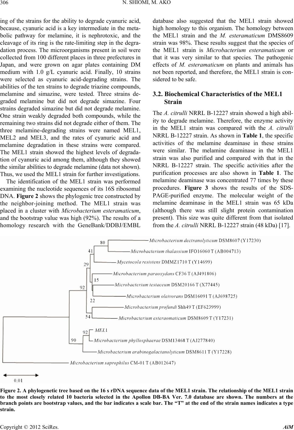
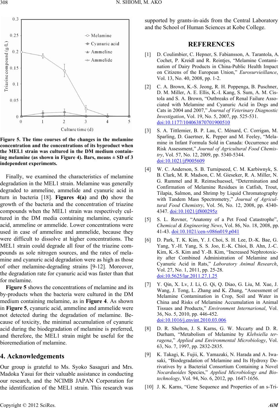
Figure 5. The time courses of the changes in the melamine
concentration and the concentrations of its byproduct when
the MEL1 strain was cultured in the DM medium contain
n. Melamine was generally
egraded to ammeline, ammelide and cyanuric acid in
tu
ning melamine, as in Figure 4. As shown
in
Sasaguri and
ble assistance in conducting
B JAPAN Corporation for
pact
,
Vol. 13, No. 40, 2008, pp. 1-2.
[2] C. A. Brown, enga, B. Puschner,
0
-
ing melamine (as shown in Figure 4). Bars, means ± SD of 3
independent experiments.
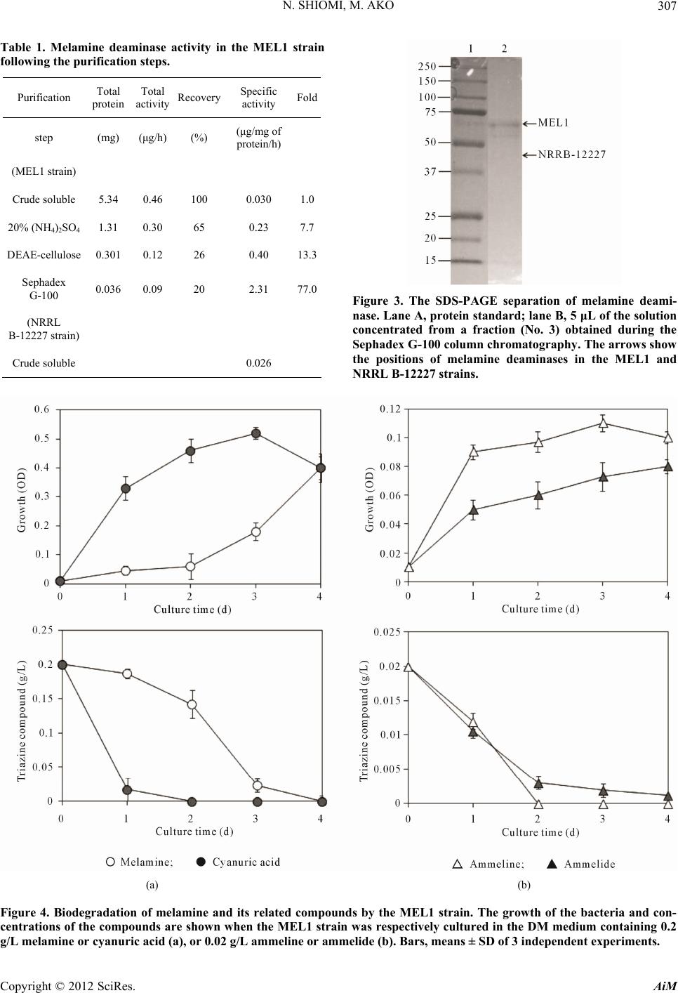
Finally, we examined the characteristics of melamine
degradation in the MEL1 strai
d
rn in bacteria [18]. Figures 4(a) and (b) show the
growth of the bacteria and the concentration of triazine
compounds when the MEL1 strain was respectively cul-
tured in the DM media containing melamine, cyanuric
acid, ammeline or ammelide. Lower concentrations were
used in case of ammeline and ammelide, because they
were difficult to dissolve at higher concentrations. The
MEL1 strain could degrade all four of the triazine com-
pounds as sole nitrogen sources, and the rates of mela-
mine and cyanuric acid degradation were as high as those
of other melamine-degrading strains [9-12]. Moreover,
the degradation rate for cyanuric acid was faster than that
for melamine.
Figure 5 shows the concentrations of melamine and its
by-products when the bacteria were cultured in the DM
medium contai
Figure 5, cyanuric acid, ammeline and ammelide were
not detected during the degradation of melamine. Be-
cause of toxicity, the minimal accumulation of cyanuric
acid during the biodegradation of melamine is preferred,
and therefore, the MEL1 strain might be useful for the
bioremediation of melamine.
4. Acknowledgements
Our group is grateful to Ms. Syoko Mrs.
Madoka Yasui for their valua
our research, and the NCIM
the identification of the MEL1 strain. This research was
REFERENCES
[1] D. Coulimbier, C. Hepner, S. Fabiansson, A. Tarantola, A.
Cochet, P. Kreidl and R. Reintjes, “Melamine Contami-
nation of Dairy Products in China-Public Health Im
on Citizens of the European Union,” Eurosurvieillance
supported by grants-in-aids from the Central Laboratory
and the School of Human Sciences at Kobe College.
K.-S. Jeong, R. H. Popp
D. M. Miller, A. E. Ellis, K.-I. Kang, S. Sum, A. M. Cis-
tola and S. A. Brown, “Outbreaks of Renal Failure Asso-
ciated with Melamine and Cyanuric Acid in Dogs and
Cats in 2004 and 2007,” Journal of Veterinary Diagnostic
Investigation, Vol. 19, No. 5, 2007, pp. 525-531.
doi:10.1177/10406387070190051
[3] S. A. Tittlemier, B. P. Lau, C. Ménard, C. Corrigan, M.
Sparling, D. Gaertner, K. Pepper and M. Feeley, “Mela-
mine in Infant Formula Sold in Canada: Occurrence and
Risk Assessment,” Journal of Agricultural Food Chemis-
try, Vol. 57, No. 12, 2009, pp. 5340-5344.
doi:10.1021/jf9005609
[4] W. C. Anderson, S. B. Turnipseed, C. M. Karbiwnyk, S.
B. Clark, M. R. Madson, C. M. Gieseker, R. A. Miller, N.
G. Rummel and R. Reimschuessel, “Determination and
Confirmation of Melamine Residues in Catfish, Trout,
Tilapia, Salmon, and Shrimp by Liquid Chromatography
with Tandem Mass Spectrometry,” Journal o
f Agricul-
tural Food Chemistry, Vol. 56, No. 12, 2008, pp. 4340-
4347. doi:10.1021/jf800295z
[5] S. L. Rovner, “Anatomy of a Pet Food Catastrophe”,
Chemical & Engineering News, Vol. 86, No. 18, 2008, pp.
41-43. doi:10.1021/cen-v086n019.p041
[6] D. Park, T. K. Kim, Y. J. Choi, S. H. Lee, D.-K. Bae, G.
Yang, Y.-H. Yang, S. S. Joo, E.-K. Chioi, B. Ahn, J.-C.
Kim, K.-S. Kim and Y.-B. Kim, “Increased Nephrotoxic-
ity after Combined Administration of Melamine and
Cyanuric Acid in Rats,” Laboratory Animal Research,
Vol. 27, No. 1, 2011, pp. 25-28.
doi:10.5625/lar.2011.27.1.25
[7] Y. Qin, X. Lv, J. Li, G. Qi, Q. Diao, G. Liu, M. Xue, J.
Wang, J. Tong, L. Zhang and K. Zhang, “Assessment of
Melamine Contamination in Crop, Soil and Water in
China and Risks of Melamine Accumulation in Animal
Tissues and Products,” Environment Internarional, Vol.
36, No. 5, 2010, pp. 446-452.
doi:10.1016/j.envint.2010.03.006
[8] D. R. Shelton, J. S. Karns, G. W. Mccarty and D. R.
Durham, “Metabolism of Melamine by Klebsiella ter-
ragena,” Applied and Environmental Microbiology, Vol.
63, No. 7, 1997, pp. 2832-2835.
[9] K. Takagi, K. Fujii, K. Yamaza
saki, “Biodegradation of Melamin
ki, N. Harada and A. Iwa-
e and Its Hydroxy De-
d Properties of an s-Tri-
rivatives by a Bacterial Consortium Containing a Novel
Nocarduoides Species,” Applied Microbiology and Bio-
technology, Vol. 94, No. 6, 2012, pp. 1647-1656.
[10] J. K. Karns, “Gene Sequence an
Copyright © 2012 SciRes. AiM
N. SHIOMI, M. AKO 309
azine Ring-Cleavage Enzyme from Pseudomonas sp.
Strain NRRLB-12227” Applied and Environmental Mi-
crobiology, Vol. 65, No. 8, 1999, pp. 3512-3517.
[11] K. Jutzi, A. M. Cook and R. Hütter, “The Degradative
Pathway of the s-Triazine Melamine,” Biochmical Jour-
ediation
nal, Vol. 208, 1982, pp. 679-684.
[12] N. Shiomi, Y. Yamaguchi, H. Nakagi, T. Fujita, T. Katusda
and S. Katoh, “Degradation of Cyanuric Acid in Soil by
Pseudomonas sp. NRRL B-12227 Using Biorem
with Self-Immobilization System,” Journal of Bioscience
and Bioengineering, Vol. 102, No. 3, 2006, pp. 206-209.
doi:10.1263/jbb.102.206
[13] N. W. Schaada, E. Postnikovaa, A. Sechlera, L. E.
Claflinb, A. K. Vidaverc, J. B. Jonesd, I. Agarkovac, A.
Ignatove, E. Dicksteind and B. A. Ramundob, “Reclassi-
fication of Subspecies of Acidovorax avenae as A. Avenae
(Manns 1905) emend., A. cattleyae (Pavarino, 1911)
comb. nov., A. citrulli (S
chaad et al., 1978) comb. nov.,
and Proposal of A. oryzae sp. nov.,” Systematic and Ap-
plied Microbi ology, Vol. 31, 2008, pp. 434-446.
doi:10.1016/j.syapm.2008.09.003
[14] N. Saitou and M. Nei, “The Neighbor-Joining Method: a
New Method for Reconstructing Phylogenetic
Molecular Biology and Evolution,
Trees,”
Vol. 4, No. 4, 1987, pp.
LAST: A New Generation of Protein Database
406-425.
[15] S. F. Altschul, T. L. Madden, A. A. Schäffer, J. Zhang, Z.
Zhang, W. Miller and D. J. Lipman, “Gapped BLAST
and PSI-B
Search Programs,” Nucleic Acids Research, Vol. 25, No.
17, 1997, pp. 3389-3402. doi:10.1093/nar/25.17.3389
[16] U. K. Laemmli, “Cleavage of Structural Proteins during
the Assembly of the Head of Bacteriophage T4,” Nature,
Vol. 227, 1970, pp. 680-685. doi:10.1038/227680a0
[17] J. L. Seffernick, M. L. de Souza, M. J. Sadowsky and L. P.
Wackett, “Melamine Deaminase and Atrazine Chlorohy-
drolase: 98 Percent Identical but Functionally Different,”
Journal of Bacteriology, Vol. 183, No. 8, 2001, pp. 2405-
2410. doi:10.1128/JB.183.8.2405-2410.2001
[18] R. W. Eaton and J. S. Karns, “Cloning and Analysis of s-
Triazine Catabolic Genes from Pseudomonas sp. Strain
NRRLB-12227,” Journal of Bacteriology, Vol. 173, No.
3, 1991, pp. 1215-1222.
Copyright © 2012 SciRes. AiM