Journal of Cosmetics, Dermatological Sciences and Applications, 2012, 2, 219-223
http://dx.doi.org/10.4236/jcdsa.2012.23041 Published Online September 2012 (http://www.SciRP.org/journal/jcdsa)
219
A Clinico-Pathological Study of Poly Morphous Light
Eruption
Prasad Pullabatla1, Kaviarasan Pitchai Kaliyaperumal2, Udhay Sidhu3
1Rajah Muthiah Medical College & Hospital, Annamalai University, Chidambaram, India; 2Department of Dermatology Venereology &
Leprosy, Rajah Muthiah Medical College, Annamalai University, Chidambaram, India; 3Rajah Muthiah Medical College, Annamalai
University, Chidambaram, India.
Email: prasaderm@hotmail.com
Received January 22nd, 2012; revised February 26th, 2012; accepted March 10th, 2012
ABSTRACT
The prevalence of PMLE varies from 5% to 15% in various studies across the world. PMLE is noticed in the first three
decades of life. The mean age in females is 33 years whereas in males it is 35 years. Females are more often affected
than males. PMLE appears to be an immunologically mediated response possibly a delayed hypersensitivity phenome-
non to a photo antigen induced or up regulated in the skin after sun exposure. One hundred new PMLE cases were in-
cluded in a two year period from 2006-2008. All patients, who had been on treatment and with concomitant dermatoses
were excluded. On histo-pathological examination 68% showed diagnostic features, 20% showed grade-II features and
12% grade-III features. Histo-pathological grading also varied with the duration of the disease. Early lesions showed
only grade-II and III picture whereas the diagnostic picture was evident with the longer duration of the disease. In the
plaque type, 14 cases showed characteristic histology after three to four months. In conclusion, PMLE is commonly
observed in females between 21 - 30 years of age. Pruritus is the chief complaint and the time required for the devel-
opment of rash increases with time of sun exposure. Most common morphological type is papule on the forearm. Pap-
ules and plaque type of lesions of PMLE of longer duration clinched the histo-pathological diagnosis.
Keywords: Polymorphous Light Eruption; Histopathology
1. Introduction
In the year 1798 Robert Willian described an erythemato-
vesicular eruption on sun exposed areas in the summer
season due to the irritation of direct sunlight and named
it as “eczema solare”. This is the first description of po-
lymorphous light eruption (PMLE). The term PMLE was
introduced by Rasch in 1990 [1]. PMLE belongs to the
idiopathic photo dermatoses together with actinic prurigo,
hydroa vacciniforme, chronic actinic dermatitis and solar
urticaria [2,3]. Some studies have claimed subsets of PM-
LE, like benign summer prurigo [4] and polymorphic
light eruption sine eruptione [5]. Even today an unambi-
guous definition for PMLE does not exist [6].
The prevalence of PMLE varies from 5% to 15% in
various studies across the world [7-10]. PMLE is noticed
in the first three decades of life. The mean age in females
is 33 years whereas in males it is 35 years [11]. Females
are more often affected than males [12]. The etiology is
not known and is likely to be multi factorial. It has a po-
lygenic mode of inheritance [13,14]. The eruption of
PMLE is induced by UVR and perhaps rarely by visible
radiation, either by sunlight or by artificial sources in-
cluding sun beds [15]. Holtze suggested that the action
spectrum may vary for the different skin lesions. It may
be due to UVB alone or to both UVA and UVB [1]. PM-
LE appears to be an immunologically mediated response
possibly a delayed hypersensitivity phenomenon to a photo
antigen induced or up regulated in the skin after sun ex-
posure [16].
The morphology of skin lesions of PMLE varies [17].
Papular and vesicular types are very common. Other va-
riants are plaque, erythema, odema, erythema multiforme
and hemorrhagic types [18]. Period of sun exposure need-
ed to trigger the eruption usually ranges from 30 minutes
to several hours [19]. Lesions generally occur symmetri-
cally and usually occur on exposed areas. In a cohort study
it was observed that 24% of patients may have a sponta-
neous clearance [19].
The histologic features of PMLE are characteristic
but not pathognomonic. The features vary according to
the age of the lesion sampled. Very early lesion shows a
mild spongiosis with focal lymphocytic exocytosis and
an occasional mild to moderate superficial and deep
perivascular, periadnexeal lympho histiocytic inflamma-
Copyright © 2012 SciRes. JCDSA
A Clinico-Pathological Study of Poly Morphous Light Eruption
220
tory infiltrate [20]. Lymphocytes are of CD4 type. The
T cell infiltrate may be accompanied by endothelial
changes [21]. Liquefactive degeneration may also be
observed. Complement deposition has also been ob-
served [22].
2. Materials and Methods
One hundred new PMLE cases were included in a two-
year period from 2006-2008. All patients, who had been
on treatment and with concomitant dermatoses were ex-
cluded. Diagnosis was based on history and clinical
findings. The diagnosis was independently confirmed by
two senior dermatologists. Details of all patients in the
study group were entered in a pre designed proforma.
Base line investigations and skin biopsy were done. Histo-
pathological examination was carried out by two patho-
logists to avoid an observer bias. Histo-pathological grad-
ing is given in Table 1.
3. Results
Out of 46,000 patients attended dermatology outpatient
department, there were 230 cases, which accounted for
0.49% of the patient population. 100 cases were selected
as per the criteria. The age and sex distribution is given
in Figure 1.
There were 63% females and 37% males. 60% were
manual laborers and 19% were students. Pruritus was
noticed in 54% of patients and in 17% there were no
symptoms. Forty one percent had papules followed by
plaques in 34%. Seven percent had only macules, 5% had
maculo papules, 10% had plaques and nodules and in 3%
all types of lesions like macules, papules and plaques
were observed. Minimum duration of the lesion was less
than one month and the maximum eight months. In 26%
of patient rash developed within one hour of sun expo-
sure. This time interval was not known in 29% of cases.
Associated symptoms were noticed in six patients. Four
patients complained of headache and two patients fever
and malaise. Face, nape of neck, back of neck, arm, fore-
arm and lower limb were the sites of involvement. Fore-
arm was the commonest site of lesions in 50% of cases.
Lesions on the lower limb were also seen in 3% of pa-
tients.
On histo-pathological examination 68% showed diag-
nostic features, 20% showed grade-II features and 12%
grade-III features. Histo-pathological grading compared
with morphological lesion and duration of the disease is
shown in Table 2.
4. Discussion
Prevalence of PMLE was found to be 0.49% in our study.
Sharma et al. showed a prevalence rate of 0.56% from an
Indian population [10]. The western population showed a
higher prevalence [7,8]. PMLE is considered to be a dis-
ease of fair skinned individuals [23]. It is less common in
India and Pakistan. The symptoms are also mild in our
study patients. This also could explain the low preva-
lence in this study.
Male, female ration in this study was 1:1.7. Women
were more frequently affected than men. This was con-
sistent with earlier findings [11,24]. Female preponder-
ance could be attributed to the recent demonstration of a
female hormone 17-β estradiol which prevents UVR in-
duced suppression of the contact hypersensitivity response
caused by the release of immuno suppressive cytokines
(IL-10) from keratinocytes [12]. Women may also be more
cognizant of their skin symptoms than men, which could
result in an over-representation of women in clinical
studies [25].
0
5
10
15
20
25
30
35
40
males
fema le s
Figure 1. Age and sex distribution.
Table 1. Histo-pathological grading of PMLE.
Grade Histo-pathological features
I-Diagnostic
Epidermal changes: Hyperkeratosis/atrophy/spongiosis
Liquefactive degeneration may or may not be present
Dermis: Dense perivascular lymphocytic infiltrate in the upper and mid dermis
II-Possible
Epidermis: Atrophy or spongiosis
No basal cell degeneration
Dermis: Lymphocytic infiltrate around the blood vessels but not dense
III-Probable
Epidermis: No marked changes
No basal cell degeneration
Dermis: Minimal lymphocytic infiltrate around the blood vessels
Copyright © 2012 SciRes. JCDSA
A Clinico-Pathological Study of Poly Morphous Light Eruption 221
Table 2. Histo-pathological grading vs. morphology of lesions and duration.
Morphology <1 month 1 - 2 3 - 4 5 - 6 7 - 8 Histo-pathological grading
Macule 3-grade-III
2 grade-II
1 grade-III 1 grade-III - -
grade I-0
grade II-3
grade III-4
Papule 6 grade-II
2 grade-III
8 grade-I
1 grade-II 12 grade-I 10 grade-I
1 grade-II 1 grade-I
grade I-31
grade II-8
grade III-2
Plaque -
1 grade-I
1 grade-III 14 grade-I 8 grade-I
2 grade-II 8 grade-I
grade I-31
grade II-2
grade III-1
Macules & papules 1 grade-II
2 grade-III 1 grade-II - 1 grade-II -
grade I-0
grade II-3
grade III-2
Papules & plaques 1 grade-III 1 grade-I
2 grade-II
2 grade-I
1 grade-II 1 grade-I 2 grade-I
grade I-6
grade II-3
grade III-1
Macules, Papules & plaques - 1 grade-III - 1 grade-II 1 grade-III
Grade I-0
Grade II-1
Grade III-2
Majority of cases in our study were in the age group of
21 - 30 years which was consistent with earlier observa-
tions [12]. Mean age in females was 27.8 years whereas
in males it was 30.5 years. Mastalier reported a mean age
in men as 46 years and in women as 28 years [11]. Sixty
percent were laborers which could be attributed to more
sun exposure everyday when compared to other occupa-
tions. Pruritus was the most common symptom. This was
also observed by Sharma et al. [10].
We found the time required to develop rash varied
from less than 1 hour to 9 hours. This also increased with
increasing working hours per day. This may be due to
“hardening effect”. The decline in severity of eruption or
rash on repeated sun exposure or as summer progresses
causes “hardening” [26]. Several authors have suggested
that the hardening effect may be the result of increased
pigmentation in the skin, epidermal thickening or immu-
nological changes [19].
The mean duration n of the disease was 3.2 months (10
days - 8 months) in our study. Boonstra [24] and
Mastalier [11] observed the mean duration as 9.2 and 6.5
years respectively. They included all cases of PMLE
whereas we excluded patients on treatment and with
other photo dermatosis, which could partly explain the
shorter duration of the disease in our study population.
Several authors have speculated that PMLE is inher-
ited as an autosomal dominant gene with reduced pene-
trance [27] but recent studies have shown a polygenic
inheritance [13,14]. We recorded family history in 4% of
patients. The heritability of PMLE varied between 6.25% -
12% in various studies [14,23].
As it is only a disease with minimal symptoms, many
patients were not aware of similar symptoms in family
members. In addition, the members of family work in
different atmospheres and varying degree of sun expo-
sure which could be responsible for the low familial in-
cidence in our report. Constitutional symptoms were ear-
lier reported by Indian studies [10].
Macules were seen as early lesion but the predominant
lesions were papules (41%) followed by plaques (34%).
This presentation was similar to other Indian studies [9,
10]. Boonstra [24] observed papules as the common pres-
entation and Mastalier [11] observed papulo vesicular
lesions.
Histo-pathology of PMLE was studied in all 100 cases.
We defined our own criteria. Diagnostic histology was
observed in 68%, possible in 20% and probable in 12%
of patients. Macules showed grade-II in 42.8% (3 cases)
and grade-III in 57% (4 cases). In this group, patients did
not show diagnostic histopathology. Papules showed diag-
nostic histo-pathology in 31 of 41 patients (75.6%) fol-
lowed by grade-II in 8 cases and grade-III in 2 cases.
Five cases in grade-I category showed a nodular collec-
tion of lymphocytes and histiocytes with claw like exten-
sion of epidermal rete ridges at the lateral boundaries of
the lesion. Overlying epidermal atrophy, exocytosis and a
superficial perivascular lymphocytic infiltrate was also
observed. These cases resembled the histo-pathology of
pin point variant of PMLE described by Bansal [27].
Out of 34 cases with plaque lesions grade-I histo-pa-
thology was detected in 91.1% (31 cases). This was fol-
lowed by grade-II in 2 cases and grade-III in 1 case. In
the eighteen patients who presented with polymorphous
eruption clinically, the results varied. Out of ten cases
Copyright © 2012 SciRes. JCDSA
A Clinico-Pathological Study of Poly Morphous Light Eruption
222
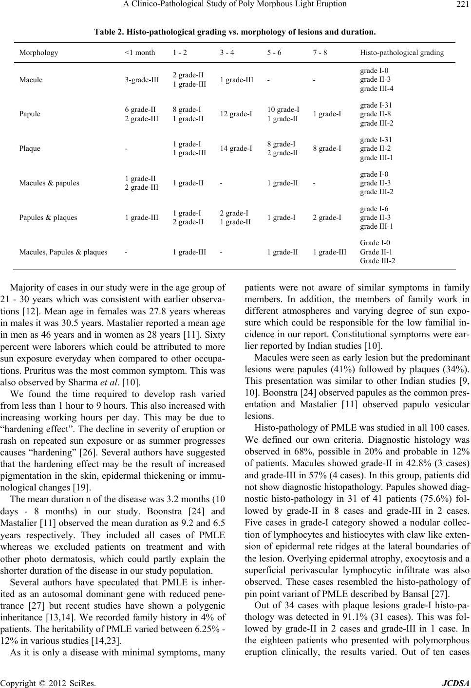
who presented with papules and plaques 60% showed
characteristic histology. In the group comprising poly-
morphous eruptions including macules, there was no
characteristic histology.
Histo-pathological grading also varied with the dura-
tion of the disease. Early lesions showed only grade-II
and III picture whereas the diagnostic picture was evi-
dent with the longer duration of the disease. In the plaque
type, 14 cases showed characteristic histology after three
to four months.
In conclusion PMLE is commonly observed in females
between 21 - 30 years of age. Pruritus is the chief com-
plaint and the time required for the development of rash
increases with time of sun exposure. Most common mor-
phological type is papule on the forearm. Papules and pla-
que type of lesions of PMLE of longer duration clinched
the histo-pathological diagnosis.
REFERENCES
[1] A. J. Stratigos, C. Antoniou and A. D. Katsambas, “Poly-
morphous Light Eruption,” Journal of the European Aca-
demy of Dermatology and Venereology, Vol. 16, No. 3,
2002, pp. 193-206.
doi:10.1046/j.1468-3083.2002.00443.x
[2] T. P. Millard and J. L. Hawk, “Photosensivity Disorders:
Cause, Effect and Management,” American Journal of
Clinical Dermatology, Vol. 3, No. 4, 2002, pp. 239-246.
doi:10.2165/00128071-200203040-00002
[3] M. Lecha, “Idiopathic Photodermatoses: Clinical, Diag-
nostic and Therapeutic Aspects,” Journal of the European
Academy of Dermatology and Venereology, Vol. 15, No.
6, 2001, pp. 499-504.
doi:10.1046/j.1468-3083.2001.00344.x
[4] M. Jeanmouqin and J. Civatte, “Benign Summer Light
Eruption: A New Entity?” Archives of Dermatology, Vol.
122, No. 4, 1988, p. 376.
[5] J. S. Dover and J. L. Hawk, “Polymorphic Light Eruption
Sine Eruption,” British Journal of Dermatology, Vol. 118,
No. 1, 1988, pp. 73-76.
doi:10.1111/j.1365-2133.1988.tb01752.x
[6] E. Ledo, “Photodermatoses,” Dermatologic Clinics, Vol.
12, 1994, pp. 797-803.
[7] W. L. Morison and R. S. Stern, “Polymorphic Light Erup-
tion: A Common Reaction Uncommonly Recognized,”
Acta Dermato-Venereologica, Vol. 62, No. 3, 1982, pp. 237-
240.
[8] C. Pao, P. G. Norris, M. Corbett and J. L. Hawl, “Poly-
morphic Light Eruption: Prevalence in Australia and Eng-
land,” British Journal of Dermatology, Vol. 130, No. 1,
1994, pp. 62-64. doi:10.1111/j.1365-2133.1994.tb06884.x
[9] P. S. Murthy, P. K. Kar, S. Grover and R. Rajagopal,
“Polymorphous Light Eruption in a Ground Crew,” In-
dian Journal of Aerospace Medicine, Vol. 50, No. 1, 2006,
pp. 39-43.
[10] L. Sharma and A. Basnet, “Clinico Epidemiological Study
of Polymorphic Light Eruption,” Indian Journal of Der-
matology, Venereology and Leprology, Vol. 74, No. 1,
2008, pp. 597-602. doi:10.4103/0378-6323.38400
[11] U. Mastalier and H. Keri, “Clinical, Laboratory, Photo-
Test and Phototherapy Findings in Polymorphic Light
Eruption: A Retrospective Study of 133 Patients,” Euro-
pean Journal of Dermatology, Vol. 8, No. 8, 1998, pp. 554-
559.
[12] F. Aubin, “Why Is Polymorphous Light Eruption so Com-
mon in Young Women?” Archives of Dermatological
Research, Vol. 296, No. 5, 2004, pp. 240-241.
doi:10.1007/s00403-004-0508-x
[13] J. M. McGregor, S. Grabezynska and R. Vaughan, “Ge-
netic Modeling of Abnormal Photo Sensivity in Families
with Polymorphic Light Eruption and Actinic Prurigo,”
Journal of Investigative Dermatology, Vol. 115, 2000, pp.
471-476. doi:10.1046/j.1523-1747.2000.00080.x
[14] T. P. Millard, V. Bataille and H. Snieder, “The Heritabil-
ity of Polymorphic Light Eruption,” Journal of Investiga-
tive Dermatology, Vol. 115, 2000, pp. 467-470.
doi:10.1046/j.1523-1747.2000.00079.x
[15] J. K. Rivers, P. G. Norris and G. M. Murphy, “UV Sun-
beds: Tanning, Photoprotection, Acute Adverse Effects
and Immunological Changes,” British Journal of Derma-
tology, Vol. 120, No. 6, 1989, pp. 767-777.
doi:10.1111/j.1365-2133.1989.tb01374.x
[16] P. G. Norris, J. N. Barker and M. H. Allen, “Adhesion
Molecule Expression in Polymorphous Light Eruption,”
Journal of Investigative Dermatology, Vol. 99, 1992, pp.
504-508. doi:10.1111/1523-1747.ep12616175
[17] A. K. Bajaj, “Polymorphic Light Eruption. What’s New
in Dermatology?” Vol. 33, 2002, pp. 2-12.
[18] E. Holzie, G. Plewig, C. Hofmann and E. Roser-Maass, “Po-
lymorphous Light Eruption,” Journal of the American Aca-
demy of Dermatology, Vol. 7, No. 1, 1982, pp. 111-125.
doi:10.1016/S0190-9622(82)80018-2
[19] J. Ferugson and S. Ibbotson, “The Idiopathic Photoder-
matoses,” Seminars in Cutaneous Medicine and Surgery,
Vol. 18, No. 4, 1999, pp. 257-273.
doi:10.1016/S1085-5629(99)80025-X
[20] J. H. Epstein, “Polymorphous Light Eruption,” Journal of
the American Academy of Dermatology, Vol. 3, No. 4,
1980, pp. 329-343. doi:10.1016/S0190-9622(80)80324-0
[21] P. G. Norris, J. Morris, D. M. McGibbon, C. Chu, J. L.
Hawk, “Polymorphic Light Eruption: An Immuno Patho-
logical Study of Evolving Lesions,” British Journal of
Dermatology, Vol. 120, No. 2, 1989, pp. 173-183.
doi:10.1111/j.1365-2133.1989.tb07781.x
[22] J. E. Muhibauer, M. C. Mihm and J. J. Harrist Jr., “Papu-
lar Polymorphic Light Eruption: Fibrin, Complement and
Immunoglobulin Detection,” Archives of Dermatology,
Vol. 120, No. 7, 1984, pp. 866-868.
doi:10.1001/archderm.1984.01650430052009
[23] A. M. Ros and G. Wennersten, “Current Aspects of Poly-
morphous Light Eruption in Sweeden,” Photodermatol-
ogy, Vol. 3, No. 5, 1986, pp. 298-302.
[24] H. E. Boonstra, H. Von Weelden, J. Toonstra and W. A.
Van Vloten, “Polymorphous Light Eruption: A Clinical,
Copyright © 2012 SciRes. JCDSA
A Clinico-Pathological Study of Poly Morphous Light Eruption
Copyright © 2012 SciRes. JCDSA
223
Photobiologic and Follow-Up Study of 110 Patients,”
Journal of the American Academy of Dermatology, Vol.
42, No. 2, 2000, pp. 199-207.
doi:10.1016/S0190-9622(00)90126-9
[25] M. Berg, “Epidemiological Studies of the Influence of
Sun Light on the Skin,” Photo-dermatology, Vol. 6, No. 2,
1989, pp. 80-85.
[26] C. T. Jansen, “The Natural History of Polymorphous
Light Eruption,” Archives of Dermatology, Vol. 115, No.
2, 1979, pp. 165-169.
doi:10.1001/archderm.1979.04010020011004
[27] I. Bansal, H. Kerr, J. J. Janiga and M. S. Quershi, “Pin-
point Papular Variant of Polymorphous Light Eruption:
Clinical and Pathological Correlation,” Journal of the
European Academy of Dermatology and Venereology,
Vol. 20, No. 4, 2006, pp. 406-411.
doi:10.1111/j.1468-3083.2006.01482.x