Journal of Cosmetics, Dermatological Sciences and Applications, 2012, 2, 126-129
http://dx.doi.org/10.4236/jcdsa.2012.23024 Published Online September 2012 (http://www.SciRP.org/journal/jcdsa)
Realization Feature of Mesenchymal Dermal Cells Tissue
Engineering Construction Response in Granulating Wound
Transplantation in Relation with Time-Frame
Elena Petersen1,2
1Moscow Scientific Research Center of Dermatovenerology and Cosmetology of Moscow City, Moscow, Russia; 2Institute of Gen-
eral Pathology and Pathophysiology RAMS, Moscow, Russia.
Email: petersen.elena.v@gmail.com
Received June 8th, 2012; revised July 11th, 2012; accepted July 25th, 2012
ABSTRACT
Derma is progenitor cells sours, which are able to differentiate further in several mesodermal lineage and neural and en-
dodermal lineage. Culture conditions, skin taking site and culture medium composition considerably contribute to it.
Spheroid cultured mesenchymal dermal cells contribution to skin regeneration in granulating wound in rat model was
estimated.
Keywords: Brief Report; 3D Cultivated Tissue Engineering Construction; Skin; Mesenchymal Dermal Cells; Sprouting
Capillary-Like Structures
1. Introduction
Derma is progenitor cells sours, that are able to different-
iate further in several mesodermal lineage (osteogenic,
chondrogenic) [1-4] and neural and endodermal lineage
(endotheliocyte, hepatocyte, islet cell) [5,6]. Culture con-
ditions, skin taking site and culture medium composition
considerably contribute to it. Spheroid cultured mesen-
chymal dermal cells contribution to skin regeneration in
granulating wound in rat model was estimated.
2. Methods
2.1. Cellular Construction
3rd - 4th passage mesenchymal dermal cells were un-
tainted in accordance with standard protocol after plastic
surgical operations on donors given the informed con-
cerned. Hanging drop method was used for spheroid cells
incubation. 25 μl (for 90 ml plates) (Corning) of suspend-
sion and 5 - 6 μl Dulbecco’s Modified Eagle Medium
(DМЕМ)/F12 (Sigma) with antibiotic Invitrogen were
added to incubate hanging drops in standard conditions
(37˚C, 5% СО2) for 7 days. Preliminarily mesenchymal
cells monolayer staining in recommended protocol was
used for transplanted cells visualization. Scores dilution
considered as minimal and not taken in account because
of Spheroid type cells resting stage reaching and short
checking point time-frame.
2.2. Animal Model
Wistar rat were used in experiment with human treatment
rule observance. Wound surface was formed with full-
thickness round incision up to fascia of 5 mm in back
area. Round rings with boarding edge were sewed against
contraction wound healing. Spheroid cells were trans-
planted after wound formation. on the 4th day Rings were
removed for wound epithelization. The evaluation on the
4th, 10th days was based on the ground of morphometry,
immunohistochemistry data, epithelization area and rate
in comparison with intact control. Standard paraffin sec-
tions histological study was used. Immunohistochemistry
study was made with primary antibodies to angiogenic
factors, to CD31, VEGF (Dako), Flk-1 (Chemicon) and
fluorescein isothiocyanate conjugated secondary antibod-
ies, FITC, (Alexa Fluor® 488). For nucleus visualization
Hoechst 33,342 (Sigma) dye was used. In immunohisto-
chemistry, cluster of differentiation 31 (CD31) also known
as platelet endothelial cell adhesion molecule (PECAM-1)
is used primarily to demonstrate the presence of endothe-
lial cells in histological tissue sections. Vascular endo-
thelial growth factor (VEGF) is a signal protein produced
by cells that stimulates vasculogenesis and angiogenesis
and, together a receptor for vascular endothelial growth
factor (Flk-1) is a marker of endothelial cells.
3. Results
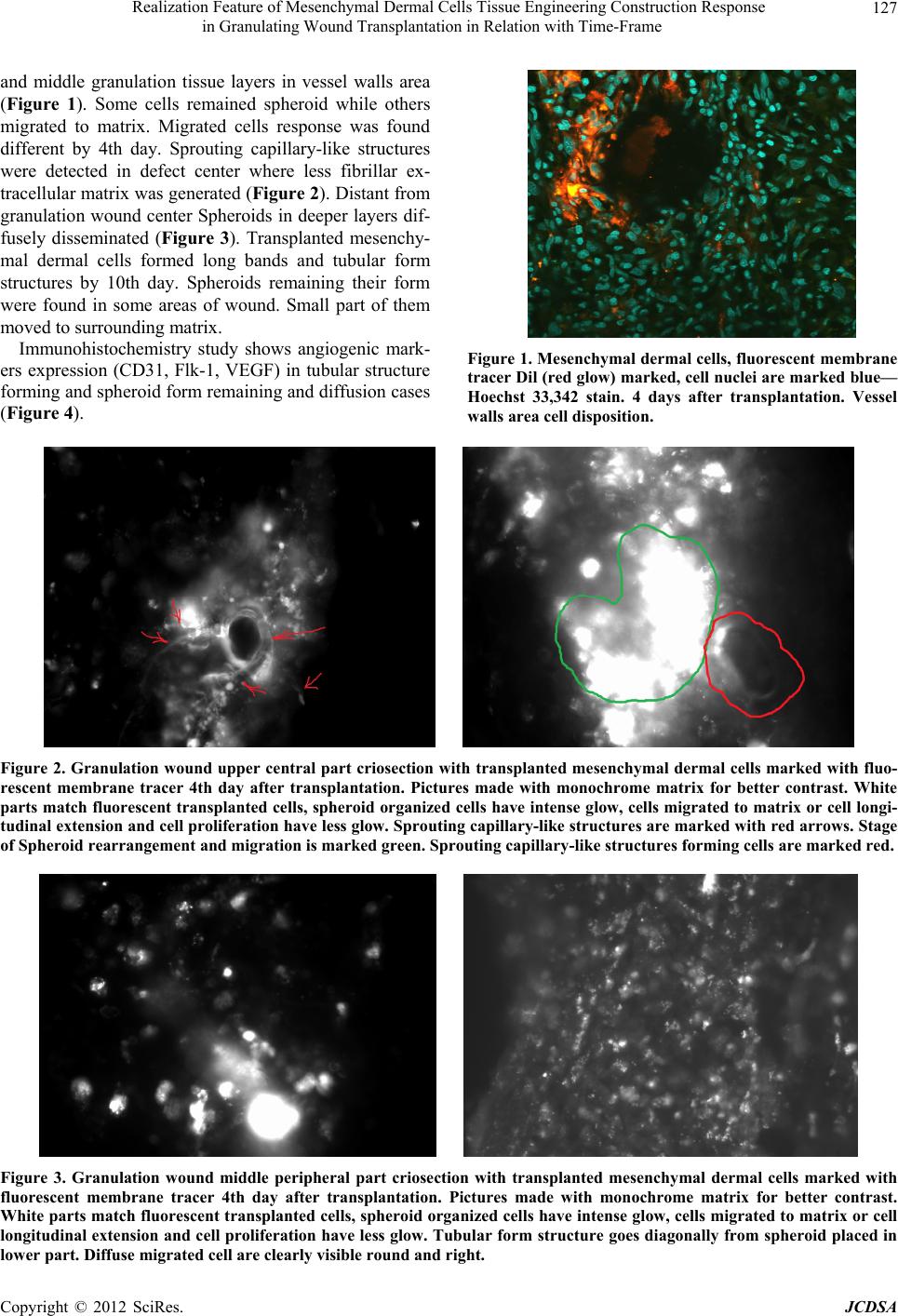
Preferential spheroid disposition was registered in upper
Copyright © 2012 SciRes. JCDSA
Realization Feature of Mesenchymal Dermal Cells Tissue Engineering Construction Response
in Granulating Wound Transplantation in Relation with Time-Frame
127
and middle granulation tissue layers in vessel walls area
(Figure 1). Some cells remained spheroid while others
migrated to matrix. Migrated cells response was found
different by 4th day. Sprouting capillary-like structures
were detected in defect center where less fibrillar ex-
tracellular matrix was generated (Figure 2). Distant from
granulation wound center Spheroids in deeper layers dif-
fusely disseminated (Figure 3). Transplanted mesenchy-
mal dermal cells formed long bands and tubular form
structures by 10th day. Spheroids remaining their form
were found in some areas of wound. Small part of them
moved to surrounding matrix.
Immunohistochemistry study shows angiogenic mark-
ers expression (CD31, Flk-1, VEGF) in tubular structure
forming and spheroid form remaining and diffusion cases
(Figure 4).
Figure 1. Mesenchymal dermal cells, fluorescent membrane
tracer Dil (red glow) marked, cell nuclei are marked blue—
Hoechst 33,342 stain. 4 days after transplantation. Vessel
walls area cell disposition.
Figure 2. Granulation wound upper central part criosection with transplanted mesenchymal dermal cells marked with fluo-
rescent membrane tracer 4th day after transplantation. Pictures made with monochrome matrix for better contrast. White
parts match fluorescent transplanted cells, spheroid organized cells have intense glow, cells migrated to matrix or cell longi-
tudinal extension and cell proliferation have less glow. Sprouting capillary-like structures are marked with red arrows. Stage
of Spheroid rearrangement and migration is marked green. Sprouting capillary-like structures forming cells are marked red.
Figure 3. Granulation wound middle peripheral part criosection with transplanted mesenchymal dermal cells marked with
fluorescent membrane tracer 4th day after transplantation. Pictures made with monochrome matrix for better contrast.
White parts match fluorescent transplanted cells, spheroid organized cells have intense glow, cells migrated to matrix or cell
longitudinal extension and cell proliferation have less glow. Tubular form structure goes diagonally from spheroid placed in
lower part. Diffuse migrated cell are clearly visible round and right.
Copyright © 2012 SciRes. JCDSA
Realization Feature of Mesenchymal Dermal Cells Tissue Engineering Construction Response
in Granulating Wound Transplantation in Relation with Time-Frame
128
Figure 4. Immunohistochemical staining for VEGF. Cells marked with lipotracer Dil have red glow, VEGF expressing cells—
green glow, combined channels are right positioned. Cell nuclei are marked blue-Hoechst 33,342 stain. Upper line—diffuse
cells, middle line—spheroid cell, lower line—tubular form structures (confocal microscopy).
4. Discussion
3D spheroid mesenchymal dermal cells actualize ephi-
thilialmesenhimal transition [7]. Upper spheroid layers
playing defense role boarder spheroid tissue microenvi-
ronment. These cell type migrates to surrounding matrix
first. Endothelial or epithelial predifferentiation of upper
layer cell can explain sprouting capillary-like structures
forming on first stages of spheroid dissemination.
Spheroids cultivated in same conditions in vitro divide
in twofast and slow matrix disseminating types. First
rapidly reflects to changed conditions in wound and ma-
trix induction signals. At first phase wound healing ex-
tracellular matrix lacks ordered fibrous proteins. That
fact and soluble signal factors during first phase trau-
matic process cause endothelial differentiation of super-
ficial migrated cell. Early in vitro experiments we showed
that VEGF-induced spheroids derived from the stromal
cells of the skin dermis, placed in Matrigel did not form
sprouting capillary-like structures, although it is known
that the induction of VEGF spheroids derived from um-
bilical cord stromal cells leads to the appearance of
sprouting capillary-like structures structures in Matrigel
[8]. Second spheroid type slowly reacts to surrounding
changes and cells migrate later. Matrix has another com-
position when cell in these spheroids are activated. As a
result migrated cell make fibroblast form phenotype and
build into matrix diffusely.
5. Conclusion
Studying 3D cultivated tissue engineering construction
helps to understand reparative regeneration processes and
create manageable induction technologies.
REFERENCES
[1] J. Toma, M. Akhavan, K. Fernandes, et al., “Isolation of
Multipotent Adult Stem Cells from the Dermis of Mam-
malian Skin,” Nature Cell Biology, Vol. 3, No. 9, 2001,
pp. 778-784.
Copyright © 2012 SciRes. JCDSA
Realization Feature of Mesenchymal Dermal Cells Tissue Engineering Construction Response
in Granulating Wound Transplantation in Relation with Time-Frame
129
[2] J. Toma, I. McKenzie, D. Bagli, et al., “Isolation and
Characterization of Multipotent Skin-Derived Precursors
from Human Skin,” Stem Cells, Vol. 23, No. 6, 2005, pp.
727-737. doi:10.1634/stemcells.2004-0134
[3] J.-F. Lavoie, J. Biernaskie, Y. Chen, et al., “Skin-Derived
Precursors Differentiate into Skeletogenic Cell Types and
Contribute to Bone Repair,” Stem Cells and Development,
Vol. 18, No. 6, 2009, pp. 893-905.
doi:10.1089/scd.2008.0260
[4] M. Sieber-Blum, M. Grim, Y. Hu and V. Szeder, “Pluri-
potent Neural Crest Stem Cells in the Adult Hair Folli-
cle,” Developmental Dynamics, Vol. 231, No. 2, 2004, pp.
258-269. doi:10.1002/dvdy.20129
[5] F. Chen, W. Zhang, D. Bi, et al., “Clonal Analysis of
Nestin Vimentin+ Multipotent Fibroblasts Isolated from
Human Dermis,” Journal of Cell Science, Vol. 120, 2007,
pp. 2875-2883. doi:10.1242/jcs.03478
[6] D. Bi, F. Chen, W. Zhang, D. Bi, et al., “Differentiation
of Human Multipotent Dermal Fibroblast into Islet-Like
Cell Cluster,” BMC Cell Biology, Vol. 11, No. 46, 2010,
pp. 1-7.
[7] I. Saburina and V. Repin, “3D-Culturing from Single Cells
to the Regeneration of Tissue (the Question about the
Phenomenon of Epithelial-Mesenchymal Plasticity),” Cell
Transplantation and Tissue Engineering, Vol. 5, No. 2,
2010, pp. 75-86.
[8] A. Gorkun, I. Saburina, N. Kosheleva, A. Pulin and V.
Repin, “Induction of Vasculogenesis in the Spheroid of
the Stromal Cells of Umbilical Cord,” Tula Journal New
Medical Technologies, 2012, in press.
Copyright © 2012 SciRes. JCDSA