M. A. ERYILMAZ ET AL.
450
the cancer. In this research, 35 (31%) of the patients, to
who biopsy was done because MR defined them as
BIRADS-4 and BIRADS-5, were examined hispatholo-
gically and the results were benign.
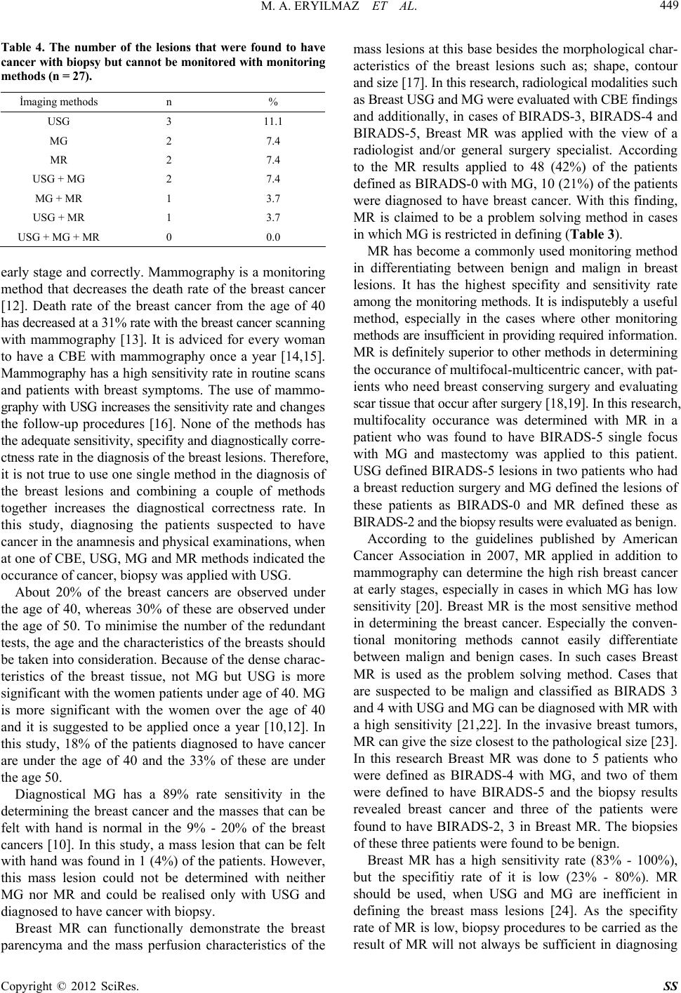
Breast MR is inefficient in defining the breast insitu
cancers and suspicious lesions under 3 mms (14). In this
research, breast insitu cancer could not be defined with
MR in one of the patients and it was defined with MG.
One of the patients was defined as BIRADS-5 with USG
and the cancer found in the biopsy was not determined in
MR (Table 4).
Cor biopsy is a more widely used method in breast
cancer diagnosis. It has many advantages such as; acquiring
adequate tissue sample, diagnosing fast, allowing receptor
use and being cheaper than the open biopsy. The imple-
mentation of the procedure accompanied with USG decr-
eases the false negativity rate to 0.2%. This application
allows breast conserving surgery and sentinel lymph node
mapping while open biopsy doesn’t, because the lympha-
tics remain unharmed [25,26]. In this research, all the
112 biopsies were done accompanied with USG and 27
(24%) of the patients were diagnosd to have cancer. All
of these patients were surgically operated.
5. Conclusion
USG and MR are prominent monitoring methods evalua-
ting the breast diseases. Breast MR is a monitoring method
that can be used in addition to USG and MG in suitable
indications. Breast MR has a sensitivity rate close to MG.
It is a problem solving method with cases of BIRADS-0
and BIRADS-4 breast lesions, where MG is inefficient.
REFERENCES
[1] A. A. Hatipoğlu and A. M. Tuncer, “Türkiyede Kanser
Kontrolü 1 (Baski),” Onur Matbacılık, Ankara, 2007.
[2] J. W. Leung, “Screening Mammography Reduced Mor-
bidity of Breast Cancer Treatment,” American Journal of
Roentgenology, Vol. 184, 2005, pp. 1508-1509.
[3] R. A. Denise, et al., “Imaging and Cancer: Research
Strategy of the American College of Radiology İmaging
Network,” Radiology, Vol. 235, 2005, pp. 741-751.
doi:10.1148/radiol.2353041760
[4] A. M. Tuncer and T. C. Sağlık, “Bakanlığı Kanserle
Savaş Dairesi Başkanlığı, Kadınlarda Meme Kanseri
Taramaları İçin Ulusal Standartlari,” 2004.
http://www.ukdk.org/pdf/meme_standart.pdf
[5] A. S. Majid, et al., “Missed Breast Carcinoma: Pitfalls
and Pearls,” Radiographics, Vol. 23, 2003, pp. 881-895.
doi:10.1148/rg.234025083
[6] M. Mahesh, “AAPM/RSNA Physics Tutorial for Resi-
dents: Digital Mammography: An Overview,” Radio-
graphics, Vol. 24, 2004, pp. 1747-1760.
doi:10.1148/rg.246045102
[7] L. E. Duijm, et al., “Value of Breast İmaging in Women
with Painful Breasts: Observational Follow Up Study,”
British Medical Journal, Vol. 317, 1998, pp. 1492-1495.
doi:10.1136/bmj.317.7171.1492
[8] T. M. Kolb, et al., “Comparison of the Performance of
Screening Mammography, Physical Examination, and
Breast US and Evaluation of Factors That İnfluence Them:
An Analysis of 27, 825 Patient Evaluations,” Radiology,
Vol. 225, 2002, pp. 165-175.
doi:10.1148/radiol.2251011667
[9] M. C. Segel, et al., “Advanced Primary Breast Cancer:
Assessment Mammography of Response to İnduction
Chemotherapy,” Radiology, Vol. 169, 1988, pp. 49-54.
[10] T. Rezanko, “Triple Test and Algorithm in Diagnosis of
Breast Tumors,” Journal of Breast Health, Vol. 3, 2008,
pp. 143-150.
[11] M. Sant, et al., “Time Trends of Breast Cancer Survival
in Europe in Relation to İncidence and Mortality,” In-
ternational Journal of Cancer, Vol. 119, No. 10, 2006, pp.
2417-2422. doi:10.1002/ijc.22160
[12] V. Özmen, “Dünya’da ve Türkiye’de Meme Kanseri
Tarama (Screening) ve Kayıt Programlari,” Journal of
Breast Health, Vol. 2, 2006, pp. 55-58.
[13] S. W. Duffy, et al., “The Swedish Two-County Trial of
Mammographic Screening: Cluster Randomisation and
End Point Evaluation,” Annals of Oncology, Vol. 14, No.
8, 2003, pp. 1196-1198. doi:10.1093/annonc/mdg322
[14] P. Boyle, “Recommendation for Mammografhic Screen-
ing after the Dust Settles,” 8th İnternational Conference:
Primary Therapy of Early Breast Canser SL, 12-15
March 2002.
[15] R. Ballard-Barbash, et al., “Exploring the Role of
Prevention, Screening and Treatment in Canser Trends in
Perry ML,” In: American Society of Clinical Oncology:
Educational Book, 2002, pp. 127-136.
[16] R. Doğan, et al., “Follow-Up Protocolof with Negative
Findings or Non-Palpabl Benign Breast Lesion: Mamo-
graphic and Ultrasonographic BI-RADS Assessment and
Ultrasonography Guided Fine Needle Aspiration Biopsy,”
Journal of Breast Health, Vol. 3, 2007, pp. 58-62.
[17] C. Kuhl, “The Current Status of Breast MR İmaging. Part
I. Choice of Technique, İmage İnterpretation, Diagnostic
Accuracy, and Transfer to Clinical Practice,” Radiology,
Vol. 244, 2007, pp. 356-378.
doi:10.1148/radiol.2442051620
[18] S. H. Heywang-Korunner, et al., “Diagnostic İmaging,”
2nd Edition, Thineme, Ludwisburg, 2001.
[19] S. G. Orel, “MR İmaging of the Breast,” Radiologic
Clinics of North America, Vol. 38, No. 4, 2000, pp. 899-
913. doi:10.1016/S0033-8389(05)70208-6
[20] D. Saslow, et al., “American Cancer Society Breast
Cancer Advisory Group. American Cancer Society Guide-
lines for Breast Screening with MRI as an Adjunct to
Mammography,” A Cancer Journal for Clinicians, Vol.
57, No. 2, 2007, pp. 75-89. doi:10.3322/canjclin.57.2.75
[21] W. A. Berg, et al., “Diagnostic Accuracy of Mam-
mography, Clinical Examination, US, and MR İmaging in
Preoperative Assessment of Breast Cancer,” Radiology,
Vol. 233, 2004, pp. 830-849.
Copyright © 2012 SciRes. SS