B. Farkas et al. / Open Journal of Obstetrics and Gynecology 2 (2012) 227-229
228
After the operation she delivered a healthy infant via
caesarian section in 1997 and she interrupted her last
pregnancy seven years ago. A D & C was carried out and
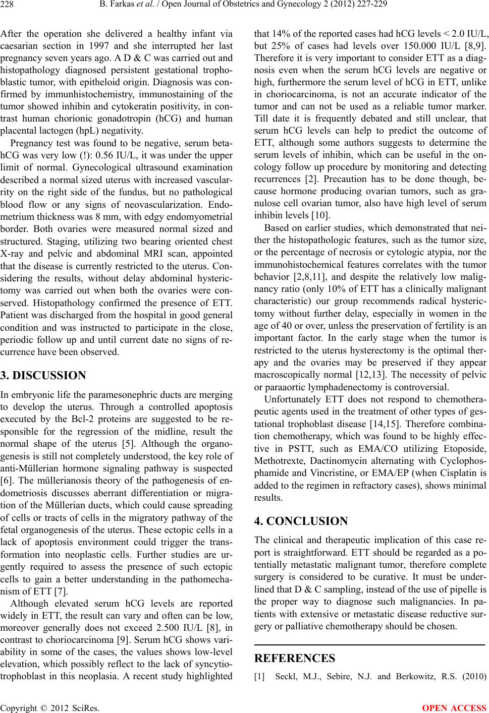
histopathology diagnosed persistent gestational tropho-
blastic tumor, with epitheloid origin. Diagno sis was con-
firmed by immunhistochemistry, immunostaining of the
tumor showed inhibin and cytokeratin positivity, in con-
trast human chorionic gonadotropin (hCG) and human
placental lactogen (hpL) negativity.
Pregnancy test was found to be negative, serum beta-
hCG was very low (!): 0.56 IU/L, it was under the upper
limit of normal. Gynecological ultrasound examination
described a normal sized uterus with increased vascular-
rity on the right side of the fundus, but no pathological
blood flow or any signs of neovascularization. Endo-
metrium thickness was 8 mm, with edgy end omyometrial
border. Both ovaries were measured normal sized and
structured. Staging, utilizing two bearing oriented chest
X-ray and pelvic and abdominal MRI scan, appointed
that the disease is currently restricted to the uterus. Con-
sidering the results, without delay abdominal hysteric-
tomy was carried out when both the ovaries were con-
served. Histopathology confirmed the presence of ETT.
Patient was discharged from the hospital in good general
condition and was instructed to participate in the close,
periodic follow up and until current date no signs of re-
currence have been observed.
3. DISCUSSION
In embryonic life the paramesonephric ducts are merging
to develop the uterus. Through a controlled apoptosis
executed by the Bcl-2 proteins are suggested to be re-
sponsible for the regression of the midline, result the
normal shape of the uterus [5]. Although the organo-
genesis is still not completely understood, the key role of
anti-Müllerian hormone signaling pathway is suspected
[6]. The müllerianosis theory of the pathogenesis of en-
dometriosis discusses aberrant differentiation or migra-
tion of the Müllerian ducts, which could cause spreading
of cells or tracts of cells in the migratory pathway of the
fetal organogenesis of the uterus. These ectopic cells in a
lack of apoptosis environment could trigger the trans-
formation into neoplastic cells. Further studies are ur-
gently required to assess the presence of such ectopic
cells to gain a better understanding in the pathomecha-
nism of ETT [7].
Although elevated serum hCG levels are reported
widely in ETT, the result can vary and often can be low,
moreover generally does not exceed 2.500 IU/L [8], in
contrast to choriocarcinoma [9]. Serum hCG shows vari-
ability in some of the cases, the values shows low-level
elevation, which possibly reflect to the lack of syncytio-
trophoblast in this neoplasia. A recent study highlighted
that 14% of the reported cases had hCG levels < 2.0 IU /L,
but 25% of cases had levels over 150.000 IU/L [8,9].
Therefore it is very important to consider ETT as a diag-
nosis even when the serum hCG levels are negative or
high, furthermore the serum level of hCG in ETT, unlike
in choriocarcinoma, is not an accurate indicator of the
tumor and can not be used as a reliable tumor marker.
Till date it is frequently debated and still unclear, that
serum hCG levels can help to predict the outcome of
ETT, although some authors suggests to determine the
serum levels of inhibin, which can be useful in the on-
cology follow up procedure by monitoring and detecting
recurrences [2]. Precaution has to be done though, be-
cause hormone producing ovarian tumors, such as gra-
nulose cell ovarian tumor, also have high level of serum
inhibin levels [10].
Based on earlier studies, which demonstrated that nei-
ther the histopathologic features, such as the tumor size,
or the percentage of necrosis or cytologic atypia, nor the
immunohistochemical features correlates with the tumor
behavior [2,8,11], and despite the relatively low malig-
nancy ratio (only 10% of ETT has a clinically malignant
characteristic) our group recommends radical hysteric-
tomy without further delay, especially in women in the
age of 40 or over, unless the preservation of fertility is an
important factor. In the early stage when the tumor is
restricted to the uterus hysterectomy is the optimal ther-
apy and the ovaries may be preserved if they appear
macroscopically normal [12,13]. The necessity of pelvic
or paraaortic lymphadenectomy is controversial.
Unfortunately ETT does not respond to chemothera-
peutic agents used in the treatment of other types of ges-
tational trophoblast disease [14,15]. Therefore combina-
tion chemotherapy, which was found to be highly effec-
tive in PSTT, such as EMA/CO utilizing Etoposide,
Methotrexte, Dactinomycin alternating with Cyclophos-
phamide and Vincristine, or EMA/EP (when Cisplatin is
added to the regimen in refractory cases), shows minimal
results.
4. CONCLUSION
The clinical and therapeutic implication of this case re-
port is straightforward. ETT should be regarded as a po-
tentially metastatic malignant tumor, therefore complete
surgery is considered to be curative. It must be under-
lined that D & C sampling, instead of the use of pipelle is
the proper way to diagnose such malignancies. In pa-
tients with extensive or metastatic disease reductive sur-
gery or palliative chemotherapy should be chosen.
REFERENCES
[1] Seckl, M.J., Sebire, N.J. and Berkowitz, R.S. (2010)
Copyright © 2012 SciRes. OPEN ACCESS