Open Journal of Synthesis Theory and Applications, 2012, 1, 13-17
http://dx.doi.org/10.4236/ojsta.2012.12003 Published Online July 2012 (http://www.SciRP.org/journal/ojsta)
Synthesis and Characterisation of Silver Nanoparticles
in Different Medium
G. Alagumuthu*, R. Kirubha
Department of Chemistry Sri Paramakalyani College, Manonmaniam Sundaranar University, Alwarkurichi, India
Email: *alagupathi@yahoo.co.in
Received April 14, 2012; revised May 30, 2012; accepted June 24, 2012
ABSTRACT
Silver nanoparticles were synthesized in different alcoholic medium such as ethylene glycol and n-butyl alcohol by
solvothermal method. The nanoparticles have been successfully synthesized by reducing silver nitrate with the above
solvents in the presence of trioctyl phosphine oxide (TOPO) as the capping agent at room temperature for 1 h. Electron
microscopy, X-ray diffraction, and absorption spectra have been used to investigate the products, and the mechanism is
proposed to interpret the con trolled synthesis of the products. The resu lts indicate that this approach provides a versatile
route to pr epare silver nanowires and nanoparticles with co ntrollable d iameters. The f ormation of nano pro ducts by th is
method is rapid, simple and stable.
Keywords: Silver Nanoparticles; TOPO; Ethylene Glycol; N-Butyl Alcohol
1. Introduction
Nanoparticles are being viewed as fundamental building
blocks of nanotechnology. The most important and dis-
tinct properties is that they exhibit larger surface area to
volume ratio. They exhibit completely new or improved
properties based on specific characteristics such as size,
distribution and morphology [1]. Advances over the past
two decades reveals that the silver nanoparticles (NPs)
possess unique optical, electrical and catalytic properties
[2,3]. During the past few years, the field of silver NPs
preparation has witnessed tremendous growth in syn-
thetic sophistication and depth of characterization [4].
Photochemical method of silver nano particles is pre-
pared by using high molecular weight carbohydrate and
carbohydrate-based dendrimers as reducing and stabiliz-
ing agent [5,6]. A number of preparation routes have
been reported for the synthesis of silver nanoparticles.
For example facile method, thermal decomposition of
silver compounds, electrochemical, sonochemical, mi-
crowave assisted process and recently via chemical route
[7,8]. The solvothermal method was also employed to
synthesize silver nanoparticle. This technique is based on
thermal decomposition of metallic compound in organic
solvent and has b een successfully applied for the synthe-
size of various types of nano sized metal oxide with large
surface area, high crystallinity and high thermal stability.
The influences of reaction conditions, viz., type of sol-
vents, concentration of precursors and reaction tempera-
ture, on the physical properties of the synthesized nano-
rods as well as mechanism were studied [9].
Different approaches have been used to synthesize sil-
ver nanowires. The template-directed approaches were
the most effective and widely used. Macroporous mem-
branes, mesoporous materials, carbon nanotubes, DNA
channels, organic nanotube arrays and silica gels have
been used as physical templates to guide the growth of
nanowires. Though above-mentioned methods can ensure
a good control over morphology of final product and
allow obtaining metal wires with high aspect ratios, the
additional removal of these physical templates may com-
plete the synthetic procedures and limit the scale at
which materials can be synthesized [10].
The purpose of this paper is to describe a facile way
for synthesis of silver nanowires and particles through
the solvothermal method by the reduction of silver nitrate
with ethylene glycol and n-butyl alcohol are an appropri-
ate medium in the presence of TOPO as an adsorption
agent and characterized herein.
2. Experimental
Silver nanoparticles were synthesized by using two dif-
ferent solvents, viz., n-butyl alcohol and ethylene glycol.
Trioctyl phosphine oxide (TOPO) was used as the cap-
ping agent. For the synthesis of silver nanoparticles, the
simple solvothermal method was used at room tempera-
ture. All the aqueous solutions were prepared using ul-
trahigh purity water which was purified by a Milli-Q
*Corresponding author.
C
opyright © 2012 SciRes. OJSTA
G. ALAGUMUTHU, R. KIRUBHA
14
system. In a typical synthesis procedure, 20 ml ethylene
glycol was added to 20 ml of 0.5 M TOPO solution and
stirred vigorously. This mixture was then injected drop
by drop into 20 ml of 0.1 M AgNO3 solution in a beak er
and placed in a magnetic stirrer that was operated at
room temperature for an hour. The solution turned col-
loidal and grey in colour, which suggested the formation
of silver nanoparticles.
Characterization of Silver Nanoparticles
A different value will be o btained depending on wh ether
a technique is sensitive to the medium size or the mean
size, and whether that median and mean is number weigh-
ed or volume weighed. In addition, nanoparticles are not
always single crystals. Many techniques viz., XRD, SEM,
FT-IR and UV-Vis spectra, yield a crystallite size as well
as the size of the aggregate particles. XRD measurements
are performed using a Philips diffractometer of ‘X’ pert
company with nano chromatized Cu K
1 (
= 1.54060 Å)
radiation, the size is determined from the width of XRD
peaks usin g Scherrer’s formul a,
0.86 cosD

where,
D = is the average crystalline size;
= is the X-ray wave length;
= is the full width and half maximum (FWHM);
= is the diffraction angle.
FT-IR measurements is undertaken in order to confirm
the formation of crystalline nanocrystals and identify
adsorbed species onto the crystal surface. Generally, FT-
IR is recorded using Nicolet FT-IR spectrometer mode
impact 400. The spectra were recorded at wave number
in the range of 400 and 4000 cm–1. The UV-Vis absorb-
ance spectra were recorded using a Shimadzu UV-1800
spectrophotometer (Japan), and 1 mm path length quartz
cuvettes were used for the measurements of visible spec-
tra. The electronic images were made on Hitachi S-4500
SEM Analyzer.
3. Results and Discussion
A drop wise addition of ethylene glycol-TOPO and n-
butyl alcohol-TOPO to silver nitrate solution resulted in
the intermediate change in colour from light brown to
grey. The size and structure of the obtained nanop articles
were confirmed by the typical XRD patterns of the
products taken with a b igger quantity of th e dried sample
and shown in Figures 1 and 2. The XRD pattern indi-
cates that the presence of three diffraction peaks, which
agreed well with (111), (200) and (220) diffractions of
face centered cubic silver(JCPDS File No. 04-0783 from
ASTM) The diameter of the nanowires is calculated by
using Debye-Scherer’s formula (11) and it was found as
9.99 nm. The final product is all composed of metallic
silver, indicating that high purity of fcc structure with all
parameters a = 4.065. Similarly the XRD patterns of sil-
ver nanoparticles synthesized using n-butyl alcohol sol-
vent in Figure 2 shows characteristic peak at 2
= 38.5,
marked with (111). It confirms the hypothesis of mono
crystallinity. The sharpening of peak clearly indicates
that the particles are in nano region. The particle size was
Figure 1. XRD Pattern of pure silver nanoparticles in ethylene glycol.
Copyright © 2012 SciRes. OJSTA
G. ALAGUMUTHU, R. KIRUBHA 15
Figure 2. XRD Pattern of silver nanopar ticles in n-butyl alcohol.
also found and the value is higher than the ethylene gly-
col solvent. Hence particle size of silver nanoparticles is
9.99 nm and 42 nm for ethylene glycol and n-butyl alco-
hol respectively. The data are compared with the reported
pure silver nitrate sample.
Optical Absorption
The optical absorption spectra of the nanoparticles were
measured using a USB—2000 UV-VIS spectrophotome-
ter. The powder material has been suspended in glycerol
using magnetic stirrer and their op tical absorp tion spectra
has been recorded at room temperature over the range 300
to 600 nm. It reveals that the absorption edges are blue-
shifted with decreasing particle size. This is confirmed
by quantum confinement effect in the nanoparticles [12].
The fundamental absorption, which corresponds to elec-
tron excitation from the valance band to conduction band,
can be used to determine the value of the optical band
gap energy. The value of optical band gap is calculated
by extrapolating the straight line portion of (αhυ)2 vs hυ
axis [13]. The obtained values are 0.095 eV and 0.046 eV
for ethylene glycol and n-butyl alcohol respectively. Th is
fact explains the bigger particle size obtained from a
slow reaction, which is associated with a more important
ripening contribution to the growth [14]. There- fore the
silver nanoparticle has larger band gap e nerg y value.
The interaction of nanoparticles obtained with the dif-
ferent medium was confirmed by FT-IR spectra. This
was shown in Figures 3 and 4. The peak observed at
3080 cm–1 corresponds to stretching vibration of –OH
bond. After the reduction with the AgNO3, the shift in
the peak at 413.21 cm–1 towards lower frequency is at-
tributed to the formation of nanoparticles. The stretching
frequency of N–O is observed at above 1385 cm–1. This
is confirmed by the changes in the peak shift of silver
nitrate and silver nanoparticles appeared in different me-
dium. In this regard, the mechanism of solvothermal
process seems to be similar to that of the polyol process
demonstrated for the synthesis of highly crystalline nano
materials from III group to V group semi conductors and
silicon [15].
SEM images of the silver nanoparticles products pre-
pared at ethylene glycol and n-butyl alcohol medium.
The image of silver nanoparticles in ethylene glycol me-
dium is shown in Figure 5. The effect of TOPO on the
morphology of silver nanoparticles products can be ex-
plained by the selective adsorption of capping agent on
the surface of the silver nanoparticles. Notably, the
TOPO and different solvents play an important role on
growth of silver nanoparticles. The silver nanoparticles
formed were predominantly cubical with uniform shape.
It is known that the shape of metal nanoparticles consid-
erably change their optical and electronic properties. This
shows that the formation of silver nano sample in the
ethylene glycol medium is significant and reveals that the
size of the silver wire is in nano form.
4. Conclusion
The present work shows that the silver nano products
were synthesized using TOPO-mediated solvothermal
Copyright © 2012 SciRes. OJSTA
G. ALAGUMUTHU, R. KIRUBHA
16
Rajan\A 1/cm
Figure 3. FTIR-Spectra of Silver nanopar t ic le s in ethyle ne glycol.
Figure 4. FTIR-Spectr a of Silver nanoparticles in n-butyl alcohol.
process. The capping agents have the capability of effec-
tive covering and stabilize the newly formed nano com-
pounds. The face centered cubic structure and use of
TOPO with proper concentration; both play an important
role in confining the growth of silver nanowires to the ID
mode. Silver nanowires with a uniform diameter of 9.99
nm were obtained. The end-to-end assemblies of silver
nanowires were formed during the reaction process, and
Copyright © 2012 SciRes. OJSTA
G. ALAGUMUTHU, R. KIRUBHA 17
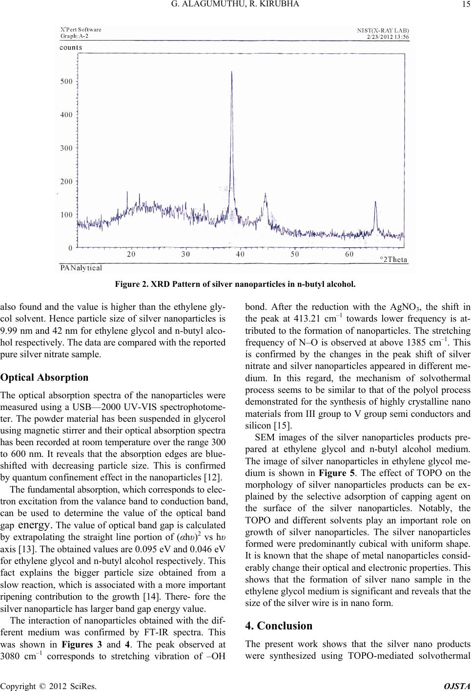
Figure 5. SEM image of Sliver nanoparticles in ethylene
glycol.
the obvious spacing between two straight silver nanowires
would gradually disappear and probab ly to be filled with
silver atoms. This method can be used to select the ap-
propriate solvent for the preparation of silver nanowires
with desired aspect ratio , indicating preferential poten tial
for applications in fabricating future nano electronic de-
vices. REFERENCES
[1] D. Jain, H. K. Daima, S. Kechhwaha and S. L. Kothani,
“Synthesis of Plant-Mediated Silver Nanoparticle Using
Papaya Fruit Extract and Evaluation of Their Antimicro-
bial Activity,” Materials Science and Applications, Vol. 4,
No. 3, 2009, p. 5573.
[2] A. Henglein, “Physicochemical Properties of Small Metal
Particles in Solution: ‘Microelectrode’ Reactions, Chemi-
sorption, Composite Metal Particles, and the Atom-to-
Metal Transition,” Journal of Physical Chemistry, Vol.
97, No. 21, 1993, pp. 5457-5471.
doi:10.1021/j100123a004
[3] Y. A. Krutyakov, A. A. Kudringkii, A. Y. Olenim and G.
V. Lisichkui, “Synthesis and Properties of Silver Nanopar-
ticles: Advances and Prospects,” Russian Chemical Re-
views, Vol. 77, No. 3, 2008, pp. 233-257.
doi:10.1070/RC2008v077n03ABEH003751
[4] A. Pal, S. Shah and S. Devi, “Preparation of Silver, Gold
and Silver-Gold Bimetallic Nanoparticles in W/O Mi-
croemulsion Containing TritonX-100,” Colloids and Sur-
faces: Physicochemical and Engineering Aspects, 302, No.
1-3, 2007, pp. 483-487.
[5] J. Kasthuri, S. Veerapandian and N. Rajendran, “Biologi-
cal Synthesis of Silver & Gold Nanoparticles Using Apiin
as Reducing Agent,” Colloids and Surfaces B: Biointer-
faces, Vol. 68, No. 1, 2009, pp. 55-60.
[6] L. Balogh and D. A. Tomalia, “Poly(Amidoamine) Den-
drimer-Templated Nanocomposites. 1. Synthesis of Ze-
rovalent Copper Nanoclusters,” Journal of the American
Chemical Society, Vol. 120, No. 29, 1998, 7355-7356.
[7] K. Patel, S. Kapoor, D. P. Daul and T. Murherjee, Jour-
nal of Chemical Sciences, Vol. 117, No. 1, 2005, pp. 53-
60. doi:10.1007/BF02704361
[8] J. J. Zhu, X. H. Liao, X. W. Zaho and H. Y. Hem, “Prepa-
ration of Silver Nanorods by Electrochemical Methods,”
Materials Letters, Vol. 49, No. 2, 2001, pp. 91-95.
doi:10.1016/S0167-577X(00)00349-9
[9] Y, G. Sun, B. Mayers, T. Herricks and Y. N. Xia, “Poly ol
Synthesis of Uniform Silver Nanowires,” Nanoletters,
Vol. 3, No. 7, 2003, pp. 955-960.
[10] Y. Xia, P. Yang, Y. Sun and Y. Wu, “One-Dimensional
Nanostructures: Synthesis, Characterization, and Applica-
tions,” Advanced Materials, Vol. 15, No. 5, 2003, pp.
353-389. doi:10.1002/adma.200390087
[11] C. Chem, B. Yu, J. Liu, Q. Dai and Y. Zhu, “Investiga-
tion of ZnO Films on Si<111> Substrate Grown by Low
Energy O+ Assisted Pulse Laser Deposited Technology,”
Materials Letters, Vol. 61, No. 14-15, 2006, pp. 2961-
2964. doi:10.1016/j.matlet.2006.10.047
[12] K. S. Ratidar, D. Patidar, Y. Janu and N. S. Saxena,
“Structural and Optical Charcterization of Che mically Syn-
thesized ZnS Nanoparticles,” Calcogenide Letters, Vol. 5,
No. 6, 2008, pp. 105-110.
[13] G. P. Jushi, N. S. Saxena, T. P. Sharma, Y. Dixit and S. C.
K. Mishra, “Bandgap Determination of Chemically Doped
Polyaniline Materials from Reflectance Measurements,”
Indian Journal of Pure & Applied Physics, Vol. 41. No. 6,
2003, pp. 462-465.
[14] M. de Dios a, F. Barroso, C. Tojoa, M.C. Blanco and M.
A. L’opez-Quintela, “Effects of the Reaction Rate on the
Size Control of Nanoparticles Synthesized in Microemul-
sions,” Colloids and Surfaces A: Physicochemical and Engi-
neering Aspects, Vol. 270-271, 2005, pp. 83-87.
[15] X. Lu, T.Hanrath, K. P. Johnton and B. A. Kargel,
“Growth of Single Crystal Silicon Nanowires in Super-
critical Solution from Tethered Gold Particles on a Sili-
con Substrate,” Nano Letters, Vol. 3, No. 1, 2003, pp. 93-
99. doi:10.1021/nl0202307
Copyright © 2012 SciRes. OJSTA