 Open Journal of Nephrology, 2012, 2, 5-18 http://dx.doi.org/10.4236/ojneph.2012.22002 Published Online June 2012 (http://www.SciRP.org/journal/ojneph) 5 Therapeutic Modalities in Diabetic Nephropathy: Future Approaches* William Brian Reeves1, Bishal B. Rawal1, Emaad M. Abdel-Rahman2, Alaa S. Awad1 1Department of Medicine, Division of Nephrology, Penn State Hershey Medical Center, Hershey, USA 2Department of Medicine, Division of Nephrology, University of Virginia, Charlottesville, USA Email: asa17@psu.edu Received February 11, 2012; revised March 19, 2012; accepted April 17, 2012 ABSTRACT Diabetes mellitus is the leading cause of end stage renal disease and is responsible for more than 40% of all cases in the United States. Several therapeutic interventions for the treatment of diabetic nephropathy have been developed and im- plemented over the past few decades with some degree of success. However, the renal protection provided by these therapeutic modalities is incomplete. More effective approaches are therefore urgently needed. Recently, several novel therapeutic strategies have been explored in treating DN patients including Islet cell transplant, Aldose reductase in- hibitors, Sulodexide (GAC), Protein Kinase C (PKC) inhibitors, Connective tissue growth factor (CTGF) inhibitors, Transforming growth factor-beta (TGF-β) inhibitors and bardoxolone. The benefits and risks of these agents are still under investigation. This review aims to summarize the utility of these novel therapeutic approaches. Keywords: Markers; Albuminuria; Diabetes; Therapy 1. Introduction Diabetes mellitus (DM) is a leading cause of morbidity and mortality in the United States. DM is often compli- cated by micro- and macrovascular involvement which contribute to damage to one or more target organs. Dia- betic nephropathy (DN) is a well-known microvascular complication of diabetes and is responsible for 40% - 50% of all cases of end stage renal disease (ESRD) in the U.S. adult population [1,2]. DN is defined by the presence of persistent pathologic albuminuria of greater than 300 mg/24 hrs (macroalbu- minuria) accompanied by abnormally elevated plasma creatinine or diminished glomerular filtration rate (GFR) [2]. Histologically, DN manifests as diffuse or nodular mesangial expansion, tubular and glomerular basement membrane thickening, as well as interstitial fibrosis. DN is usually preceded by microalbuminuria (urinary albumin excretion > 30 mg but <300 mg/24 hrs) which is the earliest clinical manifestation of renal involvement in diabetic patients [3]. The onsets of microalbuminuria and overt nephropathy are variable in DM type 1 (DMT1) and type 2 (DMT2). Patients with DMT1 have a more predictable natural history and may present with micro- albuminuria 7 - 10 years after being diagnosed with dia- betes. About 20% - 45% of these patients progress to DN over the next 10 years (almost 20 years after the diagno- sis of DMT1) [4]. On the other hand, patients with DMT2, which comprise approximately 80% of all diabetics, may have overt DN at the time of diagnosis since the duration of diabetes is often not precisely known in this popula- tion. The rate of progression of DN towards ESRD is influenced by complex interactions between genetic pre- disposition, dietary and lifestyle factors as well as thera- peutic interventions. Compared to patients with normoal- buminuria (urine albumin excretion < 30 mg/24 hrs), pa- tients with persistent macroalbuminuria (overt DN) have an almost 10-fold higher risk of developing ESRD [5]. Overt proteinuria is also an independent predictor of car- diovascular morbidity and death in diabetic patients [6]. Current therapeutic options directed at delaying the progression of diabetic nephropathy (DN) include inten- sive blood glucose control, improved blood pressure control, interruption of the RAAS using angiotensin-con- verting enzyme (ACE) inhibitors and/or angiotensin type 1 (AT1) receptor blockers (ARB) along with dietary modi- fication and cholesterol-lowering agents (for review please see: [7]). Despite aggressive multifactorial interventions, (DN) remains the single leading cause of ESRD in the United States. The cost of ESRD care for these patients exceeds $10 billion/year. Therefore, more effective ap- proaches are urgently needed. In this article, we will review several novel therapeutic strategies that have been explored recently in patients with DN which may stop or even reverse disease pro- *Conflict of interest: None. C opyright © 2012 SciRes. OJNeph  W. B. REEVES ET AL. 6 gression. 2. Diabetic Nephropathy Markers Early diagnosis of DN is crucial for its effective manage- ment. Thus, the search for reliable markers for this di- sease has been the focus of many studies. Traditionally, GFR has been considered the gold standard in the evalu- ation of overt nephropathy. Several markers have been used to measure GFR including inulin, iohexol, and io- thalamate. However these methods require complex mea- surements and are expensive, time consuming and not readily feasible in clinical practice. The clearance of en- dogenous creatinine is another index for GFR. However, it requires timed urine collections and tends to overesti- mate GFR due to tubular creatinine secretion. This has led to the development of several equations for GFR based on serum creatinine such as the Cockcroft-Gault formula (C-G) [8], the Modification of Diet in Renal Disease four-variable (MDRD-4) formula [9], and the CKD-EPI formula [10] and the measurement of serum cystatin C as an alternative to serum creatinine [11]. A study of patients with DMT2 found that serum cystatin C more accurately identified those with a GFR < 60 ml/min/1.73 m2 than did serum creatinine, MDRD-4 and C-G formulas [12]. Therefore, cystatin C may be re- garded as a superior measure of GFR, especially at lower GFR levels, than serum creatinine. Unfortunately, cys- tatin C measurements are more expensive than creatinine and may yield falsely low GFR in certain circumstances such as inflammation, steroid therapy and hyperthyroi- dism. Urine albumin excretion has received considerable at- tention and has achieved widespread clinical use as a marker of early DN and as a target for intervention. In- creased urinary albumin excretion is the hallmark of DN and estimating urinary albumin excretion is now con- sidered the most reliable marker for assessing disease progression and determining efficacy of treatment in dia- betic patients. Nonetheless, urine microalbumin mea- surements are subject to several limitations relating to specimen collection, indexing to urine creatinine or vo- lume, intrasubject variability and influences of medica- tions, diet and activity which confound its interpretation [11-15]. Thus, a prospective longitudinal study of 232 patients with DM found that the positive predictive value of microalbuminuria as a marker of risk for DN was 43% and the negative predictive value was 77% [13]. There- fore, microalbuminuria may not serve as a strong pre- dictor of DN [14]. On the other hand, macroalbuminuria (overt proteinuria) develops at advanced stage of DN when attempts to prevent progression to ESRD can be very challenging. The discovery of better markers for early detection of DN is an area of active investigation. 3. Candidate Markers in Future Recently, certain cytokines, such as connective tissue growth factor (CTGF), transforming growth factor-β (TGF-β), and tumor necrosis factor-α (TNF-α) have emerged as potential markers of progression of DN (Ta- ble 1). For instance, the number of CTGF messenger RNA positive cells in the kidney biopsy was closely re- lated to the renal biopsy fibrosis score and urinary CTGF levels in 65 subjects, three of whom had diabetes [15]. Additional studies have suggested that the urinary excre- tion of CTGF is related to both albuminuria and GFR in DMT1 [16]. Jaffa and colleagues measured the circulat- ing and urinary levels of CTGF in 1050 subjects with DMT1 from the DCCT/Epidemiology of Diabetes Inter- ventions and Complications (EDIC) study [17]. They showed that significantly higher levels of plasma CTGF are apparent in advanced kidney disease as measured by increased urinary albumin excretion rate (AER), con- cluding that plasma CTGF is a risk marker of diabetic renal and vascular disease. More recently, urinary TGF-β excretion was shown to be attenuated by ACE inhibition in DMT2 patients with nephropathy [18]. In animal models of DMT1, the renal TNF-α level (renal interstitial fluid and urinary TNF-α) showed an early rise after the induction of diabetes [19], which pre- ceded the rise in urinary albumin excretion by about 2 weeks suggesting a possible contribution of TNF-α in the complicated pathogenic process resulting in microalbu- minuria in diabetes. Further studies are necessary to as- sess value of urinary CTGF, TGF-β and TNF-α as mar- kers of DN progression. Podocyte loss, effacement, and alterations of the po- docyte cytoskeleton and structural proteins play a pivotal role in the pathogenesis of DN. Podocytes have been shown in the urine of diabetic patients with microalbu- minuria (53%) and with macroalbuminuria (80%) using immunofluorescence microscopy [20]. The number of podocytes in the urine of patients with macroalbuminuria was significantly greater than in patients with microal- Table 1. Diabetic nephropathy marker s. Current markers Candidate markers in future 1. Creatinine, Cystatin C (estimated GFR). 2. Microalbuminuria 3. Macroalbuminuria or Proteinuria 1. Urinary podocytes 2. NGAL 3. KIM-1 4. Smad 1 5. CTGF 6. TGF-β 7. TNF-α NGAL = Neutrophil Gelatinase-Associated Lipocalin; KIM-1 = Kidney In- jury Molecule 1; CTGF = Connective tissue growth factor; TGF-β = Trans- forming growth factor beta; TNF-α = Tumor necrosis factor alpha. Copyright © 2012 SciRes. OJNeph 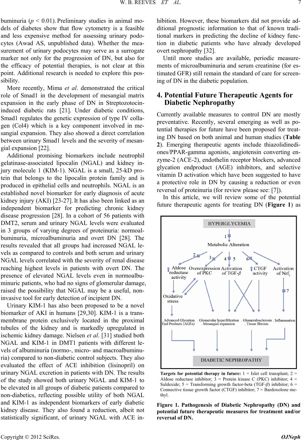 W. B. REEVES ET AL. 7 buminuria (p < 0.01). Preliminary studies in animal mo- dels of diabetes show that flow cytometry is a feasible and less expensive method for assessing urinary podo- cytes (Awad AS, unpublished data). Whether the mea- surement of urinary podocytes may serve as a surrogate marker not only for the progression of DN, but also for the efficacy of potential therapies, is not clear at this point. Additional research is needed to explore this pos- sibility. More recently, Mima et al. demonstrated the critical role of Smad1 in the development of mesangial matrix expansion in the early phase of DN in Streptozotocin- induced diabetic rats [21]. Under diabetic conditions, Smad1 regulates the genetic expression of type IV colla- gen (Col4) which is a key component involved in me- sangial expansion. They also showed a direct correlation between urinary Smad1 levels and the severity of mesan- gial expansion [22]. Additional promising biomarkers include neutrophil gelatinase-associated lipocalin (NGAL) and kidney in- jury molecule 1 (KIM-1). NGAL is a small, 25-kD pro- tein that belongs to the lipocalin protein family and is produced in epithelial cells and neutrophils. NGAL is an established novel biomarker for early diagnosis of acute kidney injury (AKI) [23-27]. It has also been linked as an independent biomarker for predicting chronic kidney disease progression [28]. In a cohort of 56 patients with DMT2, serum and urinary NGAL levels were evaluated in 3 groups of varying degrees of proteinuria: normoal- buminuria, microalbuminuria and overt DN [28]. The results revealed that all groups had increased NGAL le- vels as compared to controls and both serum and urinary NGAL levels correlated with the severity of renal disease reaching highest levels in patients with overt DN. The presence of elevated NGAL levels even in normoalbu- minuric patients, who had no signs of glomerular damage, raised the possibility that NGAL may be a useful, non- invasive tool for early detection of incipient DN. Urinary KIM-1 has also been proposed to be a novel biomarker of AKI in humans [29,30]. KIM-1 is a trans- membrane protein exclusively located in the proximal tubules of the kidney and is markedly upregulated in ischemic kidney damage. Nielsen et al. [31] studied both NGAL and KIM-1 in DMT1 patients with different le- vels of albuminuria (normo-, micro- and macroalbuminu- ria) compared to non-diabetic control subjects. They also evaluated the effect of ACE inhibition (lisinopril) on urinary NGAL excretion in patients with DN. The results of the study showed both urinary NGAL and KIM-1 to be elevated in all groups of diabetic patients compared to non-diabetics, reflecting possible utility of both NGAL and KIM-1 as independent biomarkers of early diabetic kidney disease. They also found a reduction, albeit not statistically significant, of urinary NGAL with ACE in- hibition. However, these biomarkers did not provide ad- ditional prognostic information to that of known tradi- tional markers in predicting the decline of kidney func- tion in diabetic patients who have already developed overt nephropathy [32]. Until more studies are available, periodic measure- ments of microalbuminuria and serum creatinine (for es- timated GFR) still remain the standard of care for screen- ing of DN in the diabetic population. 4. Potential Future Therapeutic Agents for Diabetic Nephropathy Currently available measures to control DN are mostly preventative. Recently, several emerging as well as po- tential therapies for future have been proposed for treat- ing DN based on both animal and human studies (Table 2). Emerging therapeutic agents include thiazolidinedi- ones/PPAR-gamma agonists, angiotensin converting en- zyme-2 (ACE-2), endothelin receptor blockers, advanced glycation endproduct (AGE) inhibitors, and selective vitamin D activation which have been suggested to have a protective role in DN by causing a reduction or even reversal of proteinuria (for review please see: [7]). In this article, we will review some of the potential future therapeutic agents for treating DN (Figure 1) as Targets for potential therapy in future: 1 = Islet cell transplant; 2 = Aldose reductase inhibitor; 3 = Protein kinase C (PKC) inhibitor; 4 = Suldexide; 5 = Transforming growth factor-beta (TGF-β) inhibitor; 6 = Connective tissue growth factor (CTGF) inhibitor; 7 = Bardoxolone me- thyl. Figure 1. Pathogenesis of Diabetic Nephropathy (DN) and potential future therapeutic measures for treatment and/or reversal of DN. Copyright © 2012 SciRes. OJNeph  W. B. REEVES ET AL. Copyright © 2012 SciRes. OJNeph 8 Table 2. Therapeutic modalities in diabetic nephr opathy. Current therapy Emerging therapy Potential therapy for future 1. Intensive glycemic control. - Pharmacologic measures. - Pancreas transplant. 2. Blood pressure control. - Drugs affecting RAAS: . ACE inhibitor . ARB . Direct renin inhibitor . Aldosterone antagonist - Drugs not affecting RAAS: . NDHP CCB . B-blocker . Diuretics 3. Lipid lowering agents. 4. Lifestyle modification. 1. Thiazolidinediones/PPAR-gamma agonists. 2. ACE-2. 3. Endothelin receptor blockers. 4. AGE inhibitors. 5. Vitamin D activation. 1. Islet cell transplant. 2. Aldose reductase inhibitors. 3. Sulodexide (GAG). 4. Protein kinase C (PKC) inhibitors. 5. Connective tissue growth factor (CTGF) inhibitors. 6. Transforming growth factor beta (TGF-B) inhibitors. 7. Bardoxolone. follows. 4.1. Islet Cell Transplant Several studies have shown that islet transplantation is associated with improved diabetic control with a possi- bility of protection against diabetic complications. War- nok et al. [33] performed a prospective crossover, cohort study of 42 patients with DM for more than 5 years and with established diabetic complications such as retino- pathy and mild nephropathy. All patients were initially enrolled in group I and treated with intensive medical therapy. Thirty one patients from group I subsequently received islet cell transplants and crossed over to group II. After almost 3 years of follow up, group II patients showed better glycemic control (HBA1c 7.5% (I) vs. 6.6% (II); p < 0.01) and less progression on retinopathy. However, both groups showed similar declines in kidney function suggesting no additional benefit of islet cell transplantation in preserving GFR (eGFR = –0.45 ml/min (I) vs. –0.12 ml/min (II); p = 0.1). While islet cells are typically obtained from a deceased organ donor, another technique involving transplant of autologous islets has been developed. The basic technique requires total pan- createctomy, fragmentation of the pancreas followed by collagenase digestion and then differential centrifugation. The isolated islets are then re-implanted in the patient’s liver via the portal vein [34]. Webb et al. [34] studied 46 patients who received auto islet transplantation. After 10 years of follow up, the median serum creatinine increased very little from 0.8 mg/dl to 0.87 mg/dl, suggesting a role for auto islet cell transplantation in possible protection against diabetic complications. 4.2. Aldose Reductase Inhibitors Aldose reductase catalyzes the first and rate-limiting step of the polyol pathway of glucose metabolism [35]. Acti- vation of the polyol pathway is implicated in diabetes induced renal dysfunction via de novo synthesis of dia- cylglycerol (DAG), activation of protein kinase C (PKC) with increased production of TGF-β, extracellular matrix proteins and prostaglandins. Increased aldose reductase activity also results in depletion of NADPH, a decrease in cellular levels of reduced glutathione, and increased oxidative stress. The complex interaction between hyper- glycemia-induced oxidative stress from aldose reductase activation, increased formation of advanced glycation endproducts (AGEs) and activation of vascular PKC iso- forms ultimately result in microvascular diabetic com- plications. Increased aldose reductase expression has been shown in DMT2 patients [36]. A number of studies have shown a decrease in urinary albumin excretion in animals treated with aldose reductase inhibitors [37-39]. For instance, the aldose reductase inhibitor, sorbinil, was found to reduce albuminuria and glomerular basement membrane thickening in STZ diabetic rats treated for five months [38]. These actions were attributed to a reduction in the renal cortical activity of glucosyl-galactosyl-hy- droxylysyl-glucohydrolase, an enzyme involved in the catabolism of collagen disaccharide units [39]. Small clinical trials have assessed the efficacy of al- dose reductase inhibitors in the treatment of DN in both DMT1 [40] and DMT2 [41]. Both studies showed re- duced urinary albumin excretion rate after aldose reduc- tase inhibitor treatment for 6 months [40] or 5 years [41]. In contrast to these results, McAuliffe et al. reported that aldose reductase inhibitors had no effect on proteinuria in  W. B. REEVES ET AL. 9 16 diabetic subjects treated for 12 months [42]. Drugs which block aldose reductase activity include spirohy- dantoins (sorbinil), carboxylic acid derivatives (tolrestat, epalrestat, ponalrestat) and flavonoids. Sorbinil and tol- restat have been withdrawn from the worldwide market because of severe toxicity (hepatotoxic). Taken together, these studies of aldose reductase inhibitors have not shown convincing evidence of benefit in the treatment of DN. 4.3. Sulodexide (GAG) Sulodexide is an oral formulation of a highly purified mixture of glycosaminoglycans. It is composed of 80% fast-moving heparin sulfate and 20% dermatan sulfate and is the most extensively studied glycosaminoglycan for diabetic patients. It bears strong chemical similarity to heparin but does not have anticoagulation properties when given orally [43]. Sulodexide has emerged as a potential treatment of DN as multiple studies have de- monstrated reductions in urinary albumin excretion with glycosaminoglycan therapy [44-47]. The precise physiology of the sulodexide-mediated renoprotection in DN is not clear, but several mecha- nisms have been proposed. Sulodexide has been shown to block heparinase-1 activity [48,49], an enzyme that is upregulated in hyperglycemia and can degrade heparin sulfate molecules of the glomerular basement membrane. As sulodexide is a mixture of glycosaminoglycans, it may help in restoring the glycoproteins present in the GBM and mesangium. Another mechanism involves re- storing the anionic heparin sulfate charge on the GBM. Finally, sulodexide may suppress high-glucose induced overexpression of TGF-β1 that is responsible for en- hanced expression of mesangial matrix and collagens [50]. In a study of the db/db mouse model of diabetes, sulodexide was shown to reduce proteinuria significantly in early stage kidney disease but not late kidney disease (12 weeks and after) [51]. The efficacy of sulodexide in diabetes was also evalu- ated in the DiNAS study [52]. DiNAS was a randomized, double blind and placebo controlled trial involving 223 patients with DMT1 or DMT2 and microalbuminuria or macroalbuminuria. Patients were randomized to receive sulodexide (50 to 200 mg daily) or placebo for 4 months. After 4 months of therapy, albuminuria decreased by as much as 74% compared with the placebo group. Four months after drug discontinuation, albuminuria remained 69% lower in those randomized to 200 mg of sulodexide compared with the placebo group. This sustained re- sponse suggests that some anatomical or structural changes had occurred with sulodexide treatment. Sulodexide was well tolerated in that study. Another study showed a sig- nificant reduction in albuminuria with long term use of oral sulodexide at a moderate dose in patients with DN [53]. In this study, thirty patients (both DMT1 and DMT2) treated with 50 mg per day of oral sulodexide for 12 months were compared with thirty matched diabetic patients in the control group. The degree of albuminuria was greatly reduced in patients treated with sulodexide at the end of 12 months but was increased in the control group (–260% and +29% respectively; p = 0.0001). Another recent study included 149 patients with DMT2 and microalbuminuria [54] who were randomized to re- ceive 200 or 400 mg of sulodexide versus placebo. The primary endpoint at 6 months was a 50% reduction in albuminuria or return to normoalbuminuria. This was achieved in 33.3% of the sulodexide 200 mg group and 18.4% of the sulodexide 400 mg group as compared to 15.4% of the placebo group (p = 0.075 and 0.781 respec- tively) [54]. Based on the experience gained from these smaller studies, two large multicenter double-blinded, ran- domized placebo controlled trials were designed to estab- lish the renoprotective potential of sulodexide. The re- sults, unfortunately, were disappointing. The first study was the Sulodexide Microalbuminuria (SUN-micro) Trial, which examined the efficacy of sulodexide given over 26 weeks in 1000 patients with DMT2, hypertension and microalbuminuria [55]. The second study was the Sulo- dexide Overt Nephropathy (SUN-macro) Trial which aimed to examine the efficacy of sulodexide in 2240 pa- tients with DMT2, hypertension and proteinuria ≥ 900 mg/24 h [55]. Both SUN-micro and SUN-macro trials used Sulodexide 200 mg daily vs. placebo in patients being treated with maximum approved or tolerated dose of ACE inhibitor or ARB in both arms. The primary outcome of the SUN-micro Trial was the conversion to normoalbuminuria and at least a 25% decrease in the urinary albumin creatinine ratio (UACR) or at least a 50% reduction in UACR. The primary outcome of the SUN-macro Trial was time to a composite end point of doubling of serum creatinine or ESRD. However, the SUN-micro trial failed to show a reduction of albumi- nuria in DN. With the failure of the SUN-micro trial, the SUN-macro trial was cancelled. 4.4. Protein Kinase C (PKC) Inhibitors Activation of PKC is one of the key metabolic pathways involved in the pathogenesis of the DN. PKC is a family of at least 12 serine-threonine protein kinases that play an important role in intracellular signal transduction [56]. Hyperglycemia-induced oxidative stress has been strongly implicated in microvascular complications from diabetes. High ambient blood glucose levels increase diacylgly- cerol levels, advanced glycation end products, and en- hance mitochondrial synthesis of reactive oxygen species, thereby activating protein kinase C (PKC), particularly in Copyright © 2012 SciRes. OJNeph  W. B. REEVES ET AL. 10 organs that are susceptible to developing diabetic micro- and macro-vascular complications [57]. Activated PKC causes kidney damage through a number of mechanisms including NADPH oxidase-dependent generation of oxi- dants, signaling TGF-β to induce extracellular matrix production and increased secretion of vasodilatory pro- stanoids which contribute to glomerular hyperfiltration [58,59]. Ruboxistaurin mesylate (RTX) (previously known as LY333531) is a bisindolylmaleimide with a high de- gree of specificity for inhibiting PKC-β1 and –β2 iso- forms [60] which has been studied in animal models of DM [61-63]. Ruboxistaurin was shown to have several positive im- pacts on the pathogenesis of DN. It was able to normalize glomerular hyperfiltration, reduce extracellular matrix pro- tein production and TGF-β1, reduce mesangial expansion, glomerulosclerosis and tubulointerstitial fibrosis and de- crease albuminuria. A randomized, double blind, placebo-controlled, mul- ticenter, pilot study was conducted to evaluate the effect of LY333531 in type 2 DN patients [64]. In this study, 123 patients with DN and macroalbuminuria were ran- domized to either 32 mg daily of RTX or placebo for 1 year [64]. Patients in both arms were continued on ACEIs or ARBs during the trial. The primary endpoint was a reduction in ACR. After one year, active treatment was associated with a reduction in albuminuria and stabiliza- tion of GFR whereas the placebo group experienced no change in albuminuria and worsening of GFR. The re- duction in albuminuria appeared as early as 1 month fol- lowing treatment initiation. Although the study showed beneficial effects of LY333531 in DN (reduction in al- buminuria and prevention in loss of eGFR), this study had some limitations. It was underpowered to detect any significant differences in albumin-creatinine ratio and eGFR. Another limitation of this study was its short du- ration of follow-up that limited conclusions about safety of RTX. Unfortunately, the PKC diabetic retinopathy study 2 (PKC-DRS 2) seemed to show an increased fre- quency of the adverse event of “diabetic nephropathy” in ruboxistaurin-treated patients compared with placebo- treated patients [65]. Furthermore, Tuttle et al. analyzed results from studies investigating the effects of ruboxis- taurin on renal outcomes and found that the rate of kid- ney outcomes was similar in ruboxistaurin-treated pa- tients and individuals receiving placebo [66]. 4.5. Connective Tissue Growth Factor (CTGF) Inhibitors CTGF is a recently identified potent profibrotic peptide that has been shown to play a role in the pathogenesis of kidney diseases, micro- and macrovascular complications of diabetes [67,68]. Several agents regulate CTGF ex- pression such as TGF-β, high glucose and fibroblast growth factor [69]. CTGF stimulates cell adhesion and migration, produc- tion and deposition of extracellular matrix (ECM) pro- teins, and angiogenesis [70,71]. Zhou et al. showed that AGE-induced CTGF expression plays a critical role in renal ECM accumulation leading to DN [72]. CTGF has been implicated in promoting tissue fibrosis in DN by activating several intracellular signaling molecules in human mesangial cells (HMC) including receptor tyro- sine kinases (TrkA) and induction of transcription factor TGF-B-inducible early gene [73]. Multiple in vitro and animal studies have demonstrated that inhibition of CTGF prevents the production of key proteins that compose scar [74] and prevents development of renal fibrosis [75, 76]. Several animal and human studies have been under- taken to evaluate the role of CTGF inhibition using a monoclonal antibody that targets CTGF (FG-3019). Fly- vbjerg and colleagues investigated the effects of FG-3019 in obese mice with DMT2 [77]. FG-3019 reduced urinary albumin excretion, GBM thickening and normalized hy- perfiltration in these mice. A similar study in rats showed that FG-3019 reduced diabetic proteinuria [78]. Likewise, encouraging results were noted in human studies examining FG-3019 as a therapeutic agent in pa- tients with DN. Adler and colleagues studied 24 micro- albuminuric subjects (21% with DMT1 and 79% with DMT2) who received 3 or 10 mg/kg FG-3019 (total 4 doses 2 weeks apart) with one year follow up. The results showed that FG-3019 was associated with a significant reduction of urinary albumin/creatinine ratio (mean pre- treatment value of 48 mg/g to a mean post-treatment value of 20 mg/g, p = 0.027) without evidence for a dose-re- sponse relationship [79]. Similarly, Schwartz and col- leagues showed that FG-3019 reduced microalbuminuria in patients with diabetes [80]. These preliminary results suggest that CTGF does play a role in the pathogenesis of DN, a role that needs further clarification. Inhibition of CTGF is promising as a therapeutic target for patients with DN. 4.6. Transforming Growth Factor-Beta (TGF-β) Inhibitors TGF-β1 is a powerful cytokine that plays several roles in the kidney; including cell proliferation, migration, dif- ferentiation, immunomodulation and ECM turnover re- gulation [81]. The role of TGF-β in diabetic nephropathy has been examined in both animal and human studies. Langham et al. [18] extracted RNA from 12 human renal biopsies taken from participants in the Diabiopsies study, a randomized controlled 2-year trial that reported a re- duction in proteinuria and cortical matrix expansion in DMT2 patients treated with perindopril (an ACE inhibi- Copyright © 2012 SciRes. OJNeph  W. B. REEVES ET AL. 11 tor) vs. placebo. The study showed a substantial diminu- tion in TGF-β mRNA gene expression (mean 83% reduc- tion, p < 0.05) in patients who had reduced proteinuria, reflecting a potential role of TGF-β inhibition in the treatment of DN. Therapeutic strategies were developed to block the production/activity of the renal TGF-β1 sys- tem to limit DN. Among these strategies are indirect ap- proaches to decrease the TGF-β effects using renin-an- giotensin inhibition, tight glycemic control, statin therapy and/or tight blood pressure control. Other direct ap- proaches decrease TGF-β effect using neutralizing anti- TGF-β antibodies and antisense or using novel antifi- brotic agents such as Pirfenidone [82]. Several animal studies have been performed to evalu- ate the therapeutic role of TGF-β blockers in DN. Sharma and colleagues administered TGF-β neutralizing anti- bodies to diabetic rats and showed that TGF-β antibodies prevented glomerular enlargement and suppressed the expression of genes encoding ECM components [83]. Ziyadeh and colleagues further showed that administra- tion of anti-TGF-β antibody could attenuate progressive diabetic kidney disease in diabetic mice by preventing pathological changes of glomerulosclerosis [84]. Pirfeni- done (PFD; 5-methyl-1-phenyl-2-(1H)-pyridone), a novel antifibrotic agent, is a low molecular weight synthetic molecule that inhibits TGF-β production and exerts anti- fibrotic properties in cell culture and various animal mo- dels of fibrosis [85,86]. Recently RamachandraRao and colleagues evaluated the therapeutic efficacy of PFD in db/db diabetic mice [87]. DN developed in the db/db mice as evidenced by albuminuria and mesangial matrix expansion by 12 to 16 wk of age. PFD was then given to the db/db mice from week 17 to week 21. Four weeks of PFD treatment led to a significant reduction in the degree of mesangial matrix expansion. They concluded that PFD can promote resolution of mesangial matrix when ad- ministered after the onset of nephropathy. They also showed that PFD did not worsen renal blood flow, lower BP, affect glycemic parameters, or cause hyperkalemia [87]. Several clinical studies have confirmed that TGF-β is increased in the kidneys of diabetic patients. Glomerular expression of TGF-β is also increased in early [85,88] and late stages [86,89] of DMT1 and DMT2 and corre- lates with the degree of glycemic control in these patients [88]. These data were the grounds for a recent clinical trial to evaluate the role of TGF-β inhibitors on the course of DN. A double blind, placebo-controlled study with 77 subjects of DN were randomized to treatment with pla- cebo, low dose (1200 mg daily) or high dose (2400 mg daily) PFD for one year [90]. Treatment with low dose, but not high dose of PFD resulted in an improvement in GFR with no change in albuminuria. The major side ef- fects were gastrointestinal symptoms and fatigue. The results of the study suggested that PFD could be a poten- tial promising therapeutic agent in patients with DN. Ad- ditional studies in larger numbers of patients, ideally with histologic assessment of the kidneys, appear to be war- ranted to support this benefit. 4.7. Anti-Inflammatory Agents—Bardoxolone Recently, an orally available synthetic triterpenoid, Bar- doxolone methyl, has shown promising results in DN. Bardoxolone methyl exerts potent anti-oxidant and anti- inflammatory activity via induction of the Nrf2 transcrip- tion factor. A Phase 2 trial of bardoloxone methyl treat- ment for 8 weeks in 20 patients with moderate-severe CKD and DMT2 demonstrated improved renal function as evidenced by increased eGFR paralleled by a signifi- cant reduction in serum creatinine and BUN [91]. A sub- sequent trial examined the effect of bardoloxone methyl (25 - 150 mg/d) administered for 52 weeks to 227 pa- tients with moderate to severe CKD and DMT2 [92]. Bardoloxone methyl produced a significant increase in GFR of 8 - 11 ml/min/1.73 m2. The improvement in GFR was evident by 8 - 12 weeks of treatment and persisted for the entire 52 week treatment period. Likewise, bar- doloxone treatment reduced the proportion of patients who experienced a 25% fall in GFR from 13% in the placebo group to only 2% in treatment group. Although hard outcomes, such as dialysis dependency and death, were not evaluated, these results are very encouraging and justify further study of bardoxolone methyl and re- lated compounds. 5. Summary and Conclusions There is clear evidence that optimal glycemic control [93-99] and blood pressure control [100-109] are of pa- ramount importance in preventing progression of DN. The renoprotective benefits of agents that block reni- nangiotensin aldosterone system (RAAS) in preventing progression of DN is well-established [110-121]. Non- dihydropyrdine CCBs (diltiazem, verapamil), that do not affect RAAS, have also been shown to reduce proteinuria and slow progression of kidney disease in diabetics [122- 127]. Similarly, lipid lowering agents (such as statins) have been shown to slow the rate of progression of DN [128], but the data supporting this are scant. Even so, it is important to understand that currently available strategies are geared towards limiting or slowing the rate of pro- gression of DN to ESRD. These modalities do not ac- tually stop the progression of DN. In this modern era of medical advancement, we are in dire need of novel strategies that can halt or even reverse the disease pro- gression. While pancreas transplantation [129] is an effective approach in preventing DN in patients with DMT1, islet Copyright © 2012 SciRes. OJNeph  W. B. REEVES ET AL. 12 cell transplantation appears to be a reasonable alternative to improve glycemic control but has questionable benefit in terms of preserving GFR in patients with established DN. Aldose-reductase is a rate-limiting enzyme in glu- cose metabolism pathway, and its activation leads to in- creased oxidative stress, formation of AGE products and activation of protein kinase C, ultimately resulting in micro-vascular complications such as DN. Although al- dose-reductase inhibitors have been shown to reduce albuminuria and prevent glomerular basement membrane thickening in animal studies, their efficacy in humans has been inconclusive. Sulodexide, an oral formulation of a purified mixture of glycosaminoglycans reduced pro- teinuria by uncertain mechanisms. It produced sustained reductions in albuminuria in both animal and human studies even after discontinuation of the drug, suggesting the possibility for anatomical or structural changes with long term protection against DN. Unfortunately, larger multi-center studies (SUN-micro and SUN-macro trials) failed to confirm these benefits. Protein kinase C inhibi- tors (Ruboxistaurin) have shown promising results with respect to improving glomerular hyperfiltration, reducing mesangial expansion and reduction of albuminuria in diabetic animal models as well as humans. They have been associated with a reduction in albuminuria and pre- vention of loss of GFR in a few randomized pilot studies, although large scale prospective studies have yet to con- firm their beneficial effects on renal outcomes. CTGF is a potent profibrotic peptide that has been implicated in extracellular matrix deposition and promotion of tissue fibrosis. Therefore, CTGF inhibition is a promising the- rapeutic target in patients with DN. FG-3019, a CTGF- inhibitor, has been shown to reduce glomerular basement membrane thickening and normalize hyperfiltration dia- betic mice and reduce albuminuria in humans. Similarly, pirfenidone is a TGF-β inhibitor that is recognized as a novel anti-fibrotic agent. In humans, at a low treatment dose, it was shown to improve GFR without improve- ment in albuminuria. Similarly, the use of bardoxalone is encouraging and justifies its further study. In summary, these data indicate that there are several promising therapeutic targets that could potentially be utilized to treat patients with established DN. While cur- rently available measures to control DN are largely pre- ventative, there is hope that therapies capable of stopping, or even reversing progression of DN will soon become available. The current cost of ESRD care exceeds $10 billion per year, much of which is devoted to care of DN. In addition, overt proteinuria in DN is a known inde- pendent risk factor for cardiovascular events, including death. Therefore, the potential ability to halt or reverse DN with these candidate markers in future may offer significant benefits in terms of minimizing cardiovascu- lar morbidity and mortality and also reducing tremendous health-care spending. However, additional human studies are warranted to prove the effectiveness of these agents in treating DN. 6. Acknowledgements This work was supported by NIH Grant DK077444. REFERENCES [1] United States Renal Data System (USRDS), “The Na- tional Institutes of Diabetes and Digestive and Kidney Diseases,” Annual Data Report, Bethesda, 2005. [2] G. Eknoyan, T. Hostetter, G. L. Bakris, L. Hebert, A. S. Levey, H. H. Parving, M. W. Steffes and R. Toto, “Pro- teinuria and Other Markers of Chronic Kidney Disease: A Position Statement of the National Kidney Foundation (NKF) and the National Institute of Diabetes and Diges- tive and Kidney Diseases (NIDDK),” Journal of Kidney Diseases, Vol. 42, No. 4, 2003, pp. 617-622. doi:10.1016/S0272-6386(03)00826-6 [3] C. E. Mogensen, “Microalbuminuria as a Predictor of Clinical Diabetic Nephropathy,” Kidney International, Vol. 31, 1987, pp. 673-689. doi:10.1038/ki.1987.50 [4] A. S. Krolewski, J. H. Warram, L. I. Rand and C. R. Kahn, “Epidemiologic Approach to the Etiology of Type I Diabetes Mellitus and Its Complications,” The New England Journal of Medicine, Vol. 317, No. 22, 1987, pp. 1390-1398. doi:10.1056/NEJM198711263172206 [5] A. M. Berhane, E. J. Weil, W. C. Knowler, R. G. Nelson and R. L. Hanson, “Albuminuria and Estimated Glome- rular Filtration Rate as Predictors of Diabetic End-Stage Renal Disease and Death,” Clinical Journal of the Ame- rican Society of Nephrology, Vol. 6, No. 10, 2011, pp. 2444-2451. doi:10.2215/CJN.00580111 [6] H. C. Gerstein, J. F. Mann, Q. Yi, B. Zinman, S. F. Din- neen, B. Hoogwerf, J. P. Halle, J. Young, A. Rashkow, C. Joyce, S. Nawaz and S. Yusuf, “Albuminuria and Risk of Cardiovascular Events, Death, and Heart Failure in Dia- betic and Nondiabetic Individuals,” The Journal of the American Medical Association, Vol. 286, No. 4, 2001, pp. 421-426. doi:10.1001/jama.286.4.421 [7] E. M. Abdel-Rahman, L. Saadulla, W. B. Reeves and A. S. Awad, “Therapeutic Modalities in Diabetic Nephropa- thy: Standard and Emerging Approaches,” Journal of General Internal Medicine, Vol. 27, No. 4, 2011, pp. 458- 468. [8] D. W. Cockcroft and M. H. Gault, “Prediction of Cre- atinine Clearance from Serum Creatinine,” Nephron, Vol. 16, No. 1, 1976, pp. 31-41. doi:10.1159/000180580 [9] A. S. Levey, J. Coresh, E. Balk, A. T. Kausz, A. Levin, M. W. Steffes, R. J. Hogg, R. D. Perrone, J. Lau and G. Eknoyan, “National Kidney Foundation Practice Guide- lines for Chronic Kidney Disease: Evaluation, Classifica- tion, and Stratification,” Annals of Internal Medicine, Vol. 139, No. 2, 2003, pp. 137-147. [10] A. S. Levey, L. A. Stevens, C. H. Schmid, Y. L. Zhang, A. F. Castro, H. I. Feldman, J. W. Kusek, P. Eggers, F. Van Lente, T. Greene and J. Coresh, “A New Equation to Es- Copyright © 2012 SciRes. OJNeph  W. B. REEVES ET AL. 13 timate Glomerular Filtration Rate,” Annals of Internal Medicine, Vol. 150, No. 9, 2009, pp. 604-612. [11] B. A. Perkins, R. G. Nelson, B. E. Ostrander, K. L. Blouch, A. S. Krolewski, B. D. Myers and J. H. Warram, “Detection of Renal Function Decline in Patients with Diabetes and Normal or Elevated GFR by Serial Mea- surements of Serum Cystatin C Concentration: Results of a 4-Year Follow-Up Study,” Journal of the American So- ciety of Nephrology, Vol. 16, No. 5, 2005, pp. 1404-1412. doi:10.1681/ASN.2004100854 [12] R. J. Macisaac, C. Tsalamandris, M. C. Thomas, E. Pre- maratne, S. Panagiotopoulos, T. J. Smith, A. Poon, M. A. Jenkins, S. I. Ratnaike, D. A. Power and G. Jerums, “The Accuracy of Cystatin C and Commonly Used Creatinine- Based Methods for Detecting Moderate and Mild Chronic Kidney Disease in Diabetes,” Diabetic Medicine, Vol. 24, No. 4, 2007, pp. 443-448. doi:10.1111/j.1464-5491.2007.02112.x [13] B. P. Tabaei, A. S. Al-Kassab, L. L. Ilag, C. M. Zawacki and W. H. Herman, “Does Microalbuminuria Predict Diabetic Nephropathy?” Diabetes Care, Vol. 24, No. 9, 2001, pp. 1560-1566. doi:10.2337/diacare.24.9.1560 [14] B. A. Perkins, L. H. Ficociello, B. E. Ostrander, K. H. Silva, J. Weinberg, J. H. Warram and A. S. Krolewski, “Microalbuminuria and the Risk for Early Progressive Renal Function Decline in Type 1 Diabetes,” Journal of the American Society of Nephrology, Vol. 18, No. 4, 2007, pp. 1353-1361. [15] Y. Ito, J. Aten, R. J. Bende, B. S. Oemar, T. J. Rabelink, J. J. Weening and R. Goldschmeding, “Expression of Con- nective Tissue Growth Factor in Human Renal Fibrosis,” Kidney International, Vol. 53, 1998, pp. 853-861. doi:10.1111/j.1523-1755.1998.00820.x [16] T. Q. Nguyen, L. Tarnow, S. Andersen, P. Hovind, H. H. Parving, R. Goldschmeding and F. A. van Nieuwenhoven, “Urinary Connective Tissue Growth Factor Excretion Correlates with Clinical Markers of Renal Disease in a Large Population of Type 1 Diabetic Patients with Dia- betic Nephropathy,” Diabetes Care, Vol. 29, No. 1, 2006, pp. 83-88. doi:10.2337/diacare.29.01.06.dc05-1670 [17] A. A. Jaffa, W. R. Usinger, M. B. McHenry, M. A. Jaffa, S. R. Lipstiz, D. Lackland, M. Lopes-Virella, L. M. Luttrell and P. W. Wilson, “Connective Tissue Growth Factor and Susceptibility to Renal and Vascular Disease Risk in Type 1 Diabetes,” The Journal of Clinical Endo- crinology & Metabolism, Vol. 93, No. 5, 2008, pp. 1893- 1900. doi:10.1210/jc.2007-2544 [18] R. G. Langham, D. J. Kelly, R. M. Gow, Y. Zhang, D. J. Cordonnier, N. Pinel, P. Zaoui and R. E. Gilbert, “Trans- forming Growth Factor- β in Human Diabetic Neph- ropathy: Effects of ACE Inhibition,” Diabetes Care, Vol. 29, No. 12, 2006, pp. 2670-2675. doi:10.2337/dc06-0911 [19] K. Kalantarinia, A. S. Awad and H. M. Siragy, “Urinary and Renal Interstitial Concentrations of TNF-α Increase Prior to the Rise in Albuminuria in Diabetic Rats,” Kid- ney International, Vol. 64, 2003, pp. 1208-1213. doi:10.1046/j.1523-1755.2003.00237.x [20] T. Nakamura, C. Ushiyama, S. Suzuki, M. Hara, N. Shi- mada, I. Ebihara and H. Koide, “Urinary Excretion of Podocytes in Patients with Diabetic Nephropathy,” Ne- phrology Dialysis Transplantation, Vol. 15, No. 9, 2000, pp. 1379-1383. doi:10.1093/ndt/15.9.1379 [21] T. Matsubara, H. Abe, H. Arai, K. Nagai, A. Mima, H. Kanamori, E. Sumi, T. Takahashi, M. Matsuura, N. Iehara, A. Fukatsu, T. Kita and T. Doi, “Expression of Smad1 Is Directly Associated with Mesangial Matrix Expansion in Rat Diabetic Nephropathy,” Laboratory Investigation, Vol. 86, 2006, pp. 357-368. doi:10.1038/labinvest.3700400 [22] A. Mima, H. Arai, T. Matsubara, H. Abe, K. Nagai, Y. Tamura, K. Torikoshi, M. Araki, H. Kanamori, T. Taka- hashi, T. Tominaga, M. Matsuura, N. Iehara, A. Fukatsu, T. Kita and T. Doi, “Urinary Smad1 Is a Novel Marker to Predict Later Onset of Mesangial Matrix Expansion in Diabetic Nephropathy,” Diabetes, Vol. 57, No. 6, 2008, pp. 1712-1722. doi:10.2337/db07-1726 [23] J. Mishra, C. Dent, R. Tarabishi, M. M. Mitsnefes, Q. Ma, C. Kelly, S. M. Ruff, K. Zahedi, M. Shao, J. Bean, K. Mori, J. Barasch and P. Devarajan, “Neutrophil Gelati- nase-Associated Lipocalin (NGAL) as a Biomarker for Acute Renal Injury after Cardiac Surgery,” The Lancet, Vol. 365, No. 9466, 2005, pp. 1231-1238. doi:10.1016/S0140-6736(05)74811-X [24] C. R. Parikh, J. Mishra, H. Thiessen-Philbrook, B. Dur- sun, Q. Ma, C. Kelly, C. Dent, P. Devarajan and C. L. Edelstein, “Urinary IL-18 Is an Early Predictive Bio- marker of Acute Kidney Injury after Cardiac Surgery,” Kidney International, Vol. 70, 2006, pp. 199-203. doi:10.1038/sj.ki.5001527 [25] M. Bennett, C. L. Dent, Q. Ma, S. Dastrala, F. Grenier, R. Workman, H. Syed, S. Ali, J. Barasch and P. Devarajan, “Urine NGAL Predicts Severity of Acute Kidney Injury after Cardiac Surgery: A Prospective Study,” Clinical Journal of the American Society of Nephrology, Vol. 3, No. 3, 2008, pp. 665-673. doi:10.2215/CJN.04010907 [26] W. Ling, N. Zhaohui, H. Ben, G. Leyi, L. Jianping, D. Huili and Q. Jiaqi, “Urinary IL-18 and NGAL as Early Predictive Biomarkers in Contrast-Induced Nephropathy after Coronary Angiography,” Nephron Clinical Practice, Vol. 108, No. 3, 2008, pp. 176-181. doi:10.1159/000117814 [27] T. L. Nickolas, M. J. O’Rourke, J. Yang, M. E. Sise, P. A. Canetta, N. Barasch, C. Buchen, F. Khan, K. Mori, J. Giglio, P. Devarajan and J. Barasch, “Sensitivity and Specificity of a Single Emergency Department Measure- ment of Urinary Neutrophil Gelatinase-Associated Lipo- calin for Diagnosing Acute Kidney Injury,” Annals of In- ternal Medicine, Vol. 148, No. 11, 2008, pp. 810-819. [28] D. Bolignano, A. Lacquaniti, G. Coppolino, V. Donato, S. Campo, M. R. Fazio, G. Nicocia and M. Buemi, “Neu- trophil Gelatinase-Associated Lipocalin (NGAL) and Progression of Chronic Kidney Disease,” Clinical Jour- nal of the American Society of Nephrology, Vol. 4, No. 2, 2009, pp. 337-344. doi:10.2215/CJN.03530708 [29] W. K. Han, V. Bailly, R. Abichandani, R. Thadhani and J. V. Bonventre, “Kidney Injury Molecule-1 (KIM-1): A Novel Biomarker for Human Renal Proximal Tubule In- jury,” Kidney International, Vol. 62, 2002, pp. 237-244. doi:10.1046/j.1523-1755.2002.00433.x Copyright © 2012 SciRes. OJNeph  W. B. REEVES ET AL. 14 [30] M. M. van Timmeren, M. C. van den Heuvel, V. Bailly, S. J. Bakker, H. van Goor and C. A. Stegeman, “Tubular Kidney Injury Molecule-1 (KIM-1) in Human Renal Di- sease,” The Journal of Pathology, Vol. 212, No. 2, 2007, pp. 209-217. doi:10.1002/path.2175 [31] S. E. Nielsen, K. J. Schjoedt, A. S. Astrup, L. Tarnow, M. Lajer, P. R. Hansen, H. H. Parving and P. Rossing, “Neu- trophil Gelatinase-Associated Lipocalin (NGAL) and Kidney Injury Molecule 1 (KIM1) in Patients with Dia- betic Nephropathy: A Cross-Sectional Study and the Ef- fects of Lisinopril,” Diabetic Medicine, Vol. 27, No. 10, 2010, pp. 1144-1150. doi:10.1111/j.1464-5491.2010.03083.x [32] S. E. Nielsen, S. Andersen, D. Zdunek, G. Hess, H. H. Parving and P. Rossing, “Tubular Markers Do Not Pre- dict the Decline in Glomerular Filtration Rate in Type 1 Diabetic Patients with Overt Nephropathy,” Kidney In- ternational, Vol. 79, No. 10, 2011, pp. 1113-1118. doi:10.1038/ki.2010.554 [33] G. L. Warnock, D. M. Thompson, R. M. Meloche, R. J. Shapiro, Z. Ao, P. Keown, J. D. Johnson, C. B. Verchere, N. Partovi, I. S. Begg, M. Fung, S. E. Kozak, S. O. Tong, K. M. Alghofaili and C. Harris, “A Multi-Year Analysis of Islet Transplantation Compared with Intensive Medical Therapy on Progression of Complications in Type 1 Dia- betes,” Transplantation, Vol. 86, No. 12, 2008, pp. 1762- 1766. doi:10.1097/TP.0b013e318190b052 [34] M. A. Webb, S. C. Illouz, C. A. Pollard, R. Gregory, J. F. Mayberry, S. G. Tordoff, M. Bone, C. J. Cordle, D. P. Berry, M. L. Nicholson, P. P. Musto and A. R. Dennison, “Islet Auto Transplantation Following Total Pancreatec- tomy: A Long-Term Assessment of Graft Function,” Pancreas, Vol. 37, No. 3, 2008, pp. 282-287. doi:10.1097/mpa.0b013e31816fd7b6 [35] D. L. Vander Jagt, B. Robinson, K. K. Taylor and L. A. Hunsaker, “Aldose Reductase from Human Skeletal and Heart Muscle. Interconvertible Forms Related by Thiol- Disulfide Exchange,” The Journal of Biological Chemis- try, Vol. 265, No. 34, 1990, pp. 20982-20987. [36] H. Kasajima, S. Yamagishi, S. Sugai, N. Yagihashi and S. Yagihashi, “Enhanced in Situ Expression of Aldose Re- ductase in Peripheral Nerve and Renal Glomeruli in Dia- betic Patients,” Virchows Archiv, Vol. 439, No. 1, 2001, pp. 46-54. doi:10.1007/s004280100444 [37] R. G. Tilton, K. Chang, G. Pugliese, D. M. Eades, M. A. Province, W. R. Sherman, C. Kilo and J. R. Williamson, “Prevention of Hemodynamic and Vascular Albumin Fil- tration Changes in Diabetic Rats by Aldose Reductase In- hibitors,” Diabetes, Vol. 38, No. 10, 1989, pp. 1258-1270. doi:10.2337/diabetes.38.10.1258 [38] W. G. Robison Jr., T. N. Tillis, N. Laver and J. H. Kino- shita, “Diabetes-Related Histopathologies of the Rat Ret- ina Prevented with an Aldose Reductase Inhibitor,” Ex- perimental Eye Research, Vol. 50, No. 4, 1990, pp. 355- 366. doi:10.1016/0014-4835(90)90136-I [39] J. P. Kassab, R. Guillot, J. Andre, N. Claperon, G. Bellon, G. Feldmann, J. Peyroux and M. Sternberg, “Renal and Microvascular Effects of an Aldose Reductase Inhibitor in Experimental Diabetes. Biochemical, Functional and Ultrastructural Studies,” Biochemical Pharmacology, Vol. 48, No. 5, 1994, pp. 1003-1008. doi:10.1016/0006-2952(94)90371-9 [40] N. Passariello, J. Sepe, G. Marrazzo, A. de Cicco, A. Peluso, M. C. Pisano, S. Sgambato, P. Tesauro and F. D’Onofrio, “Effect of Aldose Reductase Inhibitor (Tol- restat) on Urinary Albumin Excretion Rate and Glomeru- lar Filtration Rate in Iddm Subjects with Nephropathy,” Diabetes Care, Vol. 16, No. 5, 1993, pp. 789-795. doi:10.2337/diacare.16.5.789 [41] K. Iso, H. Tada, K. Kuboki and T. Inokuchi, “Long-Term Effect of Epalrestat, an Aldose Reductase Inhibitor, on the Development of Incipient Diabetic Nephropathy in Type 2 Diabetic Patients,” Journal of Diabetes and Its Complications, Vol. 15, No. 5, 2001, pp. 241-244. doi:10.1016/S1056-8727(01)00160-X [42] A. V. McAuliffe, B. A. Brooks, E. J. Fisher, L. M. Moly- neaux and D. K. Yue, “Administration of Ascorbic Acid and an Aldose Reductase Inhibitor (Tolrestat) in Diabetes: Effect on Urinary Albumin Excretion,” Nephron, Vol. 80, No. 3, 1998, pp. 277-284. doi:10.1159/000045187 [43] G. Andriuoli, R. Mastacchi and M. Barbanti, “Anti- thrombotic Activity of a Glycosaminoglycan (Sulodexide) in Rats,” Thrombosis Research, Vol. 34, No. 1, 1984, pp. 81-86. doi:10.1016/0049-3848(84)90108-7 [44] C. A. Solini A, Barzon I and Crepaldi G, “Therapy with Glycosaminoglycans Lowers Albumin Excretion Rate in Non-Insulin Dependent Diabetic Patients with Microal- buminuria,” Diabetes, Nutrition & Metabolism, Vol. 7, 1994, pp. 304-307. [45] J. Skrha, J. Perusicova, P. Pontuch and A. Oksa, “Glyco- saminoglycan Sulodexide Decreases Albuminuria in Dia- betic Patients,” Diabetes Research and Clinical Practice, Vol. 38, No. 1, 1997, pp. 25-31. doi:10.1016/S0168-8227(97)00076-4 [46] M. Velussi, et al., “Glycosaminoglycans Oral Therapy Reduces Microalbuminuria, Blood Fibrinogen Levels and Limb Arteriopathy Clinical Signs in Patients with Non- Insulin Dependent Diabetes Mellitus,” Diabetes, Nutrition & Metabolism, Vol. 9, 1996, pp. 53-58. [47] A. Solini, L. Vergnani, F. Ricci and G. Crepaldi, “Gly- cosaminoglycans Delay the Progression of Nephropathy in NIDDM,” Diabetes Care, Vol. 20, No. 5, 1997, pp. 819-823. doi:10.2337/diacare.20.5.819 [48] X. Xu, et al., “Mechanism of Action of Sulodexide-Me- diated Control of Diabetic Proteinuria: Inhibition of He- paranase-1 Activity,” Journal of the American Society of Nephrology, Vol. 16, 2005, p. 673. [49] J. B. Maxhimer, M. Somenek, G. Rao, C. E. Pesce, D. Baldwin, P. Gattuso, M. M. Schwartz, E. J. Lewis, R. A. Prinz and X. Xu, “Heparanase-1 Gene Expression and Regulation by High Glucose in Renal Epithelial Cells: A Potential Role in the Pathogenesis of Proteinuria in Dia- betic Patients,” Diabetes, Vol. 54, No. 7, 2005, pp. 2172- 2178. doi:10.2337/diabetes.54.7.2172 [50] G. Gambaro, A. O. Cavazzana, P. Luzi, A. Piccoli, A. Borsatti, G. Crepaldi, E. Marchi, A. P. Venturini and B. Baggio, “Glycosaminoglycans Prevent Morphological Re- nal Alterations and Albuminuria in Diabetic Rats,” Kid- Copyright © 2012 SciRes. OJNeph  W. B. REEVES ET AL. 15 ney International, Vol. 42, 1992, pp. 285-291. doi:10.1038/ki.1992.288 [51] M. Rossini, T. Naito, H. Yang, M. Freeman, E. Donnert, L. J. Ma, S. R. Dunn, K. Sharma and A. B. Fogo, “Sulo- dexide Ameliorates Early But Not Late Kidney Disease in Models of Radiation Nephropathy and Diabetic Neph- ropathy,” Nephrology Dialysis Transplantation, Vol. 25, No. 6, 2010, pp. 1803-1810. doi:10.1093/ndt/gfp724 [52] G. Gambaro, I. Kinalska, A. Oksa, P. Pontuch, M. Hert- lova, J. Olsovsky, J. Manitius, D. Fedele, S. Czekalski, J. Perusicova, J. Skrha, J. Taton, W. Grzeszczak and G. Crepaldi, “Oral Sulodexide Reduces Albuminuria in Mi- croalbuminuric and Macroalbuminuric Type 1 and Type 2 Diabetic Patients: The Di.N.A.S. Randomized Trial,” Journal of the American Society of Nephrology, Vol. 13, No. 6, 2002, pp. 1615-1625. doi:10.1097/01.ASN.0000014254.87188.E5 [53] A. Achour, M. Kacem, K. Dibej, H. Skhiri, S. Bouraoui and M. El May, “One Year Course of Oral Sulodexide in the Management of Diabetic Nephropathy,” Journal of Nephrology, Vol. 18, No. 5, 2005, pp. 568-574. [54] H. L. Heerspink, T. Greene, J. B. Lewis, I. Raz, R. D. Rohde, L. G. Hunsicker, S. L. Schwartz, S. Aronoff, M. A. Katz, G. M. Eisner, J. H. Mersey and T. B. Wiegmann, “Effects of Sulodexide in Patients with Type 2 Diabetes and Persistent Albuminuria,” Nephrology Dialysis Trans- plantation, Vol. 23, No. 6, 2008, pp. 1946-1954. doi:10.1093/ndt/gfm893 [55] H. J. Lambers Heerspink, et al., “Rationale for and Study Design of the Sulodexide Trials in Type 2 Diabetic, Hy- pertensive Patients with Microalbuminuria or Overt Ne- phropathy,” Diabetic Medicine, Vol. 24, No. 11, 2007, pp. 1290-1295. doi:10.1111/j.1464-5491.2007.02249.x [56] P. G. Goekjian and M. R. Jirousek, “Protein Kinase C in the Treatment of Disease: Signal Transduction Pathways, Inhibitors and Agents in Development,” Current Medici- nal Chemistry, Vol. 6, No. 9, 1999, pp. 877-903. [57] K. R. Tuttle, “Protein Kinase C-β Inhibition for Diabetic Kidney Disease,” Diabetes Research and Clinical Prac- tice, Vol. 82, Suppl. 1, 2008, pp. 70-74. doi:10.1016/j.diabres.2008.09.041 [58] S. Yamagishi, K. Fukami, S. Ueda and S. Okuda, “Mo- lecular Mechanisms of Diabetic Nephropathy and Its Therapeutic Intervention,” Current Drug Targets, Vol. 8, No. 8, 2007, pp. 952-959. doi:10.2174/138945007781386884 [59] M. Kunisaki, S. E. Bursell, F. Umeda, H. Nawata and G. L. King, “Normalization of Diacylglycerol-Protein Ki- nase C Activation by Vitamin E in Aorta of Diabetic Rats and Cultured Rat Smooth Muscle Cells Exposed to Ele- vated Glucose Levels,” Diabetes, Vol. 43, No. 11, 1994, pp. 1372-1377. doi:10.2337/diabetes.43.11.1372 [60] M. R. Jirousek, J. R. Gillig, C. M. Gonzalez, W. F. Heath, J. H. McDonald, D. A. Neel, C. J. Rito, U. Singh, L. E. Stramm, A. Melikian-Badalian, M. Baevsky, L. M. Ballas, S. E. Hall, L. L. Winneroski and M. M. Faul, “(S)13- [(Dimethylamino)methyl]-10,11,14,15-tetrahydro-4,9:16, 21-dimetheno-1H,13H-dibenzo[e,k]pyrrolo[3,4-h][1,4,13] oxadiazacyclohexadecene-1,3(2H)-dione (LY333531) and Related Analogues: Isozyme Selective Inhibitors of Pro- tein Kinase Cβ,” Journal of Medicinal Chemistry, Vol. 39, No. 14, 1996, pp. 2664-2671. doi:10.1021/jm950588y [61] H. Ishii, M. R. Jirousek, D. Koya, C. Takagi, P. Xia, A. Clermont, S. E. Bursell, T. S. Kern, L. M. Ballas, W. F. Heath, L. E. Stramm, E. P. Feener and G. L. King, “Ame- lioration of Vascular Dysfunctions in Diabetic Rats by an Oral PKC β Inhibitor,” Science, Vol. 272, No. 5262, 1996, pp. 728-731. doi:10.1126/science.272.5262.728 [62] D. Koya, M. Haneda, H. Nakagawa, K. Isshiki, H. Sato, S. Maeda, T. Sugimoto, H. Yasuda, A. Kashiwagi, D. K. Ways, G. L. King and R. Kikkawa, “Amelioration of Ac- celerated Diabetic Mesangial Expansion by Treatment with a PKC β Inhibitor in Diabetic db/db Mice, a Rodent Model for Type 2 Diabetes,” The FASEB Journal, Vol. 14, No. 3, 2000, pp. 439-447. [63] D. J. Kelly, Y. Zhang, C. Hepper, R. M. Gow, K. Jawor- ski, B. E. Kemp, J. L. Wilkinson-Berka and R. E. Gilbert, “Protein Kinase Cβ Inhibition Attenuates the Progression of Experimental Diabetic Nephropathy in the Presence of Continued Hypertension,” Diabetes, Vol. 52, No. 2, 2003, pp. 512-518. doi:10.2337/diabetes.52.2.512 [64] K. R. Tuttle, G. L. Bakris, R. D. Toto, J. B. McGill, K. Hu and P. W. Anderson, “The Effect of Ruboxistaurin on Nephropathy in Type 2 Diabetes,” Diabetes Care, Vol. 28, No. 11, 2005, pp. 2686-2690. doi:10.2337/diacare.28.11.2686 [65] L. P. Aiello, M. D. Davis, A. Girach, K. A. Kles, R. C. Milton, M. J. Sheetz, L. Vignati and X. E. Zhi, “Effect of Ruboxistaurin on Visual Loss in Patients with Diabetic Retinopathy,” Ophthalmology, Vol. 113, No. 12, 2006, pp. 2221-2230. doi:10.1016/j.ophtha.2006.07.032 [66] K. R. Tuttle, J. B. McGill, D. J. Haney, T. E. Lin and P. W. Anderson, “Kidney Outcomes in Long-Term Studies of Ruboxistaurin for Diabetic Eye Disease,” Clinical Journal of the American Society of Nephrology, Vol. 2, No. 4, 2007, pp. 631-636. doi:10.2215/CJN.00840207 [67] F. A. van Nieuwenhoven, L. J. Jensen, A. Flyvbjerg and R. Goldschmeding, “Imbalance of Growth Factor Signal- ling in Diabetic Kidney Disease: Is Connective Tissue Growth Factor (CTGF, CCN2) the Perfect Intervention Point?” Nephrology Dialysis Transplantation, Vol. 20, No. 1, 2005, pp. 6-10. doi:10.1093/ndt/gfh570 [68] N. A. Wahab, N. Yevdokimova, B. S. Weston, T. Roberts, X. J. Li, H. Brinkman and R. M. Mason, “Role of Con- nective Tissue Growth Factor in the Pathogenesis of Dia- betic Nephropathy,” Biochemical Journal, Vol. 359, Pt. 1, 2001, pp. 77-87. doi:10.1042/0264-6021:3590077 [69] L. F. Lau and S. C. Lam, “The CCN Family of Angio- genic Regulators: The Integrin Connection,” Experimen- tal Cell Research, Vol. 248, No. 1, 1999, pp. 44-57. doi:10.1006/excr.1999.4456 [70] B. L. Riser, M. Denichilo, P. Cortes, C. Baker, J. M. Grondin, J. Yee and R. G. Narins, “Regulation of Con- nective Tissue Growth Factor Activity in Cultured Rat Mesangial Cells and Its Expression in Experimental Dia- betic Glomerulosclerosis,” Journal of the American So- ciety of Nephrology, Vol. 11, No. 1, 2000, pp. 25-38. [71] S. M. Twigg, A. H. Joly, M. M. Chen, J. Tsubaki, H. S. Copyright © 2012 SciRes. OJNeph  W. B. REEVES ET AL. 16 Kim, V. Hwa, Y. Oh and R. G. Rosenfeld, “Connective tissue Growth Factor/IGF-Binding Protein-Related Pro- tein-2 Is a Mediator in the Induction of Fibronectin by Advanced Glycosylation End-Products in Human Dermal Fibroblasts,” Endocrinology, Vol. 143, No. 4, 2002, pp. 1260-1269. doi:10.1210/en.143.4.1260 [72] G. Zhou, C. Li and L. Cai, “Advanced Glycation End- Products Induce Connective Tissue Growth Factor-Me- diated Renal Fibrosis Predominantly through Transforming Growth Factor β -Independent Pathway,” American Jour- nal of Pathology, Vol. 165, No. 6, 2004, pp. 2033-2043. doi:10.1016/S0002-9440(10)63254-3 [73] N. A. Wahab, B. S. Weston and R. M. Mason, “Connec- tive Tissue Growth Factor CCN2 Interacts with and Ac- tivates the Tyrosine Kinase Receptor TrkA,” Journal of the American Society of Nephrology, Vol. 16, No. 2, 2005, pp. 340-351. doi:10.1681/ASN.2003100905 [74] P. Roestenberg, F. A. van Nieuwenhoven, J. A. Joles, C. Trischberger, P. P. Martens, N. Oliver, J. Aten, J. W. Hoppener and R. Goldschmeding, “Temporal Expression Profile and Distribution Pattern Indicate a Role of Con- nective Tissue Growth Factor (CTGF/CCN-2) in Diabetic Nephropathy in Mice,” American Journal of Physiology, Vol. 290, No. 6, 2006, pp. 1344-1354. doi:10.1152/ajprenal.00174.2005 [75] H. Yokoi, M. Mukoyama, T. Nagae, K. Mori, T. Suga- nami, K. Sawai, T. Yoshioka, M. Koshikawa, T. Nishida, M. Takigawa, A. Sugawara and K. Nakao, “Reduction in Connective Tissue Growth Factor by Antisense Treatment Ameliorates Renal Tubulointerstitial Fibrosis,” Journal of the American Society of Nephrology, Vol. 15, No. 6, 2004, pp. 1430-1440. doi:10.1097/01.ASN.0000130565.69170.85 [76] H. Okada, T. Kikuta, T. Inoue, Y. Kanno, S. Ban, T. Sugaya, M. Takigawa and H. Suzuki, “Dexamethasone Induces Connective Tissue Growth Factor Expression in Renal Tubular Epithelial Cells in a Mouse Strain-Specific Manner,” American Journal of Pathology, Vol. 168, No. 3, 2006, pp. 737-747. doi:10.2353/ajpath.2006.050656 [77] A. Flyvbjerg, et al., “Long-Term Renal Effects of a Neu- tralizing Connective Tissue Growth Factor (Ctgf)-Anti- body in Obese Type 2 Diabetic Mice,” Journal of the American Society of Nephrology, Vol. 15, 2004, p. 261. [78] Q. Wang, et al., “Amelioration of Diabetic Nephropathy (DN) Incused by Renal Ischemia-Reperfusion (IR) in Rats with Diabetes Mellitus (DM) by Treatment with FG-3019, a Monoclonal Antibody Against Connective Tissue Growth Factor (CTGF),” Journal of the American Society of Nephrology, 2004, p. 731. [79] S. G. Adler, S. Schwartz, M. E. Williams, C. Arauz- Pacheco, W. K. Bolton, T. Lee, D. Li, T. B. Neff, P. R. Urquilla and K. L. Sewell, “Phase 1 Study of Anti-CTGF Monoclonal Antibody in Patients with Diabetes and Mi- croalbuminuria,” Clinical Journal of the American Soci- ety of Nephrology, Vol. 5, No. 8, 2010, pp. 1420-1428. doi:10.2215/CJN.09321209 [80] S. Schwartz, “Phase 1 Study of FG-3019, an Anti-CTGF Monoclonal Antibody, in Type 1/2 Diabetes Mellitus with Microalbuminuria,” Diabetes, Vol. 56, 2007, p. 151. [81] W. Qi, X. Chen, P. Poronnik and C. A. Pollock, “Trans- forming Growth Factor-β/Connective Tissue Growth Factor Axis in the Kidney,” The International Journal of Biochemistry & Cell Biology, Vol. 40, No. 1, 2008, pp. 9- 13. doi:10.1016/j.biocel.2007.01.006 [82] T. A. McGowan, Y. Zhu and K. Sharma, “Transforming Growth Factor-β: A Clinical Target for the Treatment of Diabetic Nephropathy,” Current Diabetes Reports, Vol. 4, No. 6, 2004, pp. 447-454. doi:10.1007/s11892-004-0055-z [83] K. Sharma, Y. Jin, J. Guo and F. N. Ziyadeh, “Neutraliza- tion of TGF-β by Anti-TGF-β Antibody Attenuates Kid- ney Hypertrophy and the Enhanced Extracellular Matrix Gene Expression in STZ-Induced Diabetic Mice,” Diabe- tes, Vol. 45, 1996, pp. 522-530. doi:10.2337/diabetes.45.4.522 [84] F. N. Ziyadeh, B. B. Hoffman, D. C. Han, M. C. Igle- sias-de la Cruz, S. W. Hong, M. Isono, S. Chen, T. A. McGowan and K. Sharma, “Long-Term Prevention of Renal Insufficiency, Excess Matrix Gene Expression and Glomerular Mesangial Matrix Expansion by Treatment with Monoclonal Antitransforming Growth Factor- β Antibody in db/db Diabetic Mice,” Proceedings of the National Academy of Sciences, Vol. 97, No. 14, 2000, pp. 8015-8020. doi:10.1073/pnas.120055097 [85] K. Sharma, F. N. Ziyadeh, B. Alzahabi, T. A. McGowan, S. Kapoor, B. R. Kurnik, P. B. Kurnik and L. S. Weisberg, “Increased Renal Production of Transforming Growth Factor- β 1 in Patients with Type II Diabetes,” Diabetes, Vol. 46, No. 5, 1997, pp. 854-859. doi:10.2337/diabetes.46.5.854 [86] T. Yamamoto, T. Nakamura, N. A. Noble, E. Ruoslahti and W. A. Border, “Expression of Transforming Growth Factor β Is Elevated in Human and Experimental Dia- betic Nephropathy,” Proceedings of the National Aca- demy of Sciences, Vol. 90, No. 5, 1993, pp. 1814-1818. doi:10.1073/pnas.90.5.1814 [87] S. P. RamachandraRao, Y. Zhu, T. Ravasi, T. A. Mc- Gowan, I. Toh, S. R. Dunn, S. Okada, M. A. Shaw and K. Sharma, “Pirfenidone Is Renoprotective in Diabetic Kid- ney Disease,” Journal of the American Society of Ne- phrology, Vol. 20, No. 8, 2009, pp. 1765-1775. [88] M. Iwano, A. Kubo, T. Nishino, H. Sato, H. Nishioka, Y. Akai, H. Kurioka, Y. Fujii, M. Kanauchi, H. Shiiki and K. Dohi, “Quantification of Glomerular TGF- β 1 mRNA in Patients with Diabetes Mellitus,” Kidney International, Vol. 49, 1996, pp. 1120-1126. doi:10.1038/ki.1996.162 [89] T. Yamamoto, N. A. Noble, A. H. Cohen, C. C. Nast, A. Hishida, L. I. Gold and W. A. Border, “Expression of Transforming Growth Factor- β Isoforms in Human Glo- merular Diseases,” Kidney International, Vol. 49, 1996, pp. 461-469. doi:10.1038/ki.1996.65 [90] K. Sharma, J. H. Ix, A. V. Mathew, M. Cho, A. Pflueger, S. R. Dunn, B. Francos, S. Sharma, B. Falkner, T. A. McGowan, M. Donohue, S. Ramachandrarao, R. Xu, F. C. Fervenza and J. B. Kopp, “Pirfenidone for Diabetic Neph- ropathy,” Journal of the American Society of Nephrology, Vol. 22, No. 6, 2011, pp. 1144-1151. doi:10.1681/ASN.2010101049 [91] P. E. Pergola, M. Krauth, J. W. Huff, D. A. Ferguson, S. Copyright © 2012 SciRes. OJNeph  W. B. REEVES ET AL. 17 Ruiz, C. J. Meyer and D. G. Warnock, “Effect of Bar- doxolone Methyl on Kidney Function in Patients with T2D and Stage 3b-4 CKD,” American Journal of Ne- phrology, Vol. 33, No. 5, 2011, pp. 469-476. doi:10.1159/000327599 [92] P. E. Pergola, P. Raskin, R. D. Toto, C. J. Meyer, J. W. Huff, E. B. Grossman, M. Krauth, S. Ruiz, P. Audhya, H. Christ-Schmidt, J. Wittes and D. G. Warnock, “Bardoxo- lone Methyl and Kidney Function in CKD with Type 2 Diabetes,” The New England Journal of Medicine, Vol. 365, 2011, pp. 327-336. doi:10.1056/NEJMoa1105351 [93] “Retinopathy and Nephropathy in Patients with Type 1 Diabetes Four Years after a Trial of Intensive Therapy. The Diabetes Control and Complications Trial/Epidemi- ology of Diabetes Interventions and Complications Re- search Group,” The New England Journal of Medicine, Vol. 342, 2000, pp. 381-389. doi:10.1056/NEJM200002103420603 [94] “Sustained Effect of Intensive Treatment of Type 1 Dia- betes Mellitus on Development and Progression of Dia- betic Nephropathy: The Epidemiology of Diabetes Inter- ventions and Complications (EDIC) Study,” The Journal of the American Medical Association, Vol. 290, No. 16, 2003, pp. 2159-2167. doi:10.1001/jama.290.16.2159 [95] D. M. Nathan, P. A. Cleary, J. Y. Backlund, S. M. Genuth, J. M. Lachin, T. J. Orchard, P. Raskin and B. Zinman, “Intensive Diabetes Treatment and Cardiovascular Di- sease in Patients with Type 1 Diabetes,” The New Eng- land Journal of Medicine, Vol. 353, 2005, pp. 2643-2653. doi:10.1056/NEJMoa052187 [96] I. M. Stratton, A. I. Adler, H. A. Neil, D. R. Matthews, S. E. Manley, C. A. Cull, D. Hadden, R. C. Turner and R. R. Holman, “Association of Glycaemia with Macrovascular and Microvascular Complications of Type 2 Diabetes (UKPDS 35): Prospective Observational Study,” British Medical Journal, Vol. 321, 2000, pp. 405-412. doi:10.1136/bmj.321.7258.405 [97] R. R. Holman, S. K. Paul, M. A. Bethel, D. R. Matthews and H. A. Neil, “10-Year Follow-up of Intensive Glucose Control in Type 2 Diabetes,” The New England Journal of Medicine, Vol. 359, 2008, pp. 1577-1589. doi:10.1056/NEJMoa0806470 [98] UK Prospective Diabetes Study (UKPDS) Group, “Inten- sive Blood-Glucose Control with Sulphonylureas or In- sulin Compared with Conventional Treatment and Risk of Complications in Patients with Type 2 Diabetes (UKPDS 33),” The Lancet, Vol. 352, No. 9131, 1998, pp. 837-853. doi:10.1016/S0140-6736(98)07019-6 [99] A. Patel, S. MacMahon, J. Chalmers, B. Neal, L. Billot, M. Woodward, M. Marre, M. Cooper, P. Glasziou, D. Grobbee, P. Hamet, S. Harrap, S. Heller, L. Liu, G. Man- cia, C. E. Mogensen, C. Pan, N. Poulter, A. Rodgers, B. Williams, S. Bompoint, B. E. de Galan, R. Joshi and F. Travert, “Intensive Blood Glucose Control and Vascular Outcomes in Patients with Type 2 Diabetes,” The New England Journal of Medicine, Vol. 358, 2008, pp. 2560- 2572. doi:10.1056/NEJMoa0802987 [100] C. E. Mogensen, “Long-Term Antihypertensive Treat- ment Inhibiting Progression of Diabetic Nephropathy,” British Medical Journal, Vol. 285, 1982, pp. 685-688. doi:10.1136/bmj.285.6343.685 [101] R. W. Schrier, R. O. Estacio, A. Esler and P. Mehler, “Effects of Aggressive Blood Pressure Control in Nor- motensive Type 2 Diabetic Patients on Albuminuria, Re- tinopathy and Strokes,” Kidney International, Vol. 61, 2002, pp. 1086-1097. doi:10.1046/j.1523-1755.2002.00213.x [102] UK Prospective Diabetes Study Group, “Tight Blood Pressure Control and Risk of Macrovascular and Mi- crovascular Complications in Type 2 Diabetes: UKPDS 38,” British Medical Journal, Vol. 317, 1998, pp. 703- 713. doi:10.1136/bmj.317.7160.703 [103] UK Prospective Diabetes Study Group, “Cost Effective- ness Analysis of Improved Blood Pressure Control in Hypertensive Patients with Type 2 Diabetes: UKPDS 40,” British Medical Journal, Vol. 317, 1998, pp. 720- 726. doi:10.1136/bmj.317.7160.720 [104] A. Patel, S. MacMahon, J. Chalmers, B. Neal, M. Wood- ward, L. Billot, S. Harrap, N. Poulter, M. Marre, M. Cooper, P. Glasziou, D. E. Grobbee, P. Hamet, S. Heller, L. S. Liu, G. Mancia, C. E. Mogensen, C. Y. Pan, A. Rodgers and B. Williams, “Effects of a Fixed Combina- tion of Perindopril and Indapamide on Macrovascular and Microvascular Outcomes in Patients with Type 2 Diabe- tes Mellitus (the ADVANCE trial): A Randomised Con- trolled Trial,” The Lancet, Vol. 370, No. 9590, 2007, pp. 829-840. doi:10.1016/S0140-6736(07)61303-8 [105] B. E. de Galan, V. Perkovic, T. Ninomiya, A. Pillai, A. Patel, A. Cass, B. Neal, N. Poulter, S. Harrap, C. E. Mo- gensen, M. Cooper, M. Marre, B. Williams, P. Hamet, G. Mancia, M. Woodward, P. Glasziou, D. E. Grobbee, S. MacMahon and J. Chalmers, “Lowering Blood Pressure Reduces Renal Events in Type 2 Diabetes,” Journal of the American Society of Nephrology, Vol. 20, No. 4, 2009, pp. 883-892. doi:10.1681/ASN.2008070667 [106] A. V. Chobanian, G. L. Bakris, H. R. Black, W. C. Cush- man, L. A. Green, J. L. Izzo, D. W. Jones, B. J. Materson, S. Oparil, J. T. Wright and E. J. Roccella, “Seventh Re- port of the Joint National Committee on Prevention, De- tection, Evaluation and Treatment of High Blood Pres- sure,” Hypertension, Vol. 42, 2003, pp. 1206-1252. doi:10.1161/01.HYP.0000107251.49515.c2 [107] American Diabetes Association, “Standards of Medical Care in Diabetes,” Diabetes Care, Vol. 28, Suppl. 1, 2005, pp. 4-36. doi:10.2337/diacare.28.suppl_1.S4 [108] Kidney Disease Outcomes Quality Initiative (K/DOQI), “K/DOQI Clinical Practice Guidelines on Hypertension and Antihypertensive Agents in Chronic Kidney Dis- ease,” American Journal of Kidney Disease, Vol. 43, No. 5, 2004, pp. 1-290. doi:10.1053/j.ajkd.2003.11.027 [109] The ACCORD Study Group, “Effects of Intensive Blood- Pressure Control in Type 2 Diabetes Mellitus,” The New England Journal of Medicine, 2010, Vol. 362, pp. 1575- 1585. doi:10.1056/NEJMoa1001286 [110] The EUCLID Study Group, “Randomised Placebo-Con- trolled Trial of Lisinopril in Normotensive Patients with Insulin-Dependent Diabetes and Normoalbuminuria or Microalbuminuria,” Lancet, Vol. 349, No. 9068, 1997, pp. 1787-1792. doi:10.1016/S0140-6736(96)10244-0 Copyright © 2012 SciRes. OJNeph  W. B. REEVES ET AL. Copyright © 2012 SciRes. OJNeph 18 [111] M. Ravid, D. Brosh, Z. Levi, Y. Bar-Dayan, D. Ravid and R. Rachmani, “Use of Enalapril to Attenuate Decline in Renal Function in Normotensive, Normoalbuminuric Pa- tients with Type 2 Diabetes Mellitus,” Annals of Internal Medicine, Vol. 128, No. 12, 1998, pp. 982-988. [112] R. O. Estacio, B. W. Jeffers, N. Gifford and R. W. Schrier, “Effect of Blood Pressure Control on Diabetic Microvas- cular Complications in Patients with Hypertension and Type 2 Diabetes,” Diabetes Care, Vol. 23, Suppl. 2, 2000, pp. 54-64. [113] M. Ravid, H. Savin, I. Jutrin, T. Bental, B. Katz and M. Lishner, “Long-Term Stabilization of Angiotensin-Con- verting Enzyme Inhibition on Plasma Creatinine and on Proteinuria in Normotensive Type II Diabetic Patients,” Annals of Internal Medicine, Vol. 118, 1993, pp. 577- 581. [114] S. Andersen, L. Tarnow, P. Rossing, B. V. Hansen and H. H. Parving, “Renoprotective Effects of Angiotensin II Receptor Blockade in Type 1 Diabetic Patients with Dia- betic Nephropathy,” Kidney International, Vol. 57, 2000, pp. 601-606. doi:10.1046/j.1523-1755.2000.00880.x [115] B. M. Brenner, M. E. Cooper, D. de Zeeuw, W. F. Keane, W. E. Mitch, H. H. Parving, G. Remuzzi, S. M. Snapinn, Z. Zhang and S. Shahinfar, “Effects of Losartan on Renal and Cardiovascular Outcomes in Patients with Type 2 Diabetes and Nephropathy,” The New England Journal of Medicine, Vol. 345, 2001, pp. 861-869. doi:10.1056/NEJMoa011161 [116] E. J. Lewis, L. G. Hunsicker, W. R. Clarke, T. Berl, M. A. Pohl, J. B. Lewis, E. Ritz, R. C. Atkins, R. Rohde and I. Raz, “Renoprotective Effect of the Angiotensin-Receptor Antagonist Irbesartan in Patients with Nephropathy Due to Type 2 Diabetes,” The New England Journal of Medi- cine, Vol. 345, 2001, pp. 851-860. doi:10.1056/NEJMoa011303 [117] H. H. Parving, H. Lehnert, J. Brochner-Mortensen, R. Gomis, S. Andersen and P. Arner, “The Effect of Irbesar- tan on the Development of Diabetic Nephropathy in Pa- tients with Type 2 Diabetes,” The New England Journal of Medicine, Vol. 345, 2001, pp. 870-878. doi:10.1056/NEJMoa011489 [118] Y. Uresin, A. A. Taylor, C. Kilo, D. Tschope, M. Santo- nastaso, G. Ibram, H. Fang and A. Satlin, “Efficacy and Safety of the Direct Renin Inhibitor Aliskiren and Rami- pril Alone or in Combination in Patients with Diabetes and Hypertension,” Journal of the Renin-Angiotensin- Aldosterone System, Vol. 8, No. 4, 2007, pp. 190-198. doi:10.3317/jraas.2007.028 [119] H. H. Parving, F. Persson, J. B. Lewis, E. J. Lewis and N. K. Hollenberg, “Aliskiren Combined with Losartan in Type 2 Diabetes and Nephropathy,” The New England Journal of Medicine, Vol. 358, 2008, pp. 2433-2446. doi:10.1056/NEJMoa0708379 [120] M. J. Dunn, “Prostaglandins, Angiotension II, and Pro- teinuria,” Nephron, Vol. 55, Suppl. 1, 1990, pp. 30-37. doi:10.1159/000186032 [121] W. R. Melchior, V. Bindlish and L. A. Jaber, “Angio- tensin-Converting Enzyme Inhibitors in Diabetic Neph- ropathy,” The Annals of Pharmacotherapy, Vol. 27, 1993, pp. 344-350. [122] G. L. Bakris, M. R. Weir, M. Secic, B. Campbell and A. Weis-McNulty, “Differential Effects of Calcium Antago- nist Subclasses on Markers of Nephropathy Progression,” Kidney International, Vol. 65, 2004, pp. 1991-2002. doi:10.1111/j.1523-1755.2004.00620.x [123] G. Remuzzi, P. Ruggenenti and A. Benigni, “Under- standing the Nature of Renal Disease Progression,” Kid- ney International, Vol. 51, 1997, pp. 2-15. doi:10.1038/ki.1997.2 [124] H. J. Kloke, A. J. Branten, F. T. Huysmans and J. F. Wetzels, “Antihypertensive Treatment of Patients with Proteinuric Renal Diseases: Risks or Benefits of Calcium Channel Blockers?” Kidney International, Vol. 53, 1998, pp. 1559-1573. doi:10.1046/j.1523-1755.1998.00912.x [125] R. T. Gansevoort, W. J. Sluiter, M. H. Hemmelder, D. de Zeeuw and P. E. de Jong, “Antiproteinuric Effect of Blood-Pressure-Lowering Agents: A Meta-Analysis of Comparative Trials,” Nephrology Dialysis Transplanta- tion, Vol. 10, 1995, pp. 1963-1974. [126] G. L. Bakris, “Effects of Diltiazem or Lisinopril on Mas- sive Proteinuria Associated with Diabetes Mellitus,” An- nals of Internal Medicine, Vol. 112, No. 9, 1990, pp. 707-708. [127] G. L. Bakris, J. B. Copley, N. Vicknair, R. Sadler and S. Leurgans, “Calcium Channel Blockers Versus Other An- tihypertensive Therapies on Progression of NIDDM As- sociated Nephropathy,” Kidney International, Vol. 50, 1996, pp. 1641-1650. doi:10.1038/ki.1996.480 [128] G. Tonolo, M. Velussi, E. Brocco, C. Abaterusso, A. Carraro, G. Morgia, A. Satta, R. Faedda, A. Abhyankar, H. Luthman and R. Nosadini, “Simvastatin Maintains Steady Patterns of GFR and Improves AER and Expres- sion of Slit Diaphragm Proteins in Type II Diabetes,” Kidney International, Vol. 70, 2006, pp. 177-186. doi:10.1038/sj.ki.5001515 [129] R. W. Bilous, S. M. Mauer, D. E. Sutherland, J. S. Na- jarian, F. C. Goetz and M. W. Steffes, “The Effects of Pancreas Transplantation on the Glomerular Structure of Renal Allografts in Patients with Insulin-Dependent Dia- betes,” The New England Journal of Medicine, Vol. 321, No. 2, 1989, pp. 80-85. doi:10.1056/NEJM198907133210204
|