V. THAVER ET AL.
Copyright © 2012 SciRes. OJRD
62
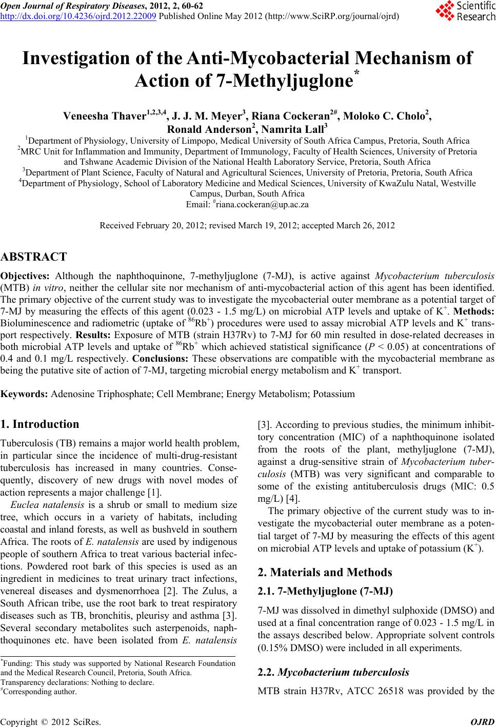
CONTROL
7-MJ [0.023]
7-MJ [0.094]
7-MJ [0.375]
7-MJ [1.5]
0
100
200
300
*
*
*
ATP levels
( % of control)
Figure 2. The effect of 7-methyljuglone (7-MJ) on the levels
of ATP in the H37Rv strain of MTB. The data shown are
that of one experiment with five replicates for each concen-
tration and are representative of 3 different experiments
showing similar trends. The results are expressed as the
mean percentages of the corresponding compound-free
control systems SD; the absolute value of the control sys-
tem is 2,306,410 relative light units. *P values < 0.05 when
compared to the solvent control.
clinical development, 7-MJ is a noteworthy exception [4].
The MIC value of this agent for MTB is 0.5 mg/L, which
is significantly lower than its IC50 value (30.0 mg/L) for
eukaryotic cell lines in vitro [4]. Nonetheless, relatively
little is known about either the primary targets or rapidity
of onset of anti-mycobacterial activity of this agent.
In the current study, a relatively brief exposure (30 -
60 min) of MTB to 7-MJ resulted in significant, dose
-related inhibition of both microbial energy metabolism
and uptake of K+, which, in both cases was maximal at
concentrations close to the MIC value. In the case of
ATP levels, exposure of MTB to 7-MJ at a concentration
of 0.0263 mg/L resulted in a significant increase in ATP,
with a progressive, dose-related decrease at higher con-
centrations. The increase may represent a stress response
to moderate oxidative trauma caused by low concentra-
tions of 7-MJ as described for other types of antimicro-
bial agents [6,7], while at higher concentrations, ire-
versible ROS-mediated toxicity predominates. Alterna-
tively, albeit speculatively, 7-MJ may interfere with the
activity of mycobacterial type 2 NADH: quinone oxi-
doreductase, an early step in the mycobacterial respira-
tory chain [8].
Inhibition of mycobacterial K+ transport by 7-MJ
closely paralleled interference with microbial energy
metabolism, and is probably secondary to ATP depletion.
MTB possesses two major K+ uptake systems. These are
the Kdp and Trk A/B systems, driven by ATP and proton
motive force, respectively [5]. The experimental condi-
tions used in the current study (low K+ medium) are
likely to favour preferential utilisation of the inducible,
high-affinity, Kdp system, accounting for the susceptibil-
ity of K+ transport to ATP depletion, which in turn may
lead to inactivation of the Trk A/B system due to dissipa-
tion of the membrane potential.
In conclusion, 7-MJ appears to target mycobacterial
energy metabolism, leading to secondary membrane dys-
function and inhibition of bacterial growth. This agent
may serve as a prototype for the development of novel
naphthoquinone-based anti-tuberculosis agents.
5. Acknowledgements
We thank Dr. Anita Mahapatra for supplying the chemi-
cal compound.
REFERENCES
[1] M. A. De Groote and G. Huitt, “Infections Due to Rap-
idly Growing Mycobacteria,” Clinical Infectious Dis-
eases, Vol. 42, No. 12, 2006, pp. 1756-1763.
doi:10.1086/504381
[2] I. Stander and C. W. van Wyk, “Toothbrushing with the
Root of Euclea natlensis,” Journal de Biologie Buccale,
Vol. 19, No. 2, 1991, pp. 167-172.
[3] L. M. van der Vijver and K. W. Gerritsma, “Naphtho-
quinones of Euclea and Diospyros Species,” Phytochem-
istry, Vol. 13, No. 10, 1974, pp. 2322-2323.
doi:10.1016/0031-9422(74)85052-1
[4] N. Lall, J. J. Meyer, Y. Wang, N. B. Bapela, C. E. J. van
Rensburg, B. Fourie and S. G. Franzblau, “Characteriza-
tion of Intracellular Activity of Antitubercular Constitu-
ents the Roots of Euclea natalensis,” Pharmaceutical Bi-
ology, Vol. 43, No. 4, 2005, pp. 353-357.
doi:10.1080/13880200590951829
[5] M. C. Cholo, H. I. Boshoff, H. C. Steel, R. Cockeran, N.
M. Matlola, K. J. Downing, V. Mizrahi and R. Anderson,
“Effects of Clofazimine on Potassium Uptake by a
Trk-Deletion Mutant of Mycobacterium tuberculosis,”
Journal of Antimicrobial Chemotherapy, Vol. 57, No. 1,
2006, pp. 79-84. doi:10.1093/jac/dki409
[6] V. M. Bulatovic, N. L. Wengenack, J. R. Uhl, L. Hall, G.
D. Roberts, F. R. Cockerill 3rd and F. Rusnak, “Oxidative
Stress Increases Susceptibility of Mycobacterium tuber-
culosis to Isoniazid,” Antimicrobial Agents and Chemo-
therapy, Vol. 46, No. 9, 2002, pp. 2665-2671.
[7] L. F. Medina, P. F. Hertz, V. Stefani, J. A. Henriques, A.
Zanotto-Filho and A. Brandelli, “Aminonaphthoquinone
Induces Oxidative Stress in Staphylococcus aureus,” Bio-
chemistry and Cell Biology, Vol. 84, No. 5, 2006, pp.
720-727. doi:10.1139/o06-087
[8] T. Yano, S. Kassovska-Bratinova, S. J. Teh, J. Winkler, K.
Sullivan, A. Isaacs, N. M. Schechter and H. Rubin, “Re-
duction of Clofazimine by Mycobacterial Type 2 NADH:
Quinone Oxidoreductase: A Pathway for the Generation
of Bactericidal Levels of Reactive Oxygen Species,”
Journal of Biological Chemistry, Vol. 286, No. 12, 2011,
pp. 10276-10287. doi:10.1074/jbc.M110.200501