Open Journal of Modern Neurosurgery, 2012, 2, 17-20
http://dx.doi.org/10.4236/ojmn.2012.22004 Published Online April 2012 (http://www.SciRP.org/journal/ojmn)
Factors Determining the Outcome of Pontine
Hemorrhage in the Absence of Surgical
Intervention
Takafumi Nishizaki, Norio Ikeda, Shigeki Nakano, Takanori Sakakura,
Masaru Abiko, Tomomi Okamura
Department of Neurosurgery, Ube Industries Central Hospital, Ube, Japan
Email: nishiza@jeans.ocn.ne.jp
Received January 17, 2012; revised February 20, 2012; accepted March 13, 2012
ABSTRACT
Objectives and Importance: Although pontine hemorrhage is very often fatal, the clinical manifestations vary accord-
ing to the location and extent of the hematoma. We investigated the prognostic factors of pontine hemorrhage by as-
sessing clinical manifestation and CT findings in relation to outcome. Materials and Methods: The outcome and
clinical features of 19 patients with pontine hemorrhage without surgical intervention were analyzed. The CT features
of the hematoma were classified into four types: massive, tegmento-basilar, transverse oval, and small unilateral. The
Glasgow Outcome Scale (GOS) was used to assess patient outcome (G, good recovery; MD, moderate disability; SD,
severe disability, V, vegetative state, D, death) at discharge. Results: The outcome was MD in 7 cases, SD in 3, and D
in 9. Eight of 9 patients with acute hydrocephalus died, whereas only one of 10 patients without hydrocephalus died (p
< 0.01). Patients who survived until discharge tended to younger than those who died (61 and 77 years, p < 0.05). Death
was more frequent among patients with a GCS score of >12, tetraparesis, or respiratory failure (p < 0.01, 0.05, 0.01,
respectively). Four of 5 patients with CT evidence of massive hemorrhage died, and another patient became vegetative.
The outcome in 6 patients with tegmento-basilar-type hematoma included D in 3, V in 2, and MD in 1, and that in 7
patients with transverse oval hematoma included D in 2, V in 1, SD in 1, and MD in 3. Five (65%) of the 8 patients with
transverse oval or small unilateral hematomas were able to walk (MD) with or without assistance, whereas only 2 (18%)
of 11 patients with tegmento-basilar-type and massive hematoma were ambulatory at discharge (p < 0.05). Conclusion:
On the basis of CT classification, the functional prognosis of transverse oval pontine hemorrhage is as favorable as that
of the small unilateral type.
Keywords: Pontine Hemorrhage; CT Findings; Prognosis
1. Introduction
Six to 7.5% of all intracranial hemorrhages occur in the
pons [1-3]. Clinical manifestations vary according to the
location or extent of the hematoma, and the causes, in-
cluding hypertension, vascular anomaly and tumor. Par-
tial pontine hematomas resulting from rupture of cryptic
vascular malformations sometimes have a better progno-
sis than those occurring due to hypertension [3]. The out-
come of hypertensive pontine hemorrhage is generally
fatal, and the clinical course is rapid, death sometimes
occurring within hours [4,5]. However, even patients
with bilateral hemorrhage occasionally have a favorable
outcome. The purpose of this study was to clarify the cli-
nical factors affecting the functional outcome of brain-
stem hemorrhage without any evidence of vascular ano-
maly or tumors, in the absence of surgical intervention,
in relation to CT findings upon presentation.
2. Materials and Methods
Nineteen patients with pontine hemorrhage were conser-
vatively treated at our hospital in the 6-year period from
2002 to 2008. Patients who underwent surgical evacua-
tion of the hematoma, or those with apparent vascular
anomaly, were excluded. The patients included 11 men
and eight women, with a median age of 68 years (range
53 - 88). Clinical characteristics of the patients examined
included age, gender, Glasgow coma scale (GCS) score,
pattern of paralysis, pupil abnormality, respiratory status,
medical history including medication, presence of hy-
drocephalus, hematoma volume, and Glasgow Outcome
Scale assessment (GOS; G, good recovery; MD, moder-
ate disability; SD, severely disability, V, vegetative state,
D, death) at discharge. Favorable outcome was defined
as GOS 2 or 3. The CT findings were classified into four
types, according to the classification reported by Russell
C
opyright © 2012 SciRes. OJMN
T. NISHIZAKI ET AL.
18
et al. [6] and Chung, et al. [7], with some modifications:
massive, tegmento-basilar, transverse oval, and small
unilateral. The massive type was defined as a hematoma
occupying both the basis and tegmentum bilaterally
(Figure 1). The tegmento-basilar type included both uni-
lateral and bilateral hematomas (Figure 2). Transverse
oval types were defined as bilateral elliptical hematomas
including the basis, tegmentum or basal-tegmental junc-
tion (Figure 3). The small unilateral type was defined as
being present exclusively in the unilateral tegmentum
(Figure 4). Several percentages were compared using
chi-square with kappa. Mean values were analyzed using
unpaired t-test. Differences at a p < 0.05 were considered
to be statistically significant.
Figure 1. Massive hematomas were defined as those occu-
pying both the basis and tegmentum bilaterally.
Figure 2. Tegmento-basilar hematomas included both uni-
lateral and bilateral types.
Figure 3. Transverse oval hematomas included elliptical he-
matomas bilaterally involving the basis, tegmentum or
basal-tegmental junction.
Figure 4. Small unilateral hematomas included those pre-
sent exclusively in the unilateral tegmentum.
3. Results
The patients’ clinical features and outcomes are listed in
Table 1. The outcome was MD in 7 cases, SD in 3, and
D in 9. With regard to hematoma type defined by CT,
four of 5 patients with massive hemorrhage died, and the
other patient became vegetative. The outcome (GOS) in
the 6 patients with tegmento-basilar hematomas was D in
3, V in 2, and MD in 1, and that in the 7 patients with
transverse oval hematomas was D in 2, V in 1, SD in 1,
and MD in 3. The outcome in the patient with a small
unilateral hematoma was MD.
Mortality: Eight (89%) of the 9 patients with acute
hydrocephalus died, whereas only one (11%) of the 10
Copyright © 2012 SciRes. OJMN
T. NISHIZAKI ET AL.
Copyright © 2012 SciRes. OJMN
19
Table 1. Clinical features and outcomes of the patients with pontine hemorrhage.
Age & gender GCS respiration hydrocephalusLocation type volume (ml) GOS
1) 46M 6 failure no pons TO 5.2 V
2) 54M 14 good no pons TB 2.3 MD
3) 55M 15 good no pons TO 0.5 MD
4) 85F 7 failure yes pons-midbrain TB 10 D
5) 68M 15 good no pons DL 0.8 MD
6) 81F 14 good no pons TO 0.8 MD
7) 62F 4 good mild pons TO 7.9 MD
8) 54F 7 failure mild pons TO 1.2 D
9) 71F 4 good no pons-vermis M 28.3 V
10) 80M 3 failure yes pons-vermis M 22.6 D
11) 59M 7 failure no pons TO 3.5 MD
12) 73F 3 failure yes pons-midbra TB 13.8 D
13) 84F 3 failure yes pons TO 2.8 D
14) 88 4 failure yes pons M 17.1 D
15) 59M 14 good no pons TB 3.9 SD
16) 53F 13 good no pons-midbra TB 5.7 V
17) 73M 12 good no pons TB 6.3 D
18) 80M 4 failure yes pons M 9.4 D
19) 58M 3 failure yes pons M 15.5 D
patients without hydrocephalus died (p < 0.01). Surviv-
ing patients tended to be younger than those who died
(61 vs. 77 years, p < 0.05), and the hematoma volumes
were 5.9 and 11.0 ml, respectively (NS). Mortality in
patients with a GCS score of >12, tetraparesis, or respi-
ratory failure on admission was higher than in the others
(p < 0.01, p < 0.05, p < 0.01, respectively). A history of
hypertension and pupil abnormality was unrelated to pa-
tient survival rate. With regard to CT classification of the
hematoma, two (25%) of 8 patients with transverse oval
or small unilateral hematomas died, while 7 (65%) of 11
patients with massive or tegmento-basilar hematomas
died (NS).
Functional outcome (MD or SD): A favorable func-
tional outcome (MD or SD) was obtained in only one of
9 patients with acute hydrocephalus, in comparison to 6
of 10 patients without hydrocephalus (p < 0.05). The
mean ages of the patients with better (MD or SD) and
poorer (V or D) outcome were 62 and 72 years, respec-
tively (NS). The hematoma volume of patients with a
better outcome was greater than that of patients with a
poor outcome (2.8 vs. 11.5 ml, p < 0.05). Patients with a
GCS score of >12, tetraparesis, or respiratory failure on
admission tended to have a better outcome than the oth-
ers (p < 0.05, p < 0.01, p < 0.05, respectively). Neither a
history of hypertension, pupil abnormality, nor age af-
fected the functional outcome of the patients. Five (65%)
of the 8 patients with transverse oval or small unilateral
hematomas were discharged as ambulatory (MD) with or
without assistance, whereas this was possible for only 2
(18%) of 11 patients with massive or tegmento-basilar
hematomas (p < 0.05).
4. Discussion
Clinical parameters and prognosis: Mangiardi et al. re-
viewed the outcome of brainstem hemorrhage by com-
paring surgically treated with conservatively managed
cases [8]. They suggested that cases with a poor outcome
were associated with hypertension, absence of vascular
malformation, older age, ventricular extension, and non-
surgical treatment. Our results were similar to their con-
clusions, in that older age or ventricular extension causing
hydrocephalus was related to a fatal outcome. Manjiardi
et al. also reported that 85% of patients treated surgically
were normal or had mild to moderate neurological deficits,
whereas only 30% of patients managed conservatively
had a similar outcome [8]. It is difficult to assess the ef-
fectiveness of surgery relative to conservative therapy,
because most patients with massive pontine hemorrhage
and in poor clinical condition are not treated surgically. In
order to clarify the natural course of pontine hemorrhage,
patients who underwent surgical intervention were ex-
cluded from this series. Patients with apparent vascular
malformations were also excluded; such lesions tend to be
focal or localized only in the subependium, and are rela-
T. NISHIZAKI ET AL.
20
tively non-fatal in comparison to hypertensive brainstem
hemorrhage [1,2]. Therefore, such patients often undergo
surgery. However, it is sometimes difficult to distinguish
patients with and without vascular malformation. Among
the present patients who survived, none of the repeated
MRI examinations revealed apparent vascular malforma-
tion during follow-up.
CT classification of pontine hemorrhage: CT classifi-
cation is an unequivocally useful tool for prognostication
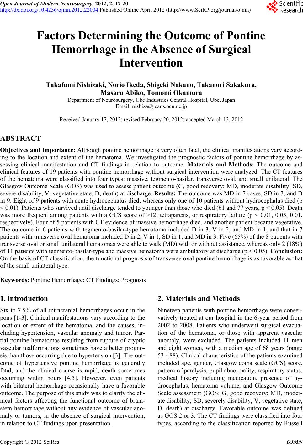
in patients with brain hemorrhage. Massive and diffuse
pontine hemorrhages are likely to be more often fatal
than those that are subependymal or focal [8]. Russell et
al. subdivided pontine hematomas into three types on the
basis of CT findings: central, tegmentobasilar and dorso-
lateral tegmental [6]. Large hematomas resulting from
systemic hypertension generally occupy the central pons,
resulting in a fatal outcome, and involve the reticular
activating system, giving rise to abrupt coma with quad-
riplegia, a decerebrate posture, or pinpoint pupils. Other
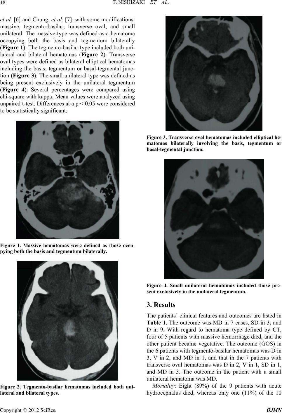
types of hematoma include partial pontine hematomas
restricted to the lateral half of the pons with sparing of
the reticular system, and these can be either tegmento-
basilar or dorsolateral tegmental.
Our present series included cases in which the CT
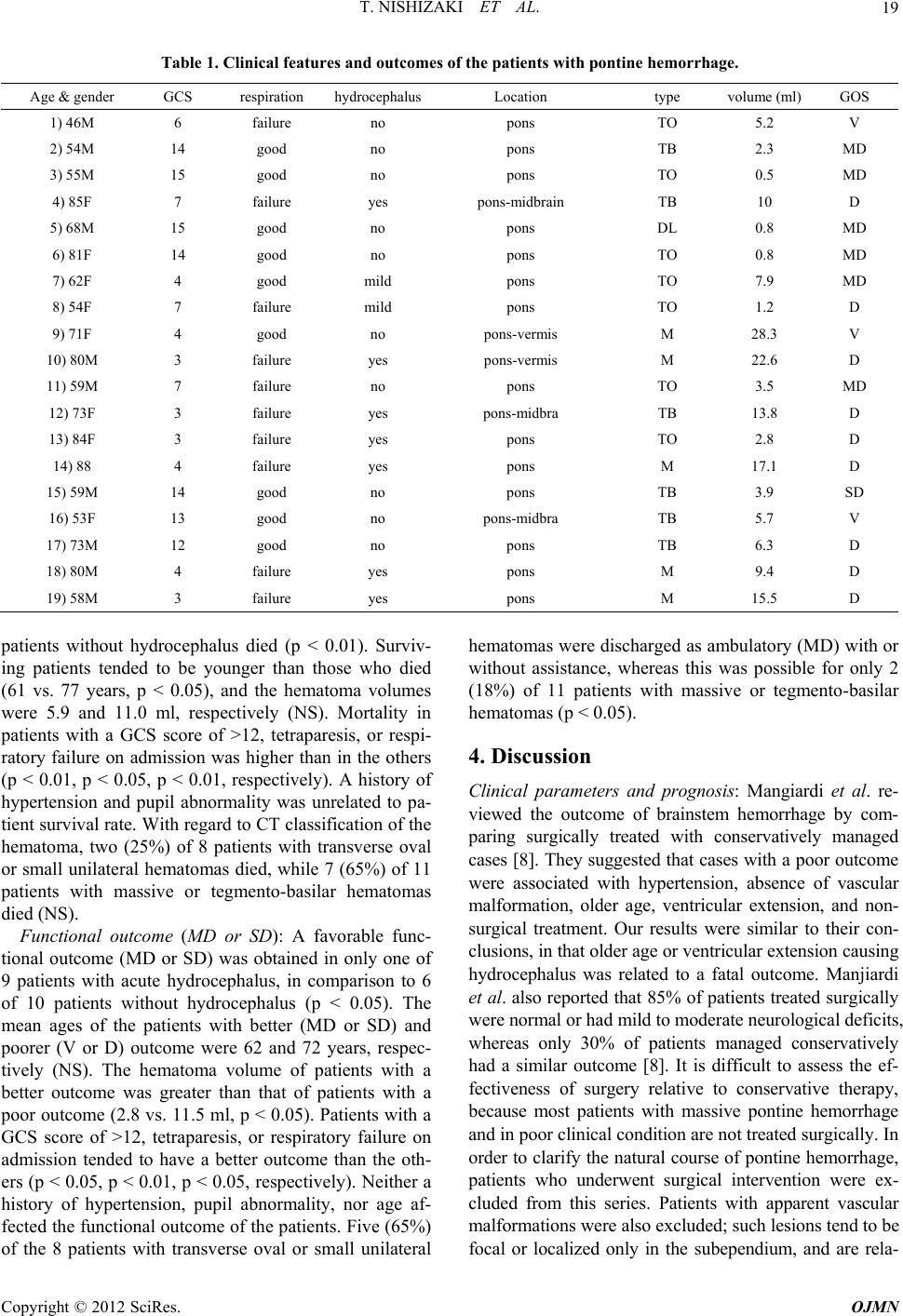
findings were difficult to classify. Transverse oval he-
matoma was defined as an elliptical hematoma with bi-
lateral involvement of the basis, tegmentum, or basal-teg-
mental junction. This category is similar to that of
Chung’s classification [7], namely that bilaterally involv-
ing the basal-tegmental junction between the basis pontis
and the tegmentum. However, in the present cases, the
hematoma sometimes involved only the basis or the teg-
mentum. Therefore, we classified such hematomas as the
transverse oval type when the basal-tegmental junction
was involved, or the basis or tegmentum alone. We ex-
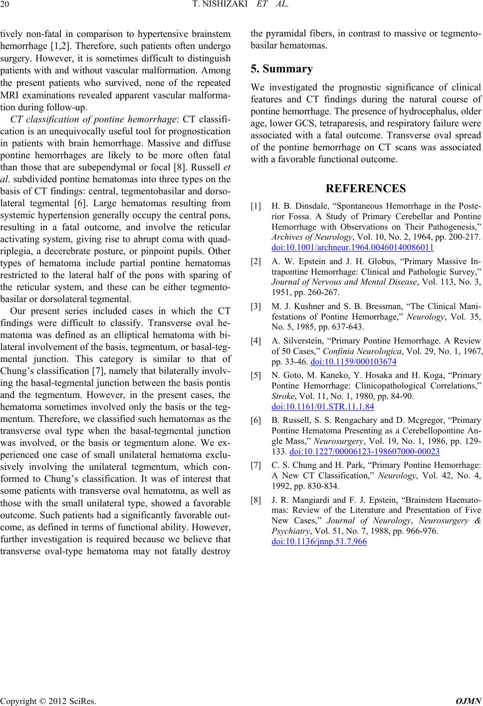
perienced one case of small unilateral hematoma exclu-
sively involving the unilateral tegmentum, which con-
formed to Chung’s classification. It was of interest that
some patients with transverse oval hematoma, as well as
those with the small unilateral type, showed a favorable
outcome. Such patients had a significantly favorable out-
come, as defined in terms of functional ability. However,
further investigation is required because we believe that
transverse oval-type hematoma may not fatally destroy
the pyramidal fibers, in contrast to massive or tegmento-
basilar hematomas.
5. Summary
We investigated the prognostic significance of clinical
features and CT findings during the natural course of
pontine hemorrhage. The presence of hydrocephalus, older
age, lower GCS, tetraparesis, and respiratory failure were
associated with a fatal outcome. Transverse oval spread
of the pontine hemorrhage on CT scans was associated
with a favorable functional outcome.
REFERENCES
[1] H. B. Dinsdale, “Spontaneous Hemorrhage in the Poste-
rior Fossa. A Study of Primary Cerebellar and Pontine
Hemorrhage with Observations on Their Pathogenesis,”
Archives of Neurology, Vol. 10, No. 2, 1964, pp. 200-217.
doi:10.1001/archneur.1964.00460140086011
[2] A. W. Epstein and J. H. Globus, “Primary Massive In-
trapontine Hemorrhage: Clinical and Pathologic Survey,”
Journal of Nervous and Mental Disease, Vol. 113, No. 3,
1951, pp. 260-267.
[3] M. J. Kushner and S. B. Bressman, “The Clinical Mani-
festations of Pontine Hemorrhage,” Neurology, Vol. 35,
No. 5, 1985, pp. 637-643.
[4] A. Silverstein, “Primary Pontine Hemorrhage. A Review
of 50 Cases,” Confinia Neurologica, Vol. 29, No. 1, 1967,
pp. 33-46. doi:10.1159/000103674
[5] N. Goto, M. Kaneko, Y. Hosaka and H. Koga, “Primary
Pontine Hemorrhage: Clinicopathological Correlations,”
Stroke, Vol. 11, No. 1, 1980, pp. 84-90.
doi:10.1161/01.STR.11.1.84
[6] B. Russell, S. S. Rengachary and D. Mcgregor, “Primary
Pontine Hematoma Presenting as a Cerebellopontine An-
gle Mass,” Neurosurgery, Vol. 19, No. 1, 1986, pp. 129-
133. doi:10.1227/00006123-198607000-00023
[7] C. S. Chung and H. Park, “Primary Pontine Hemorrhage:
A New CT Classification,” Neurology, Vol. 42, No. 4,
1992, pp. 830-834.
[8] J. R. Mangiardi and F. J. Epstein, “Brainstem Haemato-
mas: Review of the Literature and Presentation of Five
New Cases,” Journal of Neurology, Neurosurgery &
Psychiatry, Vol. 51, No. 7, 1988, pp. 966-976.
doi:10.1136/jnnp.51.7.966
Copyright © 2012 SciRes. OJMN