Materials Sciences and Applications, 2012, 3, 218-223
http://dx.doi.org/10.4236/msa.2012.34032 Published Online April 2012 (http://www.SciRP.org/journal/msa)
A Model for the Characterization and Selection of
Beeswaxes for Use as Base Substitute Tissue in Photon
Teletherapy
Rogério Matias Vidal, Divanízia do Nascimento Souza
Department of Physics, University of Sergipe, São Cristovão, Brazil.
Email: rmv.fisica@gmail.com
Received January 5th, 2012; revised February 7th, 2012; accepted March 8th, 2012
ABSTRACT
This paper presents a model for the characterization and selection of beeswaxes for use as base substitute tissue for the
manufacture of objects suitable for external radiotherapy using megavoltage photon beams. The model of characteriza-
tion was divided into three distinct stages: 1) verification of aspects related to the origin of the beeswax, the bee species,
the flora in the vicinity of the beehives and procedures to detect adulterations; 2) evaluation of physical and chemical
properties; and 3) evaluation of beam attenuation capacity. The chemical composition of the beeswax evaluated in this
study was similar to other simulators commonly used in radiotherapy. The behavior of the mass attenuation coefficient
in the radiotherapy energy range was comparable to other simulators. The proposed model is efficient and enables con-
venient assessment of the use of any particular beeswax as a base substitute tissue for radiotherapy.
Keywords: Beeswaxes; Characterization; Model; Radiotherapy
1. Introduction
Radiotherapy is a very effective tool in cancer treatment,
and relies on advanced equipment and computerized
treatment planning systems (TPS). Computerized TPS
accurately simulate the projection of the radiation field,
enabling deliverance of sufficient radiation to a tumor,
while sparing critical organs and minimizing the radia-
tion dose to healthy tissues. However, the relationship
between the dose delivered by the device and that re-
ceived by the patient cannot be easily determined [1]. To
address this issue, methods to reproduce actual clinical
treatments, by substituting the patient with an experi-
mental device that has physicochemical properties which
are similar to human tissue, must be developed. Unfor-
tunately, the equipment and accessories currently avail-
able for this purpose have differing degrees of complex-
ity and, although no similar domestically produced equip-
ment is available, all are highly taxed by the Brazilian
government, which has no specific tax legislation for
importing such equipment [2]. Then, experimentation
with alternative products which can be used with this
equipment is very important. In particular, the production
of low-cost simulators and bolus will give to small ra-
diotherapy centers the ability to investigate and develop
new treatment techniques and improve quality control. In
radiotherapy, materials that simulate human tissues are
commonly called simulators or substitute tissues. How-
ever, according to White [3], there is no simple chemical
substance capable of mimicking the atomic composition
of human tissues. Although the desired composition can
usually be obtained in aqueous mixtures or gels, these
mixtures are inconvenient, because of their toxicity and
lack of availability. To create alternative tissue substi-
tutes, an appropriate base material must be chosen, which
has scattering and attenuation properties that are similar
to the tissue to be simulated [4]. In a second stage, addi-
tional substances are added to correct any deficiencies in
the base material [5]. The official document that regu-
lates the procedures for tissue substitute characterization
is the REPORT 44 of the International Commission on
Radiation Units and Measurements (ICRU) [6]. This
document discusses the coefficients that must be ob-
tained and the quantities that should be considered during
the characterization process. REPORT 44 recommends
that in the process of evaluation and characterization of a
simulator material the following criteria should be evalu-
ated.
(Composition/purity) The elemental composition of
the material must be known to the degree of accuracy
required for the desired application; contaminants should
be avoided, especially those whose atomic number is
greater than 20 (Z > 20).
Copyright © 2012 SciRes. MSA
A Model for the Characterization and Selection of Beeswaxes for Use as Base Substitute Tissue in Photon Teletherapy 219
(Homogeneity) Inhomogeneities, due to poor compo-
nent dispersion and porosity must not introduce uncer-
tainties greater than 1% in transmission, or in the estima-
tion of radiation doses.
(Stability) The base material should be inert and
should not degrade upon repeated exposure to radiation.
(Shape) Substitute tissues should be pliable, and capa-
ble of being molded into the desired shape.
In this way, this paper proposes a model for the char-
acterization of beeswax, according to the above criteria,
for use as a substitute base tissue, to enable their use in
the manufacture of objects suitable for radiotherapy with
megavoltage photons beams.
1.1. Beeswaxes
Beeswax is a physiological product produced by bees in
their beehive. To produce beeswax, bees swallow and
digest honey, converting the food into fat; 24 hours after
honey ingestion the bees are capable of producing bees-
wax [7]. Four pairs of wax glands are located in the ven-
tral abdomen of bees and are projected side-by-side in
the last abdominal segments. The wax is expelled from
these glands in liquid form, and solidifies only when in
contact with the environmental temperature. The bees
produce wax for the construction of honeycombs and
operculation, the process by which bees close the alveoli
(the brood and honey) with a thin layer of wax. Opercu-
lum beeswax is of better quality with fewer impurities [7].
Beeswax has already been used as bolus with satisfactory
results [8,9]. In addition to being a natural product, the
market value of a kilogram of beeswax is much less than
that of other types of materials used for this purpose,
such as acrylic and polyethylene [10]. These features
make the beeswax a great alternative material to replace
the usual soft tissue substitutes. However, it is common
practice to stochastically modify their original composi-
tion [10]. In addition, other important factors [11], such
as the environmental conditions present during beeswax
production, the geographic location of the beehive, and
the bee species which produced the wax, can alter the
composition of the resulting beeswax base substitute tis-
sue. Because even small changes in the chemical struc-
ture of the wax can promote undesirable uncertainties in
radiotherapy measurements, a convenient and reliable
method for the assessment of beeswax material as a base
tissue substitute is needed. Thus, in the present study, we
propose a method to characterize beeswaxes for use as
base substitute tissue for the manufacture of objects
suitable for external radiotherapy.
1.2. Model Characterization Structure
Beeswax characterization was divided into three distinct
stages: 1) study of aspects related to the origin of the
beeswax; 2) assessment of physical and chemical proper-
ties; and 3) evaluation of beam attenuation capacity. Dur-
ing first stage (preliminary characterization stage), some
chemical properties were assessed to verify the absence
of adulterating agents. In the second stage, physical and
chemical properties were evaluated, such as moldability,
and the percentage of constituent elements. Finally, in
the third stage, practical linear and mass attenuation co-
efficients were determined. Results from these three stages
were related to the origin of the beeswax, the bee species
which produced the beeswax, the flora in the vicinity of
the beehive, and the extraction form.
2. Materials and Methods
2.1. Preliminary Characterization
The choice of beeswaxes used in the present study was
based on a published characterization of honey bee flora
from the semi-arid Brazilian state of Paraiba, performed
by Silva and Aquino [12]. Official methods for analysis
of fats and oils adopted by Brazil, the United States, the
United Kingdom and Spain were used, with minor modi-
fications, to determine the purity of the beeswax for these
experiments [13-20]. The following values were obtained:
density, saponification, acid, esters, iodine absorption,
peroxide, ash content, and melting point. Obtained re-
sults were then compared with parameters described in
the literature for pure unadulterated beeswax [10,21].
2.2. Physical and Chemical Characteristics
(Shape) The physical properties of the beeswax were
evaluated as a function of environmental temperature,
using a temperature-controlled oven. Evaluations were
performed by palpation and observation.
(Degradation by Repeated Irradiations) To evaluate
the degree of degradation associated with repeated irra-
diations, beeswax cubes (4 cm on edge) were irradiated
with a dose of 50,000 Gy using a Gammacell 220 MDS
Nordion model 220E GC No 65 R (ASSY), with a 60Co
source.
(Density) The density of the wax was measured by the
pycnometer method. At room temperature, the mass of
the empty pycnometer, m1, was measured using a digital
scale. Then, the mass of the pycnometer filled with dis-
tilled water, m2, was measured. Finally, pieces of wax
were introduced into the water filled pycnometer, and the
mass, m3, was determined. The relative density was then
calculated by the expression,
water
,
b
eeswax
.
wb
m
m
(1)
where:
water2 1,mmm
(2)
Copyright © 2012 SciRes. MSA
A Model for the Characterization and Selection of Beeswaxes for Use as Base Substitute Tissue in Photon Teletherapy
220
b
eeswax2 3.mmm (3)
The density of distilled water is .
3
water 1g/cm
Therefore, the absolute density of beeswax in g/cm3
can be calculated by,
b
w
(4)
(Composition) The chemical compositions of the bees-
waxes used in this work were characterized using three
techniques: CHN elemental analysis; energy dispersive
microanalysis (using a scanning electron microscope);
and atomic emission spectroscopy (by inductively cou-
pled plasma).
2.3. Beam Attenuation Capacity
(Practical Linear Attenuation Coefficient) REPORT 44
asserts that tissue substitutes will attenuate X-rays simi-
larly to human tissues, only if the variation of the total
attenuation coefficient as a function of incident photon
energy is identical to the tissue being simulated [6]. To
obtain linear attenuation coefficients, beeswax blocks (25
× 25 cm2, with various thicknesses) were prepared and
greased with graphite, which were then irradiated in a
SIEMENS linear accelerator (PRIMUS Mid Energy
model with a 6 MV beam energy). Measurements were
conducted using a PTW 31011 ionization chamber con-
nected to a PTW UNIDOS E electrometer as illustrated
in Figure 1.
To obtain the practical linear attenuation coefficient, the
approximation method was used [22]. Beeswax blocks
were placed at a distance of 100 cm from the head of the
machine, in order to attenuate the beam. The blocks were
placed separately in random configurations. Many meas-
urements were made with different field sizes.
The electrometer readings were introduced as I and I0
in the equation,
0
x
I
Ie
 (5)
Figure 1. Illustration of the correct positioning of a board
on the treatment table of the accelerator aligned with the
ionization chamber.
Because the thickness of the plates is known (obtained
using a micrometer), it is possible to determine the total
linear attenuation coefficients for various field sizes on
the surface of the blocks. Field sizes of 12 × 12, 10 × 10,
6 × 6, 5 × 5, 4 × 4 and 3 × 3 cm2 were tested. From the 3
× 3 cm2 field, an approximation of a Gaussian function
was used to estimate the linear attenuation coefficient for
a 0 × 0 cm2 field, by referring to the thin beam with the
same half-value layer as the 6 MV beam. Measurements
with field less than 3 × 3 cm2 were not performed, due to
the size of the cavity of the ionization chamber used.
(Practical Mass Attenuation Coefficient) The value
obtained for the linear attenuation coefficient μ is related
to the density of the beeswax used in the attenuation
process using the equation,

.
i
c
iw i

(6)
where,
c
is the mass attenuation coefficient for
the compound,
i
is the mass attenuation coeffi-
cient for each individual element and wi, is the fractional
weight of the elements in the compound. The value ob-
tained from that operation was compared with values
obtained by Hubbell and Seltzer [23], in order to deter-
mine the effective energy of the 6 MV polyenergetic
beam, and consequently, the practical simulation power
of the beeswax. These data relate the mass attenuation
coefficients for different elements and compounds with
the effective energy of the incident photon beam on the
material.
(Theoretical Mass Attenuation Coefficient) After ob-
taining the percentage of the chemical elements present
in the beeswaxes, the program Xcom [24] enables com-
parisons of the behavior of the mass attenuation coeffi-
cient with respect to other simulators and body tissues.
3. Results and Discussion
3.1. Preliminary Characterization
Bees commonly found in Brazil are a hybrid of European
honeybees: Apis mellifera mellifera, Apis mellifera ligus-
tica, Apis mellifera caucasica and Apis mellifera carniça
with the African bee Apis mellifera scutellata [10]. In
this work, beeswax obtained from bees similar to Apis
mellifera scutellata was evaluated.
We collected three quantities of beeswax during the
off-season of honey production in 2010. The first extrac-
tion (extraction A) was conducted in February from old
combs, during flowering (October-December 2009) of
Zizipos joazeiro. The second extraction (extraction B)
was conducted in March from the operculum, during
flowering (January-February 2010) of Croton sonderi-
anus. The third extraction (extraction C) was conducted
in July 2010 (also from operculum), during flowering
(March-May) of Prosopis juliflora [12]. Hereafter, bees-
Copyright © 2012 SciRes. MSA
A Model for the Characterization and Selection of Beeswaxes for Use as Base Substitute Tissue in Photon Teletherapy 221
waxes obtained by these extractions will be referred to as
beeswax A, B and C, respectively. For all samples, ex-
traction was performed by melting and filtering through
filtration screens. Contaminating agents were not de-
tected. Table 1 below shows the values measured for our
experimental beeswax samples and parameters for pure
beeswax.
3.2. Physical and Chemical Characteristics
(Shape) The document, TEC DOC1151 [25], recom-
mends that each institution have the means to ensure that
the quality of radiotherapy services offered remain within
internationally accepted limits, and the ability to correct
deviations that may lead to treatment errors.
The quality of a photon beam depends on its power of
penetration [26,27]. As outlined in TEC DOC1151 [25],
certain tests must be performed monthly to ensure beam
quality constancy for the percentage depth dose (PDD
20.10) or tissue phantom ratio (TPR 20.10), and daily to
ensure the constancy of the reference dose. All of these
measures help monitor and improve the dose of radiation
delivered to the patient, but to be performed, require spe-
cific accessories, such as simulators [28].
For this purpose, specialized companies manufacture
blocks of solid water with suitable holes for accommoda-
tion of dosimeters. The beeswaxes tested in this work
were solid at room temperature and not flexible, which is
advantageous for forming the necessary blocks. Waxes
from the operculum (types B and C) were found to be
more pliable, a characteristic that also facilitated the re-
moval of blocks from the form. In addition, Type A
beeswax (from old combs) was stickier than Types B and
C, which caused difficulties in handling.
For use as a bolus, the solidity displayed by beeswax
types A, B and C at room temperature was an obstacle,
because for this purpose greater flexibility is required,
enabling creation of custom molds of the surface and
patients contours. However, at temperatures above 39˚C,
the beeswax samples displayed greater capacity for plas-
tic deformation. Although healthy skin supports the ap-
plication of wax at this temperature, the skin of patients
undergoing treatment is already very sensitive, prevent-
ing their application as a bolus.
(Degradation due to Repeated Irradiation) Type A, B
and C beeswaxes did not display any changes after irra-
diation with 50,000 Gy. This dose corresponds to at least
25,000 radiotherapy sessions, supporting the feasibility
of using beeswax as a base tissue substitute.
(Density) The density values obtained are shown in
Table 1. Density values for beeswaxes A, B and C are
close to the density value of water 1 g/cm3, which is the
reference material normally used in dosimetric simula-
tions with megavoltage beams in radiotherapy.
(Chemical Composition) Table 2 shows results from
chemical composition analyses obtained for each type of
beeswax versus other materials used as base tissue sub-
stitutes.
Carbon, hydrogen, oxygen and nitrogen were found to
be in higher concentrations in beeswax versus the other
substitute materials. Substitute materials that most re-
semble beeswax in terms of chemical composition are
Mix D wax, paraffin and Temex, based on the quantities
of carbon and hydrogen, respectively. The low amount of
oxygen in the beeswax samples does not preclude the use
of beeswaxes as a tissue substitute, because various sub-
stitutes, such as Temex and Mix D wax, also have low
Table 1. Values measured for beeswaxes samples and pa-
rameters for pure beeswax.
Beeswax
Method A B C
Parameter
Density (g/mL) 0.94 0.923 0.923 0.92 - 0.947
Saponification (mg KOH/g)89.8 84.5 84.7 83 - 103
Acidity (mg KOH/g) 23.2 18.3 18.3 17 - 24
Esters (mg KOH/g) 81.6 74.9 74 66 - 82
Melting Point (˚C) 65.9 65.2 65 61 - 66
Iodine Absorption(g I/100 g)9.2 8.2 8.3 7.6 - 10.6
Peroxide (meq O/kg) 0.01 0.00 0.00 -
Ash Content (%) 0.035 0.032 0.036 0.000 - 0.055
Table 2. Chemical composition (percentages) obtained for
each type of beeswax compared to other materials used as
simulators.
Beeswax Other simulators
Elements A B C Water Mix DTemex
H 1 12.912.312.1 11.2 13.49.6
C 6 80.780.080.0 - 77.787
N 7 1.381.361.32 - - 0.06
O 8 2 1.991.98 88.8 3.5 0.47
Na 11<3 <3 <3 - - -
Mg 12- - - - 3.8 -
Si 14<3 <3 <3 - - -
S 16<1 <1 <1 - - 1.53
Ca 20<3 <3 <3 - - -
Ti 22- - - - 1.440.33
Fe 26<1 <1 <1 - - -
Zn 30<1 <1 <1 - - 0.45
Copyright © 2012 SciRes. MSA
A Model for the Characterization and Selection of Beeswaxes for Use as Base Substitute Tissue in Photon Teletherapy
222
concentrations of this element. The presence of zinc,
calcium and iron in the beeswax formulation can com-
pensate for the oxygen deficiency; similarly, titanium
and zinc compensates for oxygen deficiency in Temex
and Mix D wax.
3.3. Beam Attenuation Capacity
(Linear and Mass Attenuation Coefficient) Figure 2
shows a graph of the average values of linear attenuation
coefficient versus field size obtained from the apparatus
shown in Figure 1 for beeswax type B. From the 3 × 3
cm2 field, an approximation of a Gaussian function was
used to estimate the linear attenuation coefficient for a 0
× 0 cm2 field.
Figure 3 shows the variation of effective energy with
the mass attenuation coefficient obtained from the Hub-
bell and Seltzer data [23] for beeswax type B.
The triangle point in Figure 3 corresponds to the mass
attenuation coefficient value obtained during the experi-
ment. The values of attenuation coefficient and effective
energy for the other beeswaxes are presented in Table 3.
The values shown in Table 3 are in accordance with
those obtained by Robinson and Scrimger [22] for the
monoenergetic approximation of a polienergetic 6 MV
beam, demonstrating that the beeswax analyzed attenu-
ates the beam properly and does not require significant
chemical corrections to be employed as base substitute
tissue.
(Behavior of the theoretical mass attenuation coeffi-
cient) Figures 4 and 5 show variations in the mass at-
tenuation coefficients of beeswax type B with respect to
other simulators and main body tissues.
In the therapeutic energy range (i.e. energies above 1
MeV—right detail), all materials described in the graphs
(Figures 4 and 5) show similar behaviors. Type A and C
beeswax also behaved similarly (not shown). In right
detail (Figure 5) close to the 1 MeV energy the mass
attenuation coefficient curve for bone (triangle points)
slightly downward. This relatively small effect is attributed
to the difference of their chemical composition.
In this way, the necessary high accuracy demanded by
·
·
·
·
·
Field size (cm × cm)
Figure 2. Linear attenuation coefficient versus field size.
·
·
·
·
·
·
·
·
·
· ·· · ·
Figure 3. Mass attenuation coefficient versus field size for
type B beeswax.
.
.
.
.
.
.
..
.
.
Figure 4. Comparison of the behavior of the mass attenua-
tion coefficients (obtained using the program Xcom) of
beeswax B with other simulators.
.
.
.
.
.
.
..
.
.
Figure 5. Comparison of the behavior of the mass attenua-
tion coefficient (obtained using the program Xcom) of
beeswax B with main body tissues.
radiotherapy reference dosimetry may be achieved using
types A, B and C beeswaxes in the manufacturing of ob-
jects suitable for teletherapy with megavoltage photons
beams as bolus and standard phantoms.
Copyright © 2012 SciRes. MSA
A Model for the Characterization and Selection of Beeswaxes for Use as Base Substitute Tissue in Photon Teletherapy
Copyright © 2012 SciRes. MSA
223
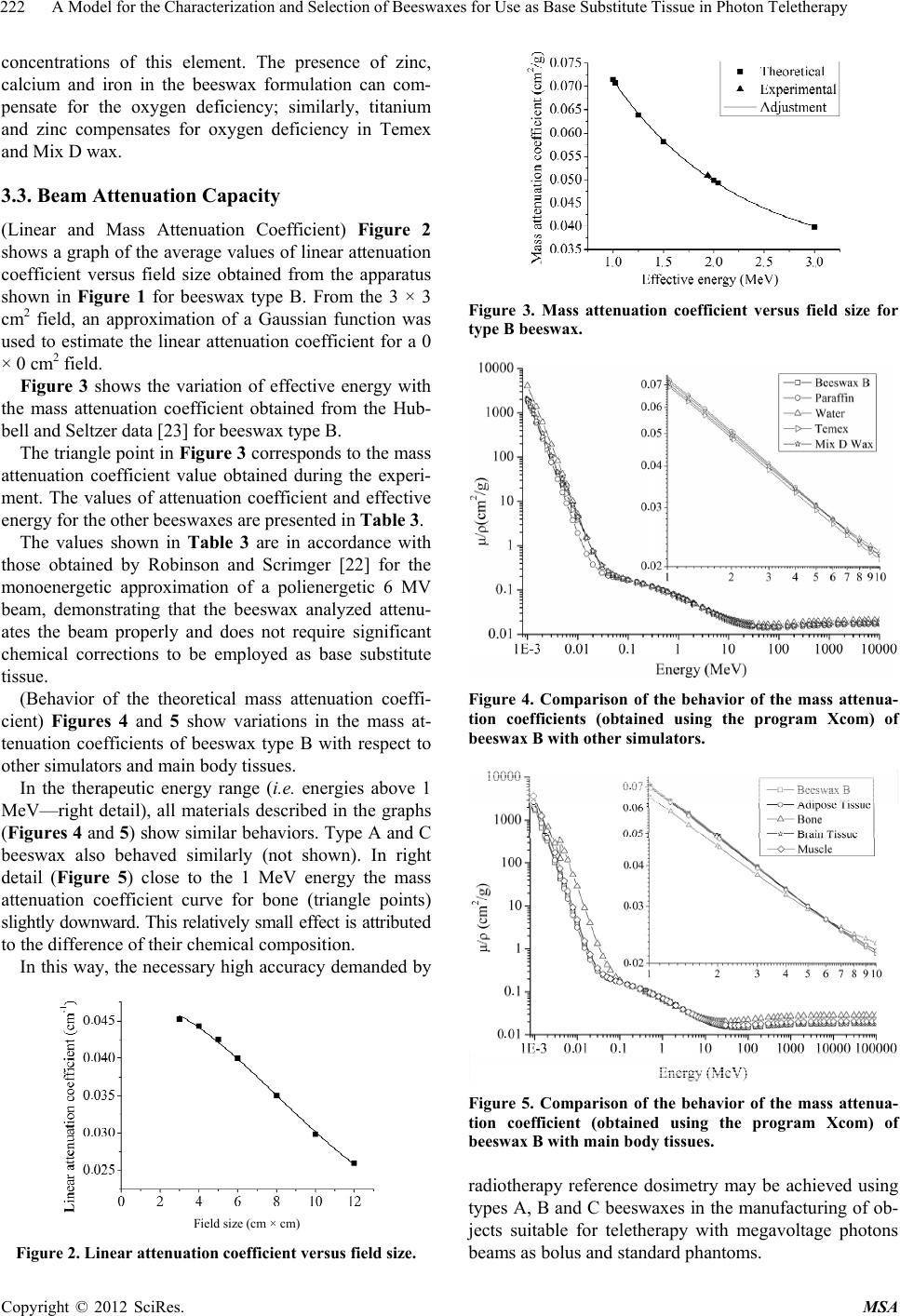
Table 3. Attenuation coefficients and effective energy.
Beeswax
Linear
attenuation
coefficient μ
Mass
attenuation
coefficient μ/ρ
Effective
energy
6 MV Eeff
Type cm–1 cm2/g MeV
A 0.0489 ± 0.0010 0.0520 ± 0.0003 1.93 ± 0.02
B 0.0479 ± 0.0054 0.0520 ± 0.0005 1.94 ± 0.03
C 0.0471 ± 0.0031 0.0510 ± 0.0002 1.89 ± 0.03
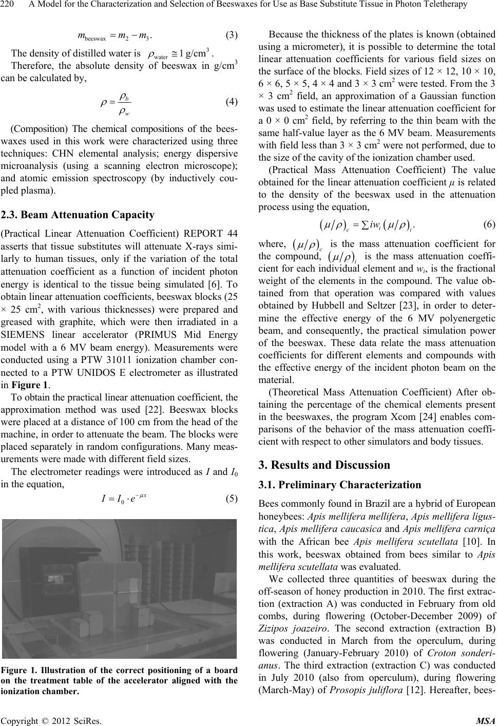
4. Conclusion
The analysis model proposed in this work is efficient and
confirms that beeswaxes represents an excellent option
for a base tissue substitute in external radiotherapy with
megavoltage photon beams. The model presented here is
convenient, and show than beeswaxes can dramatically
reduce the costs of products related to this type of appli-
cation. The major advantage of beeswax is its intrinsic
attenuation properties, which do not require significant
chemical correction.
REFERENCES
[1] E. B. Podgorsak, “Radiation Oncology Physics: A Hand-
book for Teachers and Students,” International/Atomic
Energy Agency, 1957, p. 657.
[2] “Brazilian Radiotherapy Society,” 2011.
http://www.sbradioterapia.com.br
[3] D. R. White, “Tissue Substitutes in Experimental Radia-
tion Physics,” Medical Physics, Vol. 5, No. 6, 1978, pp.
467-479. doi:10.1118/1.594456
[4] D. R. White, “The Formulation of Tissue Substitute Ma-
terials Using Basic Interaction Data,” Physics in Medicine
and Biology, Vol. 22, No. 5, 1975, pp. 22-25.
[5] D. R. White and C. Constatinou, “Anthropomorphic Phan-
tom Materials. The Progress in Medical Radiation Phys-
ics,” Plenum Press, New York, 1982, pp. 133-193.
[6] International Commission on Radiation Units and Meas-
urements, “Tissue Substitutes in Radiation Dosimetry and
Measurement,” Report. 44, 1989.
[7] R. Zovaro, “Ceras de Abelhas: Produção e Beneficia-
mento,” AEP, Vol. 1, 2007, p. 23.
[8] D. Scheidegger, “Bólus e Filtros em Cunha Feitos Com
Cera de Abelha Para Utilização em Radioterapia Com
60Co,” Publicações, CEFET, 2003.
[9] M. Tobler, “Design and Production of Wax Compensa-
tors for Electron Treatments of the Chest Wall,” Medical
Dosimetry, Vol. 21, No. 4, 2006, pp. 199-206.
doi:10.1016/S0958-3947(96)00126-4
[10] “Embrapa Meio-Norte,” 2011.
http://www.embrapa.mn.gov.br
[11] J. L. Bernal, “Physico-Chemical Parameters for the Char-
acterization of Pure Beeswax and Detection of Adultera-
tions,” European Journal of Lipid Science and Technology,
Vol. 107, No. 3, 2005, pp. 158-166.
doi:10.1002/ejlt.200401105
[12] A. R. Silva, “Characterization of the Honey Bee Flora in
the Semi-Arid of Brazilian State of Paraiba,” Archivos de
Zootecnia, Vol. 220, No. 4, 2000, pp. 427-438.
[13] Ministério da Agricultura, Pecuária e Abastecimento.
Regulamentos Técnicos de Identidade e Qualidade de
Produtos Apícolas, “Instrução Normativa No. 3, Sistema
de Consulta a Legislação—SISLEG,” 2001.
[14] United States Pharmacopoeial Convention, National Pub-
lishing, Rockville, 2000.
[15] British Prarmacopoeia Commission Secretariat, HMSO,
London, 1993.
[16] Real Farmacopea Española, “Ministerio de Sanidad y
Consumo,” 1997.
[17] R. N. Rangel, “Práticas de Físico-Química,” Blucher,
Erkrath, 2006.
[18] L. Casares, “Tratado de Analisis Químico,” Toledo, 1997.
[19] E. Moretto, “Óleos e Gorduras Vegetais: Processamento e
Análises,” UFSC, 1989.
[20] Ministério da Saúde, “Farmacopeia Brasileira,” Atheneu,
1988.
[21] J. Serra, “Características Físico-Químicas de la Cera de
Abejas Producida en España,” Alimentación, Equipos y
Tecnologia, Vol. 8, No. 3, 1989, pp. 213-216.
[22] D. M. Robinson and J. W. Scrimger, “Monoenergetic
Approximation of a Polyenergetic Beam: A Theorical
Approach,” The British Journal of Radiology, Vol. 64,
No. 761, 1991, pp. 452-454.
doi:10.1259/0007-1285-64-761-452
[23] J. H. Hubbel and S. M. Seltzer, “Tables of X-Ray Mass
Attenuation Coefficients and Mass Energy-Absorption
Coefficients 1 keV to 20 MeV for Elements Z = 1 to 92
and 48 Additional Substances of Dosimetric Interest,”
NISTIR 5632, 1995.
[24] M. J. Berger, J. H. Hubbell, S. M. Seltzer, J. S. Coursey,
and D. S. Zucker, “XCOM: Photon Cross Section Data-
base (Version 1.2),” National Institute of Standards and
Technology, Gaithersburg, 1999.
[25] IAEA, “TECDOC 1151—Aspectos Físicos de La Garantía
de Calidad en Radioterapia: Protocolo de Control de
Calidad,” International Atomic Energy Agency, Vienna,
2000.
[26] E. J. Turner, “Atoms, Radiation, and Radiation Protec-
tion,” Willey, New York, 2007.
doi:10.1002/9783527616978
[27] F. Khan, “The Physics of Radiation Therapy,” Williams
and Wilkins, Philadelphia, 1994.
[28] AIEA, “Absorbed Dose Determination in External Beam
Radiotherapy: An International Code of Practice for Do-
simetry Based on Standards of Absorbed Dose to Water,”
Technical Report Series—398, 2000.