Vol.2, No.5, 412-417 (2010)
doi:10.4236/health.2010.25062
Copyright © 2010 SciRes. Openly accessible at http://www.scirp.org/journal/HEALTH/
Health
DNA damage and cell death assessment in patients with
severe multiple trauma using comet assay
Aliy K. Zhanataev1*, Victor V. Moroz2, Andrey D. Durnev1, Maria Yu. Muravyeva2,
Vasiliy I. Reshetnyak2
1Science Research Institute of Pharmacology, Russian Academy of Medical Sciences, Moscow, Russia; *Corresponding Author:
azhanataev@yandex.ru
2Science Research Institute of General Reanimatology, Russian Academy of Medical Sciences, Moscow, Russia;
v_reshetnyak@yahoo.com
Received 9 December 2009; revised 20 February 2010; accepted 23 February 2010.
ABSTRACT
Purpose: To determine the DNA strand breaks,
oxidative DNA damage and cell death in blood
and plasma total antioxidant status (TAOS) in 22
patients with severe multiple trauma. Materials
and methods: The DNA comet assay was used
to measure DNA strand breakage, 8-oxoguanine
levels and apoptotic and necrotic nuclei in after
admission (day 0) and on days 3, 5, 7 and 15.
TAOS was determined by colorimetric method.
Results: Trauma patients had high DNA damage
at admission (p < 0.01), that further increased
with maximum value on day 5 (p < 0.001). On
day 15 the degree of DNA damage remained
significantly elevated (p < 0.01). No significant
difference in the 8-oxoguanine levels at all days
examined was found. Patients had a high per-
centage of apoptotic and necrotic comets at
admission, with maximum values on days 3 and
5. A significantly lower TAOS was observed in
patients on admission and days 3, 5, 7 and 15 (p
< 0.001 in all cases). A decreasing of TAOS on
days 7 and 15 compared to that on admission (p
< 0.05) was observed. Conclusions: Blood cells
from severe trauma patients’ display increased
DNA damage associated with apoptosis and
necrosis. Reduced plasma TAOS and a ten-
dency to increase of 8-oxoguanine in DNA was
observed.
Keywords: Apoptosis; Necrosis; Multiple Trauma;
DNA-Comet Assay; 8-Oxoguanine;
Total Antioxidant Status
1. INTRODUCTION
Severe multiple trauma remains as one of the major
problems of contemporary medicine and society throu-
ghout the world. Advances in critical care of the trauma
patient have resulted in improved outcome, but despite
these efforts, up to half of the patients with traumatic
injury die or are left with severe disability [1-3].
Multiple organ failure (MOF) is the major complica-
tion after sever multiple trauma [4-7]. Experimental
evidences suggest that MOF in the event of trauma
might be relates to stress-induced cell death by apoptosis,
but its exact mechanisms is not fully understood [8-12].
DNA damage including oxidative DNA bases modifica-
tion is one of the intrinsic signals initiating apoptosis
[13]. At present, data about DNA damage in critical ill-
ness are lacking. The Single Cell Gel Electrophoresis
assay (SCGE), or comet assay are sensitive method for
the evaluation of DNA damage from individual cells
based on the migration of denatured DNA through an
electrophoretic field [14]. The aim of this pilot study was
an assessment DNA strand breakage, 8-oxoguanine lev-
els and cellular death of white blood cells and their pos-
sible connection with total antioxidant status in patients
with severe multiple trauma.
2. MATERIALS AND METHODS
The study covered 22 patients who were admitted with
severe multiple traumas (SMT) (Table 1). The study
protocol was approved by ethics committee and in-
formed consent was obtained from all patients or their
relatives. Inclusion criteria were age older than 18 years,
admission to the trauma intensive care unit and an Acute
Physiology and Chronic Health Evaluation (APACHE II)
score higher than 12. Patients with traumatic brain injury
were excluded. The age ranged from 21 to 68 years.
Body weight ranged from 50 to 95 kg. Clinical charac-
teristics of the patients with severe multiple trauma on
admission is presented to Table 2. The length of stay in
the unit ranged from 3 to 32 days, with a mean of 10.5 ±
A. K. Zhanataev et al. / HEALTH 2 (2010) 412-417
Copyright © 2010 SciRes. http://www.scirp.org/journal/HEALTH/Openly accessible at
413
413
5.1 days. The patient’s condition on admission was an
APACHE II of 19.1 ± 5.4, blood loss 22-45 ml/kg. Blood
loss was calculated using indirect method (vital signs,
serial hematocrits measurement) and by direct intraop-
erative estimation of blood loss. Optimization of hemo-
dynamic (systolic blood pressure > 90 mm Hg) and
oxygenation (arterial oxygen saturation > 90%) was
reached during the first hours after the admission in the
intensive care unit. Single organ failure (SOF) occurred
in 12 patients (54.5%) and multiple organ failure oc-
curred in 6 patients (27.3%). All SOF was caused by
respiratory failure. Respiratory failure occurred first in
the majority of patients with multiple organ failure, fol-
lowed usually by cardiovascular insufficiency. The mor-
tality level was 23% (5 patients). Blood was collected
after admission (day 0) within 4 hours (n0 = 22), and on
days 3 (n3 = 22), 5 (n5 = 13), 7 (n7 = 11) and 15 (n15 = 5)
of admission. Blood samples (2 ml) were taken into
EDTA tubes, mixed (1:1) with RPMI-1640 medium
containing 20% DMSO as cryoprotectant and immedi-
ately frozen to –20℃. Samples stored until analysis no
more 20 days avoiding repeated freeze-thaw cycles.
Twelve subjects without acute or chronic disease were
used as control group (Table 1). Blood samples from
controls handled and stored using identical procedures.
2.1. Single Cell Gel Electrophoresis Assay
DNA damage was evaluated using alkaline comet assay
[15]. Briefly, after quick (within 1-2 min) thawing in a
water-bath at 37, 50 μl of whole blood samples were
mixed with 500 μl of 1% melted agarose (low melting
point) and layered (70 μl) onto a microscopy slide. The
slides with the agarose-embedded cells were subjected to
a lysis step (1 h at 40C in 1% N-lauroylsarcosine, 2.5M
NaCl, 100 mM Na2EDTA, 1% Triton X-100, 10%
DMSO, pH 10.0). After the lysis step slides were placed
in an ice-cold electrophoresis chamber containing alka-
line electrophoresis solution (300 mM NaOH, 1mM
Na2EDTA, pH > 13.0) for 20 min to allow DNA un-
winding. The electrophoresis was subsequently con-
ducted for 20 min at 1 V/cm and ~300 mA. At the end of
the electrophoresis the slides were fixed in 70% ethanol,
air-dried and stored in the dark at room temperature until
scored. Just prior to scoring, slides were stained with
SYBR Green I (1:10000 in TE-buffer, pH 7.4) for 20
min. Microscopical analysis was carried out at × 200
magnification using MIKMED-2 microscope (LOMO,
Russia) provided with epifluorescence and equipped
with a FITC filters. Images of 100 randomly selected
comets were captured using CCD camera (VEC-335,
EVS, Russia) and analyzed with CASP 1.1.2 image ana-
lyzer software [16] to evaluate %DNA in tail (the per-
centage of total fluorescence migrated in the tail for each
nucleus) used as a measure of DNA damage. In parallel
analysis levels of apoptotic and necrotic comets was
estimated. Comet assay in the standard alkaline version
Table 1. Demographic data of the investigated groups.
Parameter Trauma patients
(n = 22)
Healthy subjects
(n = 12)
M/F, n (%) 14 (63,6%)/8 (36,4%) 5 (41,7%)/7 (58,3%)
Age, years 41.9 ± 13.3 37.9 ± 9.6
Weight, kg 75.7 ± 6.4 67.8 ± 7.5
Motor vehicle accident18 (81,8%) -
Fall 2 (9,1%) -
Other mechanism of
injury 2 (9,1%) -
Blunt trauma 16 (72,7%) -
Penetrating trauma 6 (27,3%) -
has a fragment size resolution of 10-100 kb and early to
middle chromatin fragmentation during apoptosis can be
monitored by the technique [17]. Thus, DNA-comets of
apoptotic cells on slides clearly differ from damaged or
undamaged cells by forming comet- like structures with
spread tail and small almost invisible heads (Figure 1(b))
[14,17]. Necrotic cells displayed a characteristic view
with large nuclear remnants which reflected random
DNA degradation (Figure 1(c)).
2.2. Detection of 8-Oxoguanine and TAOS
Human 8-oxoguanine DNA Glycosylase (hOGG1)
FLARE Assay Kit (R&D Systems, Minneapolis, USA)
was used to evaluate oxidative DNA damage in blood
cells. This test uses the hOGG1 enzyme, a glycosylase
that recognizes and specifically cuts the oxidized bases
principally 8-oxoguanine (8-oxoG) from DNA, produc-
ing apurinic sites converted in breaks, which can be de-
tected by comet assay. The comet assay was carried out
as described above, with the exception that after lysis of
cells the slides were washed three times for 15 min with
FLARE™ buffer. After this time samples were incubated
with 100 μl of hOGG1 enzyme (1:400 in REC dilution
buffer) or with 100 μl of REC dilution buffer only.
Slides incubated in humidity chamber at 37 for 1 h.
DNA unwinding, electrophoresis, microscopic analysis
and comet scoring were then completed as described
above. For each blood sample the mean values of the
%DNA in tail for 100 comets from hOGG1-treated cells
(%DNAenz) and hOGG1-untreated cells (%DNAbuf)
was calculated. %DNAenz/%DNAbuf ratio (in a.u.) was
used as measure of 8-oxoG content in DNA of blood cells.
Among the methodologies used to evaluate TAOS, the
most widely used colorimetric method are 2,2’-azinobis-
3-ethylbenzothiazoline-6-sulfonic acid radical cation
(ABTS +) based methods. TAOS of plasma samples was
assayed by using a commercially available kit (Randox
Laboratories Ltd., Crumlin, UK) on an automatic Cobas
A. K. Zhanataev et al. / HEALTH 2 (2010) 412-417
Copyright © 2010 SciRes. http://www.scirp.org/journal/HEALTH/
414
Mira Plus Chemistry analyzer (Roche Diagnostics Ltd.,
USA). The results were expressed as mmol/l Trolox
equivalent.
from patients with SMT and healthy controls is pre-
sented in Figure 2(a). Tail DNA percentage in blood
cells from healthy controls ranged from 2.4 to 10.4 with
mean value 6.1 ± 2.2%. A significantly higher level of
DNA damage was seen in patients on the day of admis-
sion (10.6 ± 5.9%; p < 0.01). The further increase in
DNA damage levels was observed up to day 7 with
maximum value on day 5 (14.2 ± 4.2% DNA in tail; p <
0.001). On day 15 the degree of DNA damage returned
to the admission value, but remained significantly ele-
vated in comparison with healthy controls (6.1 ± 2.2%
vs. 10.3 ± 2.4; p < 0.01).
2.3. Statistical Analysis
Data are presented as mean ± SD. Statistical compari-
sons between controls and patients were performed by
Student’s t-test for independent samples. A Wilcoxon
matched pairs signed rank test was used to compare
paired samples. Correlations were evaluated using
Spearman’s rank correlation test. A value of p < 0.05
was considered as statistically significant.
There was no significant difference in the levels of
8-oxoG between controls and trauma patients at all days
examined (Figure 2(b)). A marked increase in 8-oxoG
levels was observed on day 7 (up to 2.1 ± 0.6 a.u.). Pair
wise comparison indicated that this increase was not
significant.
3. RESULTS
Typical DNA-comet images of blood cells taken from
healthy individuals and patients with SMT are presented
in Figure 1. Comparison of DNA damage in blood cells
(a) (b)
(c) (d)
Figure 1. Typical DNA-comet images of blood cells taken from a control subject (a) and patients with severe multiple trauma
(b,c,d). Arrows indicates DNA-comets of apoptotic (b) and necrotic (c) nuclei. Image from blood sample with high-fragmented
DNA are showed (d). Magnification × 200.
Openly accessible at
A. K. Zhanataev et al. / HEALTH 2 (2010) 412-417
Copyright © 2010 SciRes. http://www.scirp.org/journal/HEALTH/
415
415
Openly accessible at
Healthy individuals showed low level of spontane-
ously apoptosis of blood cells (0.9 ± 0.3% of apoptotic
comets; Figure 2(c)). In contrast, percentage of apop-
totic comets was found elevated in patients at day of
admission (2.7 ± 2.6%, range from 0.4 to 10.3; p < 0.01).
By day 3 mean apoptosis rate increased up to 6.9 ± 7.7%
(p < 0.001) with widely varied values from 0.7 to 32%.
From day 5 apoptotic comets score gradually decreased
to value 2.3 ± 1.1% on day 15. No necrotic DNA-comets
were found in slides from controls’ blood samples. The
mean percentage of necrotic comets in patients was 4.9 ±
5.9% at day 0 (Figure 2(d)). Further increasing to
maximum value of 9.3 ± 7.1% on day 5 was observed,
but from day 7 to day 15 necrotic DNA-comets reduced
to 5.0 ± 3.6%. No any statistical differences at pairwise
comparison were found due to high interindividual di-
versity and insufficient number of patients.
A statistically significantly lower TAOS values (Fig-
ure 2(e)) was observed in patients on admission (0.84 ±
0.22 mmol/l) and days 3, 5, 7 and 15 (0.74 ± 0.23; 0.76
± 0.26; 0.68 ± 0.25 and 0.59 ± 0.16 mmol/l, respectively)
as compared to healthy subjects (1.34 ± 0.25 mmol/l; p <
0.001 in all cases). A statistically significant decreasing
of TAOS on days 7 and 15 was also observed compared
to that on admission (p < 0.05).
day 0day 3day 5day 7day 15CON
0
5
10
15
20
25
30
**
#
*
*
**
*
a
% DNA in tail
(a)
day 0day 3day 5day 7day 15CON
0.0
0.5
1.0
1.5
2.0
2.5
3.0
3.5
4.0 b
8-oxoG (a.u.)
(b)
day 0day 3day 5day 7day 15CON
0
5
10
15
20
25
30
35
40
#
**
**
*
**
c
Apoptotic comets (%)
(c)
day 0day 3day 5day 7day 15
0
5
10
15
20
25 d
Necrotic comets (%)
(d)
day 0day 3day 5day 7day 15CON
0.0
0.5
1.0
1.5
2.0
2.5 e
#
#
** ** ** ** **
TAOS (mmol/l)
(e)
* - p < 0.01 as compared with controls; **- p < 0.001 as
compared with controls; # - p < 0.05 as compared with value
at day 0.
Figure 2. DNA damage (a), 8-oxoG levels (b), apoptotic (c)
and necrotic (d) comets percentage in blood cells and plasma
total antioxidant status (e) in healthy controls (CON) and pa-
tients with severe multiple trauma at different days of analysis.
Correlation analysis demonstrated that there is a sig-
nificant direct relationship between %DNA in tail and
percentage of necrotic comets on day 0 (r = 0.67; p <
0.001) and day 3 (r = 0.54; p < 0.01) and between
%DNA in tail and percentage of apoptotic comets on day
7 (r = 0.69; p < 0.001). A significant negative correlation
A. K. Zhanataev et al. / HEALTH 2 (2010) 412-417
Copyright © 2010 SciRes. http://www.scirp.org/journal/HEALTH/Openly accessible at
416
between %DNA in tail and 8-oxoG levels on day 0 (r =
–0.71; p < 0.001), day 3 (r = –0.62; p < 0.001) and day 5
(r = –0.74; p < 0.001) was found. No any correlations
between TAOS and DNA damage, 8-oxoG, apoptotic or
necrotic comets were found
4. DISCUSSION
The DNA comet assay is sensitive method for the
evaluation of DNA damage from individual cells based
on the migration of denatured DNA through an electro-
phoretic field [14]. Under alkaline conditions, comet
assay detects single and double strand breaks, alkali-
labile and abasic sites, and DNA cross-links. By adding
modifying enzymes can be investigated specific types of
DNA damage, such as oxidative damage of DNA bases
[14]. Also, comet assay enable the discrimination of
apoptotic and necrotic nuclei on the basis of their char-
acteristic signature of DNA fragmentation patterns [14,
17]. Our data represent the first examination of the DNA
damage in blood cells of patients with severe multiple
trauma. A significantly higher level of DNA strand
breaks, apoptotic and necrotic nuclei in blood cells of
patients versus controls, beginning from day of admis-
sion were found. The analysis of the data allows assum-
ing that the main processes connected with DNA damage
and cell death at trauma occur from day 3 to day 5. In
our trial trauma has served as a model for investigation
because the interval between triggering event and de-
velopment of organ failure can be assessed precisely and
primary organ injury can be easily separated from re-
mote organ dysfunctions.
Moderate hypoxia and hypoperfusion (Table 2), organ
and soft tissue injuries, fractures, as well as ische-
mia/reperfusion probably induced disbalance in pro- and
antioxidative system and as result oxidative stress in
patients. The low level of TAOS in patients beginning
Table 2. Clinical characteristics of the patients with severe
multiple trauma on admission.
Parameter Value
PaO2/FiO2 267.54 ± 44.32
Glasgo coma score 13.5 ± 1.3
Mean arterial pressure, mm Hg 64.1 ± 23.2
Crystalloid infusion in 4 hours, ml 4826 ± 259
Lactate, mmol/l 3.7 ± 1.45
Creatinin, mg/dl 0.82 ± 0.48
Bilirubin, mg/dl 0.96 ± 0.32
Initial hemoglobin level, g/l 112.3 ± 15.4
Platelets, × 103/ml 210.7 ± 38.2
from day of admission with a progressive 42% reduction
during the next two weeks was observed. Several studies
indicate that oxidative stress occurs in critical illness
[9,19,20]. The presence of 8-oxoG in DNA is considered
a marker of oxidative DNA damage. Using HPLC with
electrochemical detection elevated oxidative DNA dam-
age in trauma patients as measured by estimating 8-
oxodG:dG ratios in DNA of blood cells was shown [20].
However, in this study using comet FLARE assay no
differences in 8-oxoG levels were observed, despite a
markedly reduced TAOS. On the one hand it might be
connected with intensive DNA repair. Alkaline comet
assay detects strand breaks, alkali-labile sites, as well as
abasic sites-excision repair sites missing either a pyrimi-
dine or purine nucleotide, and the observed high level of
DNA breaks might reflect the high repair activity. The
presence of an inverse relationship between DNA breaks
and 8-oxoG levels observed on days 0, 3 and 5 may
support this hypothesis. On the other hand elimination of
cells with oxidatively damaged DNA through apop-
tosis/necrosis can lead to an underestimation of 8-oxoG.
So, necrotic and apoptotic comets have been excluded
from 8-oxoG analysis due to methodological features.
Data will allow assuming that evaluation of repair prod-
uct of 8-oxoG-8-hydroxy-2-deoxyguanosine (8-oxodG)
in plasma and/or 8-oxoG content in circulating plasma
cell-free DNA in future may be a more useful approach
for estimating oxidative DNA damage in critical ill pa-
tients. Also we found that there were no significant cor-
relations between DNA damage and TAOS. These re-
sults indicate that DNA damage at trauma may be caused
not only by oxidative stress but also by other pathways.
In eight patients for days 3 and/or 5 the massive DNA
fragmentation revealed on slides as a diffuse distributed
high-fragmented DNA was observed (Figure 1(d)),
complicating the analysis and a scoring of DNA comets.
It is not clear, whether it is a DNA of necrotic or apop-
totic cells. Pachl et al. showed that in critically ill pa-
tients both apoptotic and necrotic DNA contributed to
total plasma DNA and its increase predicted future de-
velopment of multiple organ failure and death [21]. The
question remains open with regard to whether is this
high-fragmented DNA a consequence apoptosis/necrosis
of blood cells or this DNA eliminated in blood from
damaged tissues and/or organs due to intensive cell
death. Apoptosis in thymus, liver, lung, intestine and
spleen after major trauma combined with shock has been
demonstrated in animal experiments [22]. Non-lethal
mechanical trauma causes significant TNF-alpha pro-
duction that in turn stimulates myocardial apoptosis via
oxidative/nitrative stress [23]. In patients with major
trauma, early apoptosis was detected in lymphoid tissues
[12]. It is necessary to notice that the exception of the
analysis of the samples with high-fragmented DNA can
explain absence of statistically significant differences at
A. K. Zhanataev et al. / HEALTH 2 (2010) 412-417
Copyright © 2010 SciRes. http://www.scirp.org/journal/HEALTH/
417
417
the analysis, despite obvious tendencies.
Continuing studies with a larger number of patients
are required. Our results underscore the need to use the
comet assay in two variants, the alkali and the neutral
assays that allows a more complete study of DNA dam-
age. Also, parallel analysis of plasma/serum DNA and
DNA repair rate of oxidative DNA damage may help
evaluate interrelations of different factors and its role in
cellular death mechanisms and development MOF.
Openly accessible at
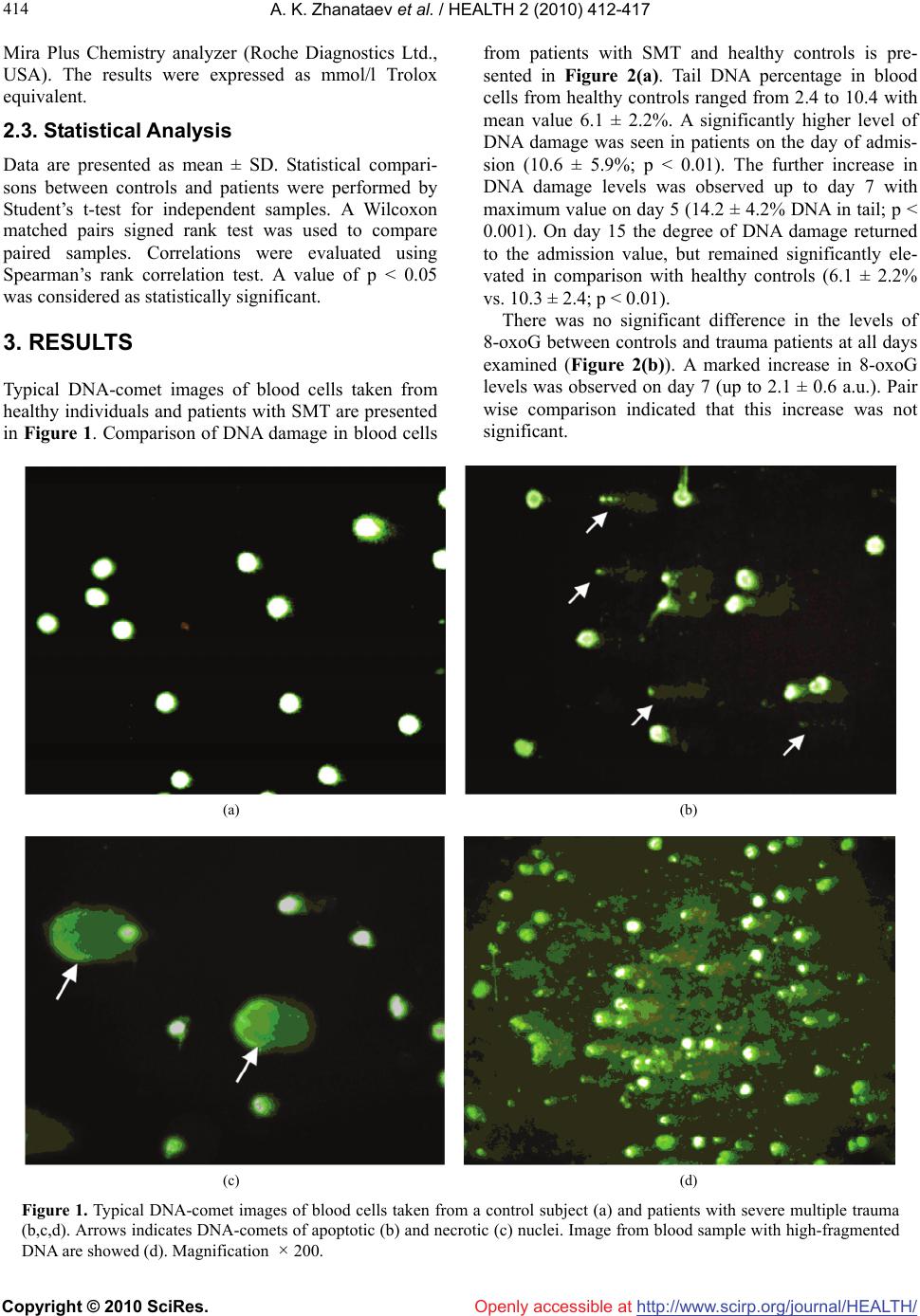
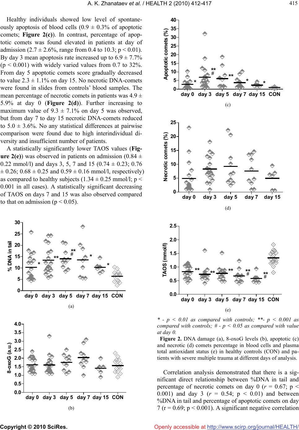
In conclusion, blood cells from severe trauma pa-
tients’ display increased DNA damage associated with
apoptosis and necrosis. Reduced plasma TAOS and a
tendency to increase of 8-oxoguanine in DNA was ob-
served.
REFERENCES
[1] Holtslag, H.R., van Beeck, E.F., Lindeman, E., et al.
(2007) Determinants of long-term functional conse-
quences after major trauma. Journal of Trauma, 62(4),
919-927.
[2] Soberg, H.L., Bautz-Holter, E., Roise, O., et al. (2007)
Long-term multidimensional functional consequences of
severe multiple injuries two years after trauma: A pro-
spective longitudinal cohort study. Journal of Trauma,
62(2), 461-470.
[3] Stalp, M., Koch, C., Ruchholtz, S., et al. (2002) Stan-
dardized outcome evaluation after blunt multiple injuries
by scoring systems: A clinical follow-up investigation 2
years after injury. Journal of Trauma, 52(6), 1160-1168.
[4] Nast-Kolb, D., Aufmkolk, M., Rucholtz, S., et al. (2001)
Multiple organ failure still a major cause of morbidity
but not mortality in blunt multiple trauma. Journal of
Trauma, 51(5), 835-842.
[5] Durham, R.M., Moran, J.J., Mazuski, J.E., et al. (2003)
Multiple organ failure in trauma patients. Journal of
Trauma, 55(4), 608-616.
[6] Antonelli, M. and Caricato, A. (2007) Post-injury multi-
ple organ failure and late outcome. Is it just an associa-
tion? Critical Care, 11(5), 166.
[7] Walsh, C.R. (2005) Multiple organ dysfunction syndrome
after multiple trauma. Orthopedic Nursing, 25( 5), 324-333.
[8] Keel, M. and Trentz, O. (2005) Pathophysiology of poly-
trauma. Injury, 36(6), 691-709.
[9] Goodyear-Brunch, C. and Pierce, J.D. (2002) Oxidative
stress in critically ill patients. American Journal of Critical
Care, 11, 543-553.
[10] Cobb, P.J., Buchman, T.G., Karl, I.E., et al. (2000) Mo-
lecular biology of multiple organ dysfunction syndrome:
Injury, adaptation and apoptosis. Surgical Infection
(Larchmt), 1(3), 207-215.
[11] Papathanassoglou, E.D., Moynihan, J.A. and Ackerman,
M.H. (2000) Does programmed cell death (apoptosis)
play a role in the development of multiple organ dys-
function in critically ill patients? A review and a theo-
retical framework. Critical Care Medicine, 28(2), 537-
549.
[12] Hotchkiss, R.S., Schmieg, R.E., Swanson, P.E., et al.
(2000) Rapid onset of intestinal epithelial and lympho-
cyte apoptotic cell death in patients with trauma and
shock. Critical Care Medicine, 28(9), 3207-3217.
[13] Norbury, C.J. and Zhivotovsky, B. (2004) DNA dam-
age-induced apoptosis. Oncogene, 23, 2797-2808.
[14] Collins, A.R. (2004) The comet assay for DNA damage
and repair: principles, applications, and limitations. Mo-
lecular Biotechnology, 26(3), 249-261.
[15] Tice, R.R., Agurell, E., Anderson, D., et al. (2000) Single
cell gel/comet assay: Guidelines for in vitro and in vivo
genetic toxicology testing. Environmental and Molecular
Mutagenesis, 35(3), 206-221.
[16] Konca, K., Lankoff, A. and Banasik, A. (2003) A cross-
platform public domain PC image-analysis program for
the comet assay. Mutation Research, 534(1-2), 15-20.
[17] Morley, N., Rapp, A., Dittmar, H., et al. (2006) UVA-
induced apoptosis studied by the new apo/necro-Comet-
assay which distinguishes viable, apoptotic and necrotic
cells. Mutagenesis, 21(2), 105-114.
[18] Barzilai, A. and Yamamoto, K. (2004) DNA damage
responses to oxidative stress. DNA Repair (Amst), 3(8-9),
1109-1115.
[19] Valko, M., Leibfritz, D., Moncol, J., et al. (2007) Free
radicals and antioxidants in normal physiological func-
tions and human disease. International Journal of Bio-
chemistry & Cell Biology, 39(1), 44-84.
[20] Oldham, K.M., Wise, S.R., Chen, L., et al. (2002) A lon-
gitudinal evaluation of oxidative stress in trauma patients.
Journal of Parenteral and Enteral Nutrition, 26(3), 189-
197.
[21] Pachl, J., Duska, F., Waldauf, P., et al. (2005) Apoptosis
as an early event in the development of multiple organ
failure? Physiological Research, 54, 697-699.
[22] Guan, J., Jin, D.D., Jin, L.J., et al. (2002) Apoptosis in
organs of rats in early stage after polytrauma combined
with shock. Journal of Trauma, 52(1), 104-111.
[23] Li, S., Tao, L., Jiao, X., et al. (2007) TNFalpha-initiated
oxidative/nitrative stress mediates cardiomyocyte apop-
tosis in traumatic animals. Apoptosis, 12(10), 1795-1802.