Vol.1, No.3, 97-102 (201
doi:10.4236/oji.2011.13012
C
opyright © 2011 SciRes. Openly accessible at http://www.scirp.org/journal/oji/
1) Open Journal of Immunology
TLR4 is involved in mediating fatal murine pneumonia
due to Burkholderia cenocepacia
Viviane Balloy1,2#, Heidi Nagel1,2#, Reuben Ramphal3, Mustapha Si-Tahar1,2#,
Michel Chignard1,2#*
1Unité de Défense Innée et Inflammation, Institut Pasteur, Paris, France;
2INSERM U874, Paris, France; *Corresponding Author: chignard@pasteur.fr
3Department of Medicine, University of Florida, Gainesville, Florida, USA.
Received 14 April 2011; revised 15 July 2011; accepted 11 August 2011.
ABSTRACT
Background: We previously showed that MyD88
knocked out mice were protected from death
due to B. cenocepacia pneumonia implying that
a toll-like receptor(s) (TLR) was involved in me-
diating death. The aim of the present study w as
to determine which TLR(s) was involved in trig-
gering the inflammatory response responsible
for the pathogenesis. We specifically focus on
the TLRs 4 a nd 5, as th e s e two recept ors are t he
main ones involved in the recognition of P. ae-
ruginosa, a flagellated Gram-bacterium similar
to B. cenocepacia. Methods: Mice were inf-
ected intratracheally with a suspension of B. ce-
nocepacia. Animals were then observed daily
for signs of morbidity. Alternatively, bronchoal-
veolar lavages (BAL) w ere collected at different
time points to further determine cytokine con-
centrations and the number of CFU of B. ceno-
cepacia. Results: The data clearly indicate that
the innate immune response of the host to B.
cenocepacia lung infection was due to TLR4
that senses the pathogen while TLR5 does not
do so in vivo. As with the MyD88-/- strain, TLR4-/-
mice were protected from death and cytokine
and chemokine synthesis to infection were re-
duced. The only paradoxical observation was the
reduced pathogen burden in the case of TLR4-/-
mice compared to the enhanced (but transient)
pathogen burden observed with MyD88-/- mice,
suggesting that another TLR was involved in
bacterial clearance. Conclusion: The dat a clearly
demonstrate a deleterious implication of TLR4
in the host to B. cenocepacia lung infection.
Keywords: Rodent; Bacterial; Inflammation;
Innate Immunity; Lung
1. INTRODUCTION
Burkholderia cepacia complex (Bcc) strains have
emerged as problematic opportunistic pathogens causing
severe infections in patients with immune suppression,
chronic granulomatous disease and cystic fibrosis [1-3].
Infections due to Bcc are specially a serious concern to
CF patients due to their inherent antibiotic resistance and
high potential for patient-to-patient transmission [4]. At
least 17 Bcc species have been recovered from respira-
tory secretions of CF patients in many countries [5-6],
but B. cenocepacia is the most common species isolated
[5,7-8]. The clinical outcomes of Bcc infections range
from asymptomatic carriage to a fulminant and fatal
pneumonia, the so-called “cepacia” syndrome [9]. There
is increasing evidence that the acute pulmonary deterio-
ration associated with B. cenocepacia infection is the con-
esquence of a marked inflammatory host response [10].
This could be due to lipopolysaccharide (LPS) as it has
been shown that LPS purified from Bcc species exhibits
potent proinflammatory activity mediated through Toll-
like receptor (TLR) 4 [11], and may account, at least in
part, for the sepsis-like cepacia syndrome [10-12].
TLR recognize conserved pathogen-associated mo-
lecular patterns and trigger the activation of genes in-
volved in innate defense. Four main TLRs are involved
in sensing bacteria, TLR2 in tandem with TLR1 or 6
detects the presence of lipopeptides and peptidoglycan,
TLR4 in association with CD14 and MD2 recognizes
LPS, TLR5 detects the presence of flagellin, and TLR9
recognizes hypomethylated DNA [13-15].
Numerous groups have established the importance of
TLRs in pulmonary host defense and this laboratory has
demonstrated their involvement during influenza A [16],
Aspergillus fumigatus [17,18], and Pseudomonas aeru-
ginosa [19-21] infections. Recently, we showed that
# These authors contributed equally to the work.
V. Balloy et al. / Open Journal of Immunology 1 (2011) **-**
Copyright © 2011 SciRes. Openly accessible at http://www.scirp.org/journal/oji/
98
MyD88, a key downstream adapter for most of the TLRs,
was involved the death due to B. cenocepacia pneu mo-
nia suggesting a role of TLRs in the pathogenesis [22].
Thus, MyD88 knocked out mice (MyD88-/-) were pro-
tected from death following B. cenocepacia lung infec-
tion. We demonstrated that the pathogenesis was due to a
hyperinflammatory host response mediated by TNF-
.
The aim of the present study was to determine which
TLR(s) is playing a role in the recognition of B. cenoce-
pacia and triggering of hyperinflammation. We specifi-
cally focus on the TLR4 and 5, knowing that these two
receptors are the main actors in the recognition of P.
aeruginosa, a flagellated Gram- bacterium which like B.
cenocepacia is a highly problematic pathogen for indi-
vidual with CF [19,20,23-25].
2. METHODS
2.1. Bacterial Strain and Growth Conditions
B. cenocepacia of the epidemic ET12 lineage (strain
J2315) was provided by the Pasteur Institute microor-
ganisms depository. Bacteria were grown on tryptic soy
agar (TSA) at 33˚C for 48 h. Single colonies removed
from the plate were grown in 5 mL of tryptic soy broth
at 33˚C with shaking for 16 h - 18 h, corresponding to
midlog phase. Bacteria were harvested by centrifugation
(3,000 ×g for 15 min), resuspended in saline and optical
densities of the suspensions were adjusted to give the
desired bacterial concentration which was verified by
serial dilutions and plating on TSA.
2.2. Mouse Strains
TLR4-/- and TLR5-/- mice were obtained from S.
Akira (Osaka University, Osaka, Japan). All mice
were backcrossed at least eight times with C57BL/6
to ensure similar genetic backgrounds. Double knock-
ed-out TLR4,5-/- mice were generated by breeding
TLR4-/- mice and TLR5-/- mice. C57/BL6 mice from
which these mice were derived were used as the control
mice. These latter mice were supplied by the Centre
d’Elevage R. Janvier, Le Genest Saint-Isle, France and
used at about 8 weeks of age. Upon arrival for experi-
mentation mice were fed normal mouse chow and
water ad libitum and were housed under standard
conditions with air filtration. Mice were cared for in
accordance with Pasteur Institute guidelines in com-
pliance with the European animal welfare regulation.
2.3. Immunosuppressive Treatment and
Infection
As mice are relatively resistant to B. cenocepacia and
generally do not die from pulmonary infection unless
encased in agar beads, we developed a lethal model in
the neutropenic mouse [22], where wild type mice die
after an intratracheal challenge with about 4 - 5 × 10⁷ cfu.
Chemotherapy-induced neutropenia was achieved by the
intravenous administration of 5 mg/kg of the antineo-
plastic drug vinblastine (Cell Pharm GmbH, Germany)
66 h before infection. Under these conditions, the hae-
matological profile of the mice after vinblastine and
during the infection displayed total polymorphonuclear
cell depletion from day 0 (the day of infection) until day
2 for both wild-type and TLR-/- mice. Of note, in sepa-
rate investigations, we previously demonstrated that with
this vinblastine regimen, mouse survival and cytokine
production are similar to those obtained when neutro-
penia is induced with the antigranulocyte monoclonal
antibody RB6-8C5, [26]. Animals were infected intra-
tracheally under general anesthesia achieved with a
mixture of ketamine (40 mg/kg) and xylazine (8 mg/kg)
administered via the intramuscular route and infected as
previously described [19] with a 50-μl suspension of B.
cenocepacia suspension (4 × 10⁷
cfu/mouse). Mice were
then observed daily for signs of morbidity. Alternatively,
mice were killed at different time points (6 h or 24 h) by
intraperitoneal injection of 300 mg/kg sodium pentobar-
bital. Airways were washed twice with 1 ml saline, and
the bronchoalveolar lavage (BAL) was collected [27] to
further determine cytokine concentrations using DuoSet
ELISA kits (R&D Systems). One hundred microliters of
the BAL were diluted and plated on TSA plates to deter-
mine the number of CFU of B. cenocepacia.
2.4. Statistics
Survival of wild-type and TLR-/- animals was com-
pared using Kaplan-Meier analysis log-rank test. In-
flammatory mediators levels and bacterial counts were
expressed as the mean ± SEM. Differences between
groups were assessed for statistical significance using the
Kruskal-Wallis ANOVA test, followed by the Mann-
Whitney U test. A value of p < 0.05 was considered sta-
tistically significant.
3. RESULTS
3.1. Effect of TLR4 and TLR5 Deletion on
Mouse Survival
The survival of WT, TLR4-/-, TLR5-/- and TLR4,5
double knockout neutropenic mice was followed for 10
days following intratracheal challenge with B. cenoce-
pacia. The survival curves are shown in Figure 1. It was
noted that only 22% of WT mice survived during the 10
day observation whereas 84% of the mice that had TLR4
deleted (p < 0.0001 compared to WT), and 75% of the
V. Balloy et al. / Open Journal of Immunology 1 (2011) **-**
Copyright © 2011 SciRes. Openly accessible at http://www.scirp.org/journal/oji/
9999
Wild-type, TLR5-/-, TLR4-/- and TLR4,5-/- mice treated with vinblastine
and infected by 4 × 10⁷ cfumouse B. cenocepacia. Survivals were checked
every 24 h. Logrank (Mantel-Cox) test for comparisons of Kaplan-Meier
survival curves indicated a significant difference in the survival of
TLR4-deficiente mice (n = 19 for TLR4-/-; p < 0.0001; and n = 24 for TLR
4,5-/-; p < 0.0005) and no significant difference in the survival of TLR5-/-
mice (n = 34 for TLR5-/-; p > 0.05) compared to that of wild-type animals
(n = 36).
Figure 1. Mice survival upon infection by B. cenocepacia as a
function of TLR4 or 5 expression.
mice that had both TLR4 and TLR5 deleted (p < 0.0005
compared to WT) survived. Mortality among TLR5-/-
mice was non significantly different from that of WT
mice, both in terms of percentage mortality and time to
death. Collectively, these data suggested that the TLR4
response to B. cenocepacia was indeed deleterious as
has been noted for the response to LPS challenge [10,12]
and that the TLR5 response did not play a significant
role either in protecting the mice or inducing a deleteri-
ous inflammatory response.
3.2. Role of TLR in the Clearance of B.
Cenocepacia from the Airw ays
In order to examine whether there was a correlation
between bacterial clearance, survival and TLR function,
we examined bacterial clearance from the airways of
WT and mutant mice by culturing of BAL fluid (Figure
2). At 24 h hours post-infection, both WT and TLR5-/-
mice had retained similar amounts of the challenge dose.
Thus, clearance of this bacterium did not appear to be
affected by the absence of a TLR5 response, which was
not surprising as the TLR4 arm of the innate immune
response was intact. However, the number of bacteria
remaining in the airways of TLR4-/- (p < 0.01) and
TLR4,5-/- (p < 0.01) was significantly lower than that
seen in WT mice despite the latter mice not having the
two major TLRs that are involved in defense against
flagellated gram negative bacteria. Thus the response
mediated through TLR4 appears to both lead to death
Wild-type, TLR5-/-, TLR4-/- and TLR4,5-/- mice were treated with vin-
blastine and infected by 4 × 10⁷ cfumouse B. cenocepacia. BAL were per-
formed at 24 h p.i. for the measurements of bacterial loads determined by
serial dilutions and plating on TSA. Data are the mean ± SEM values ob-
tained from four animals. **, p < 0.01 when compared with the corre-
sponding WT values. ns = non significant.
Figure 2. Pathogen burden of the lung upon infection by B.
cenocepacia as a function of TLR4 and 5 expression.
and to inhibit bacterial clearance, since in its absence
there is significantly better survival and bacterial clear-
ance from the airways compared to WT mice. This find-
ing is consistent with enhanced survival seen with
MyD88-/- mice [22] where multiple MyD88-/- depend-
ent TLRs are not functional. One salient difference
however is that the bacterial burden in MyD88-/- mice at
24 h was much greater than that seen in WT mice [22],
suggesting that another MyD88 dependent factor, active
in TLR4, 5-/- mice, functions to assist in the early clear-
ance of B. cenocepacia.
3.3. Role of TLR in the Induction of
Cytokine/ Chemokine Secretion during
B. Cenocepacia Lung Infection
Using the BAL fluids collected to examine bacterial
clearance from the airways, we measured several cyto-
kines/chemokines responses of the mice to explain the
sometimes paradoxical data obtained for survival and
bacterial clearance. TNF
, which we had previously
shown to be involved in the death of mice infected with
this bacterium [22], was significantly lower in TLR4-/-
mice than in WT mice but just as high in TLR5-/- mice
as in WT mice (Figure 3), confirmaing a significant role
for this cytokine in LPS induced inflammation. Similarly
all other cytokines/chemokines measured showed low
levels in the absence of TLR4 and similar levels in WT
and TLR5-/- mice (Figure 4).
4. DISCUSSION
The innate immune response of the host to B. ceno-
cepacia lung infection clearly indicates that TLR4
senses the pathogen while TLR5 does not do so in vivo.
In the absence of the former, the host response in terms
V. Balloy et al. / Open Journal of Immunology 1 (2011) **-**
Copyright © 2011 SciRes. Openly accessible at http://www.scirp.org/journal/oji/
100
Wild-type, TLR5-/-, TLR4-/- and TLR4,5-/- mice were treated with vin-
blastine and infected by 4 × 10⁷ cfumouse B. cenocepacia. BAL were per-
formed at 6 and 24 h p.i. for the measurements of TNF- concentrations.
Data are the mean ± SEM values obtained from eight animals. **, p < 0.01
when compared with the corresponding WT values. ns = non significant.
Figure 3. Induction of TNF-
synthesis upon infection by B.
cenocepacia as a function of TLR4 and 5 expressions.
of cytokine and chemokine synthesis to infection is re-
duced, although not totally ablated, indicating the par-
ticipation of other signaling pathway(s). Moreover, as
with the MyD88-/- strain, TLR4-/- mice were protected
from death. We thus deduce that activation of TLR4 by
B. cenocepacia triggers a hyperinflammatory state that is
responsible for the observed pathogenesis [22]. The only
paradoxical observation is the reduced pathogen burden
in the case of TLR4-/- mice compared to the enhanced
(but transient) pathogen burden observed with MyD88-/-
mice. How TLR4 inhibits bacterial clearance was not
ascertainable, but the observation appears to be consis-
tent for the two different groups of mice with the TLR4-/-
deletion. It is also of note that the in vivo data do not fit
with our previous in vitro data [28]. Using respiratory
epithelial cells isolated from TLR4-/- mice or cells over-
expressing a functional form of TLR5, we established
that TLR5, but not TLR4, mediated B. cenocepacia-
induced lung epithelial inflammatory response. Such a
discrepancy between in vivo and in vitro results suggests
that the role of respiratory epithelial cells in lethal B.
cenocepacia pneumonia is limited. We have however
previously noted differences between in vivo and in vitro
data. Thus, with P. aeruginosa, we observed the lack of
involvement of TLR2 and 4 in the induction of IL-6
synthesis in vivo [19] while the absence of expression of
the very same TLRs by respiratory epithelial cells in-
fected in vitro reduced the synthesis of IL-6 considerably
[21]. Altogether, it is clear that examination of epithelial
cells in tissue culture is prone to variations and may not
always be reflective of infection.
The observations reported here also do not concur with
those of Urban et al. [29] who used an agar bead model of
infection to examine the role of flagella in B. cenocepacia
chronic lung infection. They observed reduced mortality
Wild-type, TLR5-/-, TLR4-/- and TLR4,5-/- mice were treated with vin-
blastine and infected by 4 × 10⁷ cfumouse B. cenocepacia. BAL were
performed at 6 and 24 h p.i. for the measurements of IL-6, KC and G-CSF
concentrations. Data are the mean ± SEM values obtained from at least
eight animals. *, p < 0.05 and **, p < 0.01 when compared with the corre-
sponding WT values. ns = non significant.
Figure 4. Induction of IL-6, KC and G-CSF synthesis upon
infection by B. cenocepacia as a function of TLR4 and 5 ex-
pressions.
and noted significant reductions of KC, a murine ho-
molog of IL-8 both in BAL fluid and serum,when wild
type mice were challenged with a flagellin mutant, indi-
cating that there is normally a TLR5 mediated response
in that model of infection. However, their data in vitro
showing that flagellin does stimulate a response in cells
through TLR5 is similar to ours [28]. Rather than dis-
miss a role for TLR5, this discordance raises the ques-
V. Balloy et al. / Open Journal of Immunology 1 (2011) **-**
Copyright © 2011 SciRes. Openly accessible at http://www.scirp.org/journal/oji/
101101
tion whether B. cenocepacia expresses flagellin in vivo
during an acute infection versus the agar bead model of
infection. This possibility is not with precedent, as
members of the genus Bord etella, which are responsible
for a variety of acute bronchial infections repress flagel-
lum production upon entry into the airways [30]. Lastly,
while we have chosen to examine the role of LPS medi-
ated inflammation in death, it is likely that other viru-
lence factors may also be involved, as some TLR4-/-
mice do still die. This organism is known to have at least
four protein secretion systems, any or all of which may
also be involved in death. Analysis of these factors is
however beyond the scope of this study.
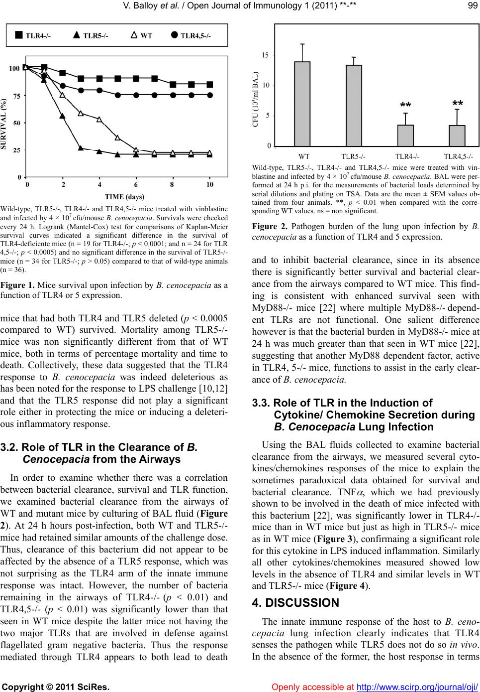
It is concluded that B. cenocepacia induces a hyperin-
flammatory state mediated through its very potent LPS
[10,12]. However, suppression of the LPS-TLR4 inter-
action and the consequent down-regulation of lung in-
flammation still leaves an effective innate immune re-
sponse that is capable of controlling bacterial growth.
5. ACKNOWLEDGEMENTS
This work is supported by Institut Pasteur, Inserm and Legs Poix.
REFERENCES
[1] Mahenthiralingam, E., Baldwin A. and Vandamme, P.
(2002) Burkholderia cepacia complex infection in pa-
tients with cystic fibrosis. Journal of Medical Microbi-
ology, 51, 533-538.
[2] Coenye, T. and Vandamme, P. (2003) Diversity and sig-
nificance of Burkholderia species occupying diverse
ecological niches. Environmental Microbiology, 5, 719-
729. doi:10.1046/j.1462-2920.2003.00471.x
[3] Vandamme, P., Holmes, B., Coenye, T., Goris, J., Ma-
henthiralingam, E., LiPuma, J.J. and Govan J.R. (2003)
Burkholderia cenocepacia sp. nov.—a new twist to an
old story. Research in Microbiology, 154, 91-96.
doi:10.1016/S0923-2508(03)00026-3
[4] Mahenthiralingam, E. and Vandamme, P. (2005) Taxon-
omy and pathogenesis of the Burkholderia cepacia com-
plex. Chronic Respiratory Disease, 2, 209-217.
doi:10.1191/1479972305cd053ra
[5] Reik, R., Spilker. T. and Lipuma, J.J. (2005) Distribution
of Burkholderia cepacia complex species among isolates
recovered from persons with or without cystic fibrosis.
Journal of Clinical Microbiology, 43, 2926-2928.
doi:10.1128/JCM.43.6.2926-2928.2005
[6] Vanlaere, E., Baldwin, A., Gevers, D., Henry, D., De
Brandt E., LiPuma, J.J., Mahenthiralingam, E., Speert,
D.P., Dowson, C., Vandamme, P. and Taxon, K. (2009) A
complex within the Burkholderia cepacia complex, com-
prises at least two novel species, Burkholderia contami-
nans sp. nov. and Burkholderia lata sp. nov. rnational
Journal of Systematic and Evolutionary Microbiology, 59,
102-111.
[7] LiPuma, J.J., Spilker, T., Gill, L.H., Campbell, P.W. 3rd,
Liu, L. and Mahenthiralingam, E. (2001) Disproportion-
ate distribution of Burkholderia cepacia complex species
and transmissibility markers in cystic fibrosis. American
Journal of Respiratory and Critical Care Medicine, 164,
92-96.
[8] Speert, D.P., Henry, D., Vandamme, P., Corey, M. and
Mahenthiralingam E. (2002) Epidemiology of Burk-
holderia cepacia complex in patients with cystic fibrosis,
Canada. Emerging Infectious Diseases, 8, 181-187.
[9] Mahenthiralingam, E., Urban, T.A. and Goldberg, J.B.
(2005) The multifarious, multireplicon Burkholderia ce-
pacia complex. Nature Reviews Microbiology, 3, 144-
156. doi:10.1038/nrmicro1085
[10] De Soyza, A., Ellis, C.D., Khan, C.M., Corris, P.A. and
Demarco de Hormaeche, R. (2004) Burkholderia ceno-
cepacia lipopolysaccharide, lipid A, and proinflamma-
tory activity. American Journal of Respiratory and Cri-
tical Care Medicine, 170, 70-77.
doi:10.1164/rccm.200304-592OC
[11] Bamford, S., Ryley, H. and Jackson S. (2007) Highly
purified lipopolysaccharides from Burkholderia cepacia
complex clinical isolates induce inflammatory cytokine
responses via TLR4-mediated MAPK signalling path-
ways and activation of NFkappaB. Cellular Microbiology,
9, 532-543. doi:10.1111/j.1462-5822.2006.00808.x
[12] Zughaier, S.M., Ryley, H.C. and Jackson, S.K. (1999)
Lipopolysaccharide (LPS) from Burkholderia cepacia is
more active than LPS from Pseudomonas aeruginosa and
Stenotrophomonas maltophilia in stimulating tumor ne-
crosis factor alpha from human monocytes. Infection and
Immunity, 67, 1505-1507.
[13] Iwamura, C. and Nakayama, T. (2008) Toll-like receptors
in the respiratory system: their roles in inflammation.
Current Allergy and Asthma Reports, 8, 7-13.
doi:10.1007/s11882-008-0003-0
[14] Bauer, S., Müller, T. and Hamm, S. (2009) Pattern recog-
nition by Toll-like receptors. Advances in Experimental
Medicine and Biology, 653, 15-34.
doi:10.1007/978-1-4419-0901-5_2
[15] Kumar, H., Kawai, T and Akira, S. (2009) Toll-like re-
ceptors and innate immunity. Biochemical and Biophysi-
cal Research Communications, 388, 621-625.
doi:10.1016/j.bbrc.2009.08.062
[16] Le Goffic, R., Balloy, V., Lagranderie, M., Alexopoulou,
L., Escriou, N., Flavell, R., Chignard, M. and Si-Tahar,
M. (2006) Detrimental contribution of the Toll-like re-
ceptor (TLR)3 to influenza A virus-induced acute pneu-
monia. PLoS Pathogens, 2, e53.
doi:10.1371/journal.ppat.0020053
[17] Balloy, V., Si-Tahar, M., Takeuchi, O., Philippe, B., Na-
hori, M.A., Tanguy, M., Huerre, M., Akira, S., Latgé, J.P.
and Chignard, M. (2005) Involvement of toll-like recep-
tor 2 in experimental invasive pulmonary aspergillosis.
Infection and Immunity, 73, 5420-5425.
doi:10.1128/IAI.73.9.5420-5425.2005
[18] Balloy, V. and Chignard, M. (2009) The innate immune
response to Aspergillus fumigatus. Microbes and Infec-
tion, 11, 919-927. doi:10.1016/j.micinf.2009.07.002
[19] Ramphal, R., Balloy, V., Huerre, M., Si-Tahar, M. and
Chignard, M. (2005) TLRs 2 and 4 are not involved in
hypersusceptibility to acute Pseudomonas aeruginosa
lung infections. Journal of Immunology, 175, 3927-3934.
[20] Ramphal, R., Balloy, V., Jyot, J., Verma, A., Si-Tahar, M.
V. Balloy et al. / Open Journal of Immunology 1 (2011) **-**
Copyright © 2011 SciRes. http://www.scirp.org/journal/oji/ Openly accessible at
102
and Chignard, M. (2008) Control of Pseudomonas aeru-
ginosa in the lung requires the recognition of either lipo-
polysaccharide or flagellin. Journal of Immunology, 181,
586-592.
[21] Raoust, E., Balloy, V., Garcia-Verdugo, I., Touqui, L.,
Ramphal, R. and Chignard, M. (2009) Pseudomonas ae-
ruginosa LPS or flagellin are sufficient to activate
TLR-dependent signaling in murine alveolar macro-
phages and airway epithelial cells. PLoS One, 4, e7259.
doi:10.1371/journal.pone.0007259
[22] Ventura, G.M., Balloy, V., Ramphal, R., Khun, H., Huerre,
M., Ryffel, B., Plotkowski, M.C., Chignard, M. and
Si-Tahar, M. (2009) Lack of MyD88 protects the immu-
nodeficient host against fatal lung inflammation triggered
by the opportunistic bacteria Burkholderia cenocepacia.
Journal of Immunology, 183, 670-676.
doi:10.4049/jimmunol.0801497
[23] Feuillet, V., Medjane, S., Mondor, I., Demaria, O., Pagni,
P.P., Galán, J.E., Flavell, R.A. and Alexopoulou, L. (2006)
Involvement of Toll-like receptor 5 in the recognition of
flagellated bacteria. Proceedings of the National Acad-
emy of Sciences of the United States of America, 103,
12487-12492. doi:10.1073/pnas.0605200103
[24] Skerrett, S.J., Wilson, C.B., Liggitt, H.D. and Hajjar,
A.M. (2007) Redundant Toll-like receptor signaling in
the pulmonary host response to Pseudomonas aeruginosa.
American Journal of Physiology—Lung Cellular and
Molecular Physiology, 292, L312-L322.
doi:10.1152/ajplung.00250.2006
[25] Morris, A.E., Liggitt, H.D., Hawn, T.R. and Skerrett, S.J.
(2009) Role of Toll-like receptor 5 in the innate immune
response to acute P. aeruginosa pneumonia. American
Journal of Physiology—Lung Cellular and Molecular
Physiology, 297, L1112-L1119.
doi:10.1152/ajplung.00155.2009
[26] Balloy, V., Sallenave, J.M., Crestani, B., Dehoux, M. and
Chignard, M. (2003) Neutrophil DNA contributes to the
antielastase barrier during acute lung inflammation.
American Journal of Respiratory Cell and Molecular Bi-
ology, 28, 746-753. doi:10.1165/rcmb.2002-0119OC
[27] Gonçalves de Moraes, V.L., Singer, M., Vargaftig, B.B.
and Chignard, M. (1998) Effects of rolipram on cyclic
AMP levels in alveolar macrophages and lipopolysac-
charide-induced inflammation in mouse lung. British
Journal of Pharmacology, 123, 631-636.
doi:10.1038/sj.bjp.0701649
[28] de C Ventura, G.M., Le Goffic, R., Balloy, V., Plotkowski,
M.C., Chignard, M. and Si-Tahar, M. (2008) TLR 5, but
neither TLR2 nor TLR4, is involved in lung epithelial
cell response to Burkholderia cenocepacia. FEMS Im-
munology & Medical Microbiology, 54, 37-44.
doi:10.1111/j.1574-695X.2008.00453.x
[29] Urban, T.A., Griffith, A., Torok, A.M., Smolkin, M.E.,
Burns, J.L. and Goldberg, J.B. (2004) Contribution of
Burkholderia cenocepacia flagella to infectivity and in-
flammation. Infection and Immunity, 72, 5126-5134.
doi:10.1128/IAI.72.9.5126-5134.2004
[30] Akerley, B.J., Cotter, P.A. and Miller, JF. (1995) Ectopic
expression of the flagellar regulon alters development of
Bordetella-host interaction. Cell, 80, 611-620.
doi:10.1016/0092-8674(95)90515-4
LIST OF ABBREVIATIONS USED
BAL, bronchoalveolar lavage;
CF, cystic fibrosis;
MyD, myeloid-differentiation;
p.i., post-infection;
TLR, Toll-like receptor.