Paper Menu >>
Journal Menu >>
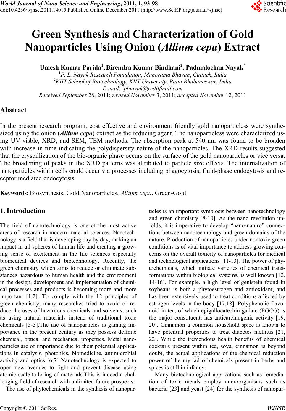 World Journal of Nano Science and Engineering, 2011, 1, 93-98 doi:10.4236/wjnse.2011.14015 Published Online December 2011 (http://www.SciRP.org/journal/wjnse) Copyright © 2011 SciRes. WJNSE Green Synthesis and Characterization of Gold Nanoparticles Using Onion (Allium cepa) Extract Umesh Kumar Parida1, Birendra Kumar Bindhani2, Padmalochan Nayak* 1P. L. Nayak Research Foundation, Manorama Bhavan, Cuttack, India 2KIIT School of Biotechnology, KIIT University, Patia Bhubaneswar, India E-mail: *plnayak@rediffmail.com Received September 28, 2011; revised November 3, 2011; accepted November 12, 2011 Abstract In the present research program, cost effective and environment friendly gold nanoparticless were synthe- sized using the onion (Allium cepa) extract as the reducing agent. The nanoparticless were characterized us- ing UV-visble, XRD, and SEM, TEM methods. The absorption peak at 540 nm was found to be broaden with increase in time indicating the polydispersity nature of the nanoparticles. The XRD results suggested that the crystallization of the bio-organic phase occurs on the surface of the gold nanoparticles or vice versa. The broadening of peaks in the XRD patterns was attributed to particle size effects. The internalization of nanoparticles within cells could occur via processes including phagocytosis, fluid-phase endocytosis and re- ceptor mediated endocytosis. Keywords: Biosynthesis, Gold Nanoparticles, Allium cepa, Green-Gold 1. Introduction The field of nanotechnology is one of the most active areas of research in modern material sciences. Nanotech- nology is a field that is developing day by day, making an impact in all spheres of human life and creating a grow- ing sense of excitement in the life sciences especially biomedical devices and biotechnology. Recently, the green chemistry which aims to reduce or eliminate sub- stances hazardous to human health and the environment in the design, development and implementation of chemi- cal processes and products is becoming more and more important [1,2]. To comply with the 12 principles of green chemistry, many researches tried to avoid or re- duce the uses of hazardous chemicals and solvents, such as using natural materials instead of traditional toxic chemicals [3-5].The use of nanoparticles is gaining im- portance in the present century as they possess definite chemical, optical and mechanical properties. Metal nano- particles are of importance due to their potential applica- tions in catalysis, photonics, biomedicine, antimicrobial activity and optics [6,7] Nanotechnology is expected to open new avenues to fight and prevent disease using atomic scale tailoring of materials.This is indeed a chal- lenging field of research with unlimited future prospects. The use of phytochemicals in the synthesis of nanopar- ticles is an important symbiosis between nanotechnology and green chemistry [8-10]. As the nano revolution un- folds, it is imperative to develop “nano-naturo” connec- tions between nanotechnology and green domains of the nature. Production of nanoparticles under nontoxic green conditions is of vital importance to address growing con- cerns on the overall toxicity of nanoparticles for medical and technological applications [11-13]. The power of phy- tochemicals, which initiate varieties of chemical trans- formations within biological systems, is well known [12, 14-16]. For example, a high level of genistein found in soybeans is both a phytoestrogen and antioxidant, and has been extensively used to treat conditions affected by estrogen levels in the body [17,18]. Polyphenolic flavo- noid in tea, of which epigallocatechin gallate (EGCG) is the major constituent, has anticarcinogenic activity [19, 20]. Cinnamon a common household spice is known to have potential properties to treat diabetes mellitus [21, 22]. While the tremendous health benefits of chemical cocktails present within tea, soya, cinnamon is beyond doubt, the actual applications of the chemical reduction power of the myriad of chemicals present in herbs and spices is still in infancy. Many biotechnological applications such as remedia- tion of toxic metals employ microorganisms such as bacteria [23] and yeast [24] for the synthesis of nanopar- 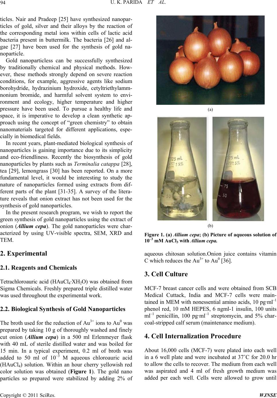 U. K. PARIDA ET AL. Copyright © 2011 SciRes. WJNSE 94 ticles. Nair and Pradeep [25] have synthesized nanopar- ticles of gold, silver and their alloys by the reaction of the corresponding metal ions within cells of lactic acid bacteria present in buttermilk. The bacteria [26] and al- gae [27] have been used for the synthesis of gold na- noparticle. Gold nanoparticless can be successfully synthesized by traditionally chemical and physical methods. How- ever, these methods strongly depend on severe reaction conditions, for example, aggressive agents like sodium borohydride, hydrazinium hydroxide, cetyltriethylamm- nonium bromide, and harmful solvent system to envi- ronment and ecology, higher temperature and higher pressure have been used. To pursue a healthy life and space, it is imperative to develop a clean synthetic ap- proach using the concept of “green chemistry” to obtain nanomaterials targeted for different applications, espe- cially in biomedical fields. In recent years, plant-mediated biological synthesis of nanoparticles is gaining importance due to its simplicity and eco-friendliness. Recently the biosynthesis of gold nanoparticles by plants such as Terminalia catappa [28], tea [29], lemongrass [30] has been reported. On a more fundamental level, it would be interesting to study the nature of nanoparticles formed using extracts from dif- ferent parts of the plant [31-35]. A survey of the litera- ture reveals that onion extract has not been used for the synthesis of gold nanoparticles. In the present research program, we wish to report the green synthesis of gold nanoparticles using the extract of onion (Allium cepa). The gold nanoparticles were char- acterized by using UV-visible spectra, SEM, XRD and TEM. 2. Experimental 2.1. Reagents and Chemicals Tetrachloroauric acid (HAuCl4·XH2O) was obtained from Sigma Chemicals. Freshly prepared triple distilled water was used throughout the experimental work. 2.2. Biological Synthesis of Gold Nanoparticles The broth used for the reduction of Au3+ ions to Au0 was prepared by taking 10 g of thoroughly washed and finely cut onion (Allium cepa) in a 500 ml Erlenmeyer flask with 40 mL of sterile distilled water and was boiled for 15 min. In a typical experiment, 0.2 ml of broth was added to 50 ml of 10–3 M aqueous chloroauric acid (HAuCl4) solution. Within an hour cherry yellowish red color solution was obtained (Figure 1). The gold nano particles so prepared were stabilized by adding 2% of (a) (b) Figure 1. (a) Allium cepa; (b) Picture of aqueous solution of 10–3 mM AuCl3 with Allium cepa. aqueous chitosan solution.Onion juice contains vitamin C which reduces the Au3+ to Au0 [36]. 3. Cell Culture MCF-7 breast cancer cells and were obtained from SCB Medical Cuttack, India and MCF-7 cells were main- tained in MEM with nonessential amino acids, 10 pg·ml–1 phenol red, 10 mM HEPES, 6 ngml-1 insulin, 100 units ml–1 penicillin, 100 pg·ml–1 streptomycin, and 5% char- coal-stripped calf serum (maintenance medium). 4. Cell Internalization Procedure About 16,000 cells (MCF-7) were plated into each well in a 6 well plate and were incubated at 37˚C for 20.0 hr to allow the cells to recover. The medium from each well was aspirated and 4 ml of fresh growth medium was added per each well. Cells were allowed to grow until 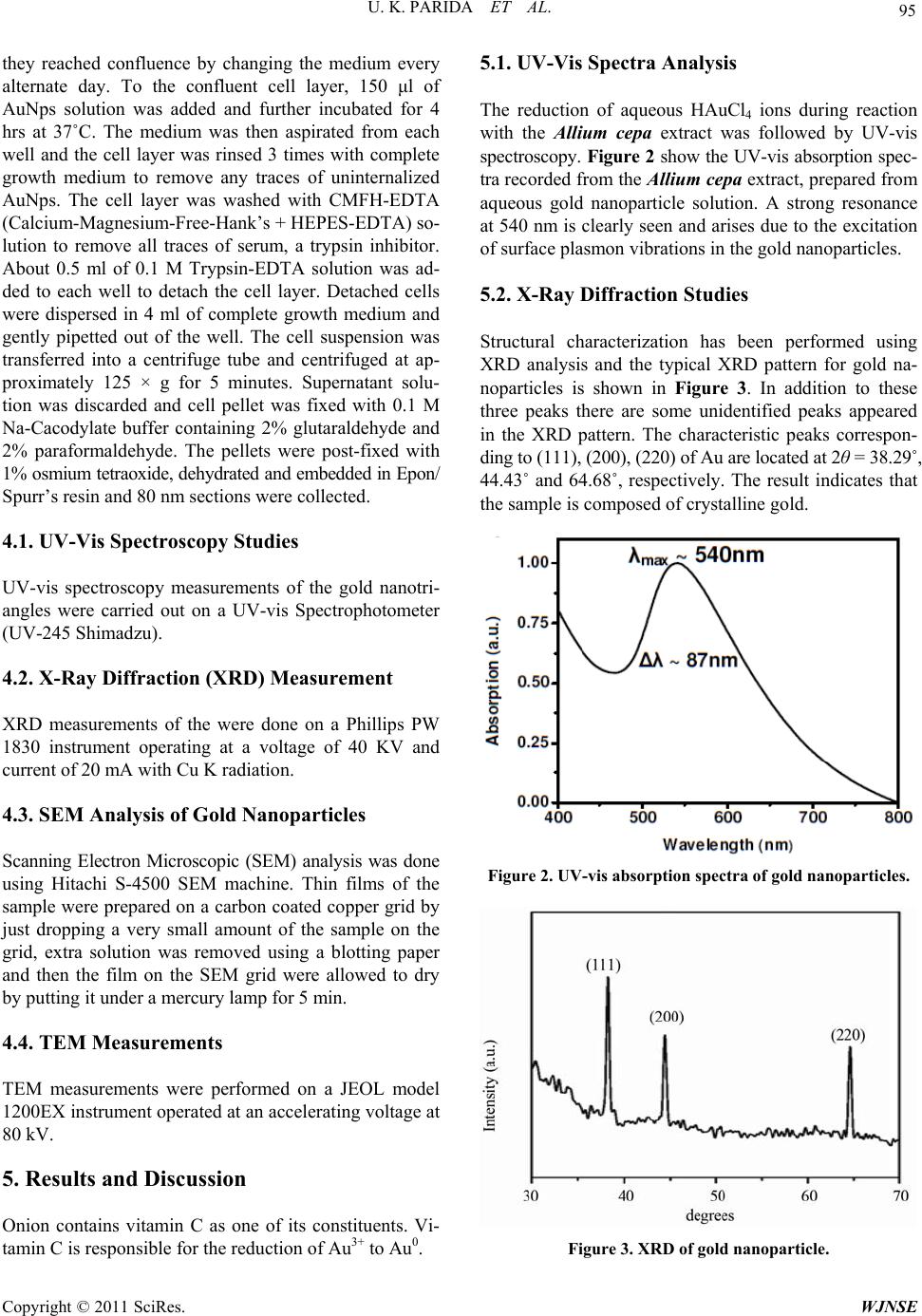 U. K. PARIDA ET AL. Copyright © 2011 SciRes. WJNSE 95 they reached confluence by changing the medium every alternate day. To the confluent cell layer, 150 μl of AuNps solution was added and further incubated for 4 hrs at 37˚C. The medium was then aspirated from each well and the cell layer was rinsed 3 times with complete growth medium to remove any traces of uninternalized AuNps. The cell layer was washed with CMFH-EDTA (Calcium-Magnesium-Free-Hank’s + HEPES-EDTA) so- lution to remove all traces of serum, a trypsin inhibitor. About 0.5 ml of 0.1 M Trypsin-EDTA solution was ad- ded to each well to detach the cell layer. Detached cells were dispersed in 4 ml of complete growth medium and gently pipetted out of the well. The cell suspension was transferred into a centrifuge tube and centrifuged at ap- proximately 125 × g for 5 minutes. Supernatant solu- tion was discarded and cell pellet was fixed with 0.1 M Na-Cacodylate buffer containing 2% glutaraldehyde and 2% paraformaldehyde. The pellets were post-fixed with 1% osmium tetraoxide, dehydrated and embedded in Epon/ Spurr’s resin and 80 nm sections were collected. 4.1. UV-Vis Spectroscopy Studies UV-vis spectroscopy measurements of the gold nanotri- angles were carried out on a UV-vis Spectrophotometer (UV-245 Shimadzu). 4.2. X-Ray Diffraction (XRD) Measurement XRD measurements of the were done on a Phillips PW 1830 instrument operating at a voltage of 40 KV and current of 20 mA with Cu K radiation. 4.3. SEM Analysis of Gold Nanoparticles Scanning Electron Microscopic (SEM) analysis was done using Hitachi S-4500 SEM machine. Thin films of the sample were prepared on a carbon coated copper grid by just dropping a very small amount of the sample on the grid, extra solution was removed using a blotting paper and then the film on the SEM grid were allowed to dry by putting it under a mercury lamp for 5 min. 4.4. TEM Measurements TEM measurements were performed on a JEOL model 1200EX instrument operated at an accelerating voltage at 80 kV. 5. Results and Discussion Onion contains vitamin C as one of its constituents. Vi- tamin C is responsible for the reduction of Au3+ to Au0. 5.1. UV-Vis Spectra Analysis The reduction of aqueous HAuCl4 ions during reaction with the Allium cepa extract was followed by UV-vis spectroscopy. Figure 2 show the UV-vis absorption spec- tra recorded from the Allium cepa extract, prepared from aqueous gold nanoparticle solution. A strong resonance at 540 nm is clearly seen and arises due to the excitation of surface plasmon vibrations in the gold nanoparticles. 5.2. X-Ray Diffraction Studies Structural characterization has been performed using XRD analysis and the typical XRD pattern for gold na- noparticles is shown in Figure 3. In addition to these three peaks there are some unidentified peaks appeared in the XRD pattern. The characteristic peaks correspon- ding to (111), (200), (220) of Au are located at 2θ = 38.29˚, 44.43˚ and 64.68˚, respectively. The result indicates that the sample is composed of crystalline gold. Figure 2. UV-vis absorption spectra of gold nanoparticles. Figure 3. XRD of gold nanoparticle.  U. K. PARIDA ET AL. Copyright © 2011 SciRes. WJNSE 96 5.3. Scanning Electron Microscopy of Gold Nano Particles The SEM photograph of gold nanoparticles are shown in Figure 4. SEM photograph of gold nanoparticles clearly indicates that in the room temperature synthesized sam- ples the size the average size of the nanoparticles is ~100 nm, with spherical and cubic shape. 6. Cellular Internalization Studies TEM images of breast tumor (MCF-7) cells treated with AuNPs unequivocally validated our hypothesis. Signifi- cant internalization of nanoparticles via endocytosis within the MCF-7 was observed (Figures 5). The inter- nalization of nanoparticles within cells could occur via processes including phagocytosis, fluid-phase endocyto- sis, and receptor mediated endocytosis. The viability of Figure 4. SEM image of gold nanoparticles. Figure 5. TEM Images of different MCF-7 cells showing uptake of Allium cepa—AuNps. and MCF-7 cells post-internalization suggests that the phytochemical coating renders the nanoparticles non- toxic to cells. Such a harmless internalization of AuNps will provide new opportunities for probing cellular proc- esses via nanoparticulate-mediated imaging. 7. Conclusions The rapid green synthesis of gold nanoparticles using Allium cepa extract has been demonstrated. The nanopar- ticles were stabilized using aqueous chitosan solution. The reduction of gold nanoparticles takes place because of the presence of vitamin C in onion extract. The UV- visible spectra measurements were carried out at 540 nm with sharp peak. The surface of the nanoparticle was as- certained from the SEM and crystallinity was observed from the XRD spectra. The viability of and MCF-7 cells post-internalization suggests that the phytochemical coat- ing renders the nanoparticles non-toxic to cells. 8. References [1] M. Poliakoff and P. Anastas, “A Principled Stance,” Nature, Vol. 413, 2001, p. 257. doi:10.1038/35095133 [2] M. Poliakoff, J. M. Fitzpatrick, T. R. Farren and P. T. Anastas, “Green Chemistry: Science and Politics of Change,” Science, Vol. 297, No. 5582, 2002, pp. 807-810. doi:10.1126/science.297.5582.807 [3] R. A. Gross and B. Kalra, “Biodegradable Polymers for the Environment,” Science, Vol. 297, No. 5582, 2002, pp. 803-807. doi:10.1126/science.297.5582.803 [4] J. M. DeSimone, “Practical Approaches to Green Sol- vents,” Science, Vol. 297, No. 5582, 2002, pp. 799-803. doi:10.1126/science.1069622 [5] P. Raveendran, J. Fu and S. L. Wallen, “Completely ‘Green’ Synthesis and Stabilization of Metal Nanoparticles,” Jour- nal of American Chemical Society, Vol. 125, No. 46, 2003, pp. 13940-13941. doi:10.1021/ja029267j [6] K. Govindraju, V. Kiruthiga and G. Singaravelu, “Evalua- tion of Biosynthesized Silver Nanoparticles against Fun- gal Pathogens of Mulberry Morus Indica,” Journal of Bio- pesticides, Vol. 1, 2008, pp. 101-104. [7] K. Govindraju, V. Kiruthiga, K. V. Ganesh and G. Sin- garavelu, “Extracellular Synthesis of Silver Nanoparticles by a Marine Alga, Sargassum wightii Grevilli and Their Antibacterial Effects,” Journal of Nanoscience and Nano- technology, Vol. 9, No. 9, 2009, pp. 5497-5501. doi:10.1166/jnn.2009.1199 [8] J. Huang, Q. Li, D. Sun, Y. Lu, Y. Su, X. Yang, H. Wang, Y. Wang, W. Shao, N. J. Hong and C. Chen, “Biosynthe- sis of Silver and Gold Nanoparticles by Novel Sundried Cinnamomum camphora Leaf,” Nanotechnology, Vol. 18, No. 10, 2007, pp. 105104-105115. doi:10.1088/0957-4484/18/10/105104 [9] L. Jorge, G. Torresdey, E. Gomez, J. R. Peralta-Videa, J.  U. K. PARIDA ET AL. Copyright © 2011 SciRes. WJNSE 97 G. Parsons, H. Troiani and M. J. Yacaman, “Phytore- mediation of Heavy Metals and Study of the Metal Coor- di- nation by X-Ray Absorption Spectroscopy,” Lang- muir, Vol. 19, 2003, p. 1357. [10] J. L. Gardea-Torresdey, K. J. Tiemann, J. G. Parsons, G. Gamez, I. Herrera and M. Jose Yacaman, “Investigation into the Mechanism(s) of Au (III) Binding and Reduction by Alfalfa Biomass,” Microchemical Journal, Vol. 71, No. 2-3, 2002, pp. 193-204. doi:10.1016/S0026-265X(02)00011-5 [11] R. Hardman, “A Toxicologic Review of Quantum Dots: Toxicity Depends on Physicochemical and Environmen- tal Factors,” Environmantal Health Perspectives, Vol. 114, No. 2, 2006, pp. 165-172. doi:10.1289/ehp.8284 [12] J. Curtis, M. Greenberg, J. Kester, S. Phillips and G. Krie- ger, “Nanotechnology and Nanotoxicology: A Primer for Clinicians,” Toxicological Reviews, Vol. 25, No. 4, 2006, pp. 245-260. doi:10.2165/00139709-200625040-00005 [13] N. Lewinski, V. Colvin and R. Drezek, “Cytotoxicity of Nanoparticles,” Small, Vol. 4, No. 1, 2008, pp. 26-49. doi:10.1002/smll.200700595 [14] J. C. Espín, M. T. García-Conesa and F. A. Tomás-Barberán, “Nutraceuticals: Facts and Fiction,” Phytochemistry, Vol. 68, No. 22-24, 2007, pp. 2986-3008. doi:10.1016/j.phytochem.2007.09.014 [15] S. Rochfort and J. Panozzo, “Phytochemicals for Health, the Role of Pulses,” Journal of Agricultural and Food Chemistry, Vol. 55, No. 20, 2007, pp. 7981-7994. doi:10.1021/jf071704w [16] K. D. R. Setchell, N. M. Brown, P. Desai, L. Zimmer-Ne- chemias, B. E. Wolfe, W. T. Brashear, A. S. Kirschner, A. Cassidy and J. E. Heubi, “Bioavailability of Pure Isofla- vones In Healthy Humans and Analysis of Commercial Soy Isoflavone Supplements,” Journal of Nutrition, Vol. 131, No. 4, 2001, pp. 1362S-1275S. [17] P. J. Magee and I. R. Rowland, “Phyto-Oestrogens, Their Mechanism of Action: Current Evidence for a Role in Breast and Prostate Cancer,” British Journal of Nutrition, Vol. 91, No. 4, 2004, pp. 513-520. doi:10.1079/BJN20031075 [18] J. L. Limer and V. Speirs, “Phyto-Oestrogens and Breast Cancer Chemoprevention,” Breast Cancer Research, Vol. 6, 2004, pp. 119-127. doi:10.1186/bcr781 [19] O. J. Bandele and N. Osheroff, “(-)-Epigallocatechin Gal- late, “A Major Constituent of Green Tea, Poisons Human Type II Topoisomerases” Chemical Research in Toxicol- ogy, Vol. 21, No. 4, 2008, pp. 936-943. doi:10.1021/tx700434v [20] S. Shankar, S. Ganapathy and R. K. Srivastava, “Green Tea Polyphenols: Biology and Therapeutic Implications in Cancer,” Front Biosci, Vol. 12, 2007, pp. 4881-4899. doi:10.2741/2435 [21] K. Dannemann, W. Hecker, H. Haberland, A. Herbst, A. Galler, T. Schäfer, E. Brähler, W. Kiess and T. M. Ka- pellen, “Use of Complementary and Alternative Medicine in Children with Type 1 Diabetes Mellitus—Prevalence, Patterns of Use, and Costs,” Pediatr Diabetes, 2008. [22] S. Suppapitiporn and N. Kanpaksi, “The Effect of Cin- namon Cassia Powder in Type 2 Diabetes Mellitus,” Journal of the Medical Association of Thailand, Vol. 89 Supplement 3, 2006, p. 200. [23] J. R. Stephen and S. J. Maenaughton, “Fungus-Mediated Synthesis of Silver Nanoparticles and Their Immobiliza- tion in the Mycelial Matrix: A Novel Biological Ap- proach to Nanoparticle Synthesis,” Current Opinon in Biotechnology, Vol. 10, No. 3, 1999, p. 230. doi:10.1016/S0958-1669(99)80040-8 [24] R. K. Mehra and D. R. Wingre, “Methabenzthiazuron on Oxygen Evolution and Cell Growth,” Journal of Cellular Biochemistry, Vol. 45, No. 1, 1991, p. 30. doi:10.1002/jcb.240450109 [25] B. Nair and T. Pradeep, “Preparation of Gold Nanoparti- cles from Mirabilis Jalapa Flowers,” Crystal Growth De- sign, Vol. 2, No. 4, 2002, pp. 293-298. doi:10.1021/cg0255164 [26] G. Southam and T. J. Beveridge, “The Use of Microor- ganisms for the Formation of Metal Nanoparticles,” Geo- chimica et Cosmochimica Acta, Vol. 60, No. 22, 1996, p. 4369. doi:10.1016/S0016-7037(96)00235-9 [27] M. G. Robinson, L. N. Brown and D. Beverley, “Effect of Gold (III) on the Fouling Diatom Amphora Coffeae- for- mis: Uptake, Toxicity and Interactions with Copper,” Biofouling, Vol. 11, No. 1, 1997, pp. 59-79. doi:10.1080/08927019709378320 [28] B. Ankamwar, “Biosynthesis of Gold Nanoparticles (Green- Gold) Using Leaf Extract of Terminalia Catappa,” E-Jour- nal of Chemistry, Vol. 7, No. 4, 2010, pp. 1334-1339. [29] S. K. Nune, N. Chanda, R. Shukla, K. Katti, R. R. Kul- karni, S. Thilakavathy, S. Mekapothula, R. Kannan and K. V. Katti, “Green Nanotechnology from Tea: Phytochemi- cals in Tea as Building Blocks for Production of Biocom- patible Gold Nanoparticles,” Journal of Materials Chem- istry, Vol. 10, 2009, p. 1039. [30] S. S. Shiv, A. Rai, B. Ankamwar, A. Singh, A. Ahmad and M. Sastry, “Biological Synthesis of Triangular Gold Na- noprisms,” Nature Materials, Vol. 3, 2004, pp. 482-488. doi:10.1038/nmat1152 [31] F. E. Kandil, A. M. Soliman, S. R. Skodack and T. J. Ma- bry, “In Vitro Antibacterial Activity of the Extracts De- rived from Terminalia catappa,” Asian Journal of Che- mistry, Vol. 11, 1999, pp. 1001-1004. [32] S. P. Pawar and S. C. Pal, “Antimicrobial Activity of Ex- tracts of Terminalia catappa Root,” Indian Journa of Me- dical Sciences, Vol. 56, No. 6, 2002, pp. 276-278. [33] T. F. Ko, Y. M. Weng and R. Y. Chiou, “Antimutagenic- ity of Supercritical CO2 Extracts of Terminalia catappa Leaves and Cytotoxicity of the Extracts to Human Hepa- toma Cells,” Journal of Agricultural and Food Chemistry, Vol. 11, No. 9, 2002, pp. 5343-5348. doi:10.1021/jf0203500 [34] C. C. Lin, Y. F. Hsu and T. C. Lin, “Evaluation of Anti- oxidant Activity of Eleven Thai Medicinal Herbs,” Anti- cancer Research, Vol. 21 No. 1A, 2001, pp. 237-243. [35] C. C. Chyou, S. Y. Tsai, P. T. Ko and J. L. Mau, “Ter-  U. K. PARIDA ET AL. Copyright © 2011 SciRes. WJNSE 98 minalia catappa Linn. (Combretaceae),” Food Chemistry, Vol. 78. No. 4, 2002, pp. 483-488. [36] J. Saffi, L. Sonego, Q. D. Varela and M. Salvador “Anti- oxidant Activity of L-Ascorbic Acid in Wild Type and Superoxide Dismutase Deficient Strains of Saccharo- myces Cerevisiae,” Redox Report, Vo1. 11, 2006, pp. 179- 184. |