Eosinophilic Granuloma Arising from the Sacrum: A Case Report7
(a) (b) (c)
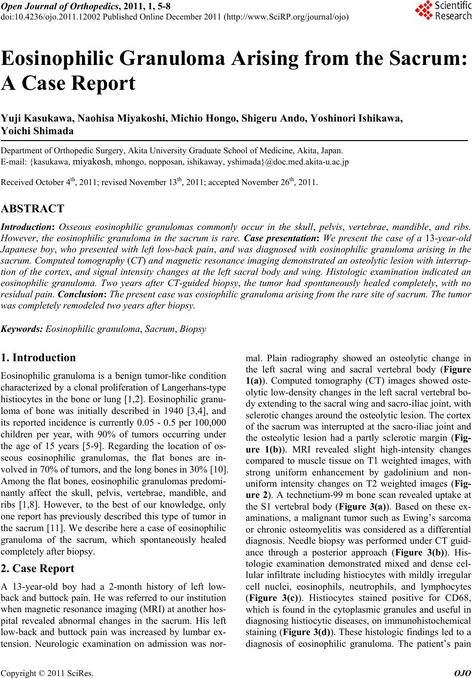
Figure 4. MRI and CT images two years after biopsy. (a) Axial CT image 2 years after biopsy demonstrating remodeled tra-
becular bone in the sacral body and wing, without interruption of cortex. The lesion demonstrated higher intensity compared
to normal bone marrow in the axial T1-weighted (b) and T2-weighted images (c) 2 years after biopsy.
of the vertebra, known as “vertebra plana”. The thoracic
spine is the most common site, followed by the lumbar
and cervical spine, respectively [6,12]. To the best of our
knowledge, the present patient represents the first re-
ported case of eosinophilic granuloma in the sacrum.
Although eosinophilic granuloma bone lesions are u-
sually asymptomatic, osteolytic lesions sometimes cause
fractures or pain as a result of swelling, deformity, and
soft tissue components [6]. The chief complaint in the
current case was low-back pain; however, the osteolytic
lesion in the sacrum was poorly defined by plain radiog-
raphy. MRI of the lumbar spine, however, revealed in-
tensity changes in the sacrum. MRI thus provides a
highly sensitive, but nonspecific, modality for detecting
bone marrow involvement and soft tissue mass in eosi-
nophilic granulomas [8].
Eosinophilic granulomas can be divided in to acute and
chronic phases, depending on their status [6,8]. Th e acute
phase includes destructive, osteolytic lesions with poor
margins, which are difficult to differentiate from malig-
nant tumors such as Ewing’s sarcoma, or acute osteo-
myelitis [8]. CT is useful for evaluating osseous eosino-
philic granuloma lesions, and for determining the extent
of cortical destruction and soft tissue involvement [8].
CT-guided biopsy is also useful for diagnosing eosino-
philic granuloma as a cause of vertebral osteolysis or
vertebra plana [9], and was helpful for diagnosing eosi-
nophilic granuloma in the sacrum with cortical destruc-
tion in the present case.
Treatment options include aggressive resection, biopsy,
combined chemotherapy and radiotherapy, and conserva-
tive treatment. Radiation therapy is associated with a risk
of secondary malignancy [13] as well as effects on the
pelvic viscera, and should be avoided, especially in chil-
dren, because it may destroy the growth potential of the
endochondral plates [14,15]. Ando et al. reported eosi-
nophilic granulomas in the pelvis with different radi-
ologic features and clinical courses [16]. They reported
that osteolytic t umor lesions with sclerotic ma rgins he ale d
spontaneously after b iopsy, while tumors with poor oste-
olytic margins progressed after biopsy. The present case
was consistent with this prev ious report in that p art of th e
osteolytic lesion in the sacrum rev ealed sclerotic margins
on CT, and subsequently spontaneously healed comple-
tely. Surgical treatment, especially in adolescents, should
be reserved for specific cases with neurologic defects or
instability.
4. Conclusions
In conclusion, this patient represents the case of eosino-
philic granuloma arising in the rare site of sacrum, which
spontaneously and completely healed 2 years after biopsy.
Biopsy is a better choice to decide a therapeutic strategy
of the osteolytic tumor in the sacrum. If the tumor is
eosinophilic granuloma, the tumor has a chance to heal
spontaneously. REFERENCES
[1] F. Plasschaert, C. Craig, R. Bell, W. G. Cole, J. S. Wun-
der and B. A. Alman, “Eosinophilic Granuloma: A Dif-
ferent Behavior in Children Than in Adults,” Journal of
Bone and Joint Surgery, Vol. 84, No. 6, 2002, pp. 870-
872. doi:10.1302/0301-620X.84B6.12585
[2] D. L. Muscolo, G. Slullitel, M. Ranalletta, L. A. Aponte-
Tinal and M. A. Ayerza “Spontaneous Remission of Mas-
sive Solitary Eosinophilic Granuloma of the Femur,” Jour-
nal of Pediatric Orthopaedics, Vol. 23, No. 6, 2003, pp.
763-765. doi:10.1097/01241398-200311000-00014
[3] S. Otani and J. G. Ehrlich, “Solitary Granuloma of Bone
Simulating Primary Neoplasm,” American Journal of Pa-
thology, Vol. 16, No. 4, 1940, pp. 479-490.
[4] L. Lichtenstein and H. L. Jaffe, “Eosinophilic Granuloma
of Bone. With Report of a Case,” American Journal of
Pathology, Vol. 16, No. 5, 1940, pp. 595-604.
[5] P. Grundy and R. Ellis, “Hisiocytosis X: A Review of the
Etiology, Pathology, Staging, and Therapy,” Medical and
Pediatric Oncology, Vol. 14, No.1, 1986, pp. 45-50.
[6] R. David, R. A. Oria, R. Kumar, E. B. Singleton, M. M. Lin-
dell, A. Shirkhoda and J. E. Madewell, “Radiologic Fea-
tures of Eosinophilic Granuloma of Bone,” American Jour-
nal of Roen tgen ology, Vo l. 1 53 , No . 5 , 1 9 89 , pp . 1021-1026.
Copyright © 2011 SciRes. OJO