Open Journal of Veterinary Medicine
Vol.2 No.4(2012), Article ID:25427,3 pages DOI:10.4236/ojvm.2012.24039
Morphology of the Temporomandibular Joint (TMJ) of Sheep (Ovis aries)
Department of Orthodontics and Dentofacial Orthopedics, Bharati Vidyapeeth Dental College and Hospital, Pune, India
Email: *amolp66@yahoo.com, gunitbindra@gmail.com
Received September 10, 2012; revised October 26, 2012; accepted November 5, 2012
Keywords: Sheep; TMJ; Condyle
ABSTRACT
The detailed anatomy of TMJ of sheep was explored so that it could be used as an experimental animal for study of condylar growth. The experimental animal was a 3-month-old sheep, head of which was procured from a local abettor. The results showed that the sheep is an excellent experimental model for the study of condylar growth, with and without the use of functional appliances, because of similarity in anatomy related to size, shape and position of the condyle to that of human beings. Thus, it is concluded that the study will help future investigators in the field of dentistry to consider the sheep as an experimental animal for further research.
1. Introduction
The oro-facial system (including stomatognathic system, maxilla-mandibular apparatus and masticatory system) is a functional unit composed of numerous parts. Among these parts, central position is occupied by the temporomandibular joint (TMJ), structure of which shows very close coordination with shape features, including individual tooth positions. TMJ is a diarthrodial articulation between the condylar processes of the mandibles and glenoid fossa of the temporal bones. Ligaments on the lateral sites of the TMJ attach dorsally to the zygomatic arches and retroarticular processes, while ventrally to the mandibular neck. These ligaments are incorporated into the joint capsules and stabilize the intra-articular discs [1,2].
While in vitro studies provide vital information for the development of the mandibular condyle, obvious next logical step is to test the hypothesis in an animal joint. Unfortunately, the choice of an animal and experimental design is not as straightforward as considered. Due to physiological and anatomical differences between the human temporomandibular joint (TMJ) and that of the experimental animals, there is no animal model that is applicable per se to human beings. In addition, many other variables such as gender, age, size and depth/age of the defect, (i.e. intra-articular environment), postoperative treatment, etc need consideration while designing the experiments. Therefore, an investigator is faced with the challenging task of choosing the most appropriate defect model and treatment protocol to answer a specific question. Although large animal models such as dogs, sheep, and horses may closely resemble the human beings compared to smaller animal models such as rabbits, it is usually neither practical to conduct initial experiments in larger animals nor fiscally feasible [3]. Therefore, it is well accepted to choose a small animal model for initial phase of investigation and large animal model like sheep for final preclinical evaluation/confirmation. Although sheep has been routinely used as an experimental animal model, very little information is available about the anatomy of its TMJ [4].
The use of primates changed dramatically as a function of research period [4]. During 1973-82, studies on monkey accounted for 37% of TMJ work, but during 1998-2003, this use dwindled to 6%. Work on dogs and cats also declined from 18% to 6%. The drop in use of monkeys and dogs has been substituted by rats whose use increased from 22% to 37%, rabbits from 6% to 22% and surprisingly, artiodactyl ungulates i.e. pigs [5,6], sheep [7,8], goats [9] and cattle increased from approximately 10% to 24%. While the most likely reason for this change over seems to be pressure from animal protection activist, knowledge of the detailed anatomy of TMJ from large animals like sheep appeared essential for research aiming to study condylar growth. The experimental animals were five sheep (Ovis aries) 3-month-old, heads of which was procured from a local abettor as shown in Figure 1.
The main components of TMJ are condylar process, glenoid fossa and interposing articular disc.
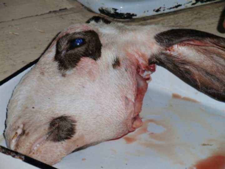
Figure 1. Experimental animal—sheep.
2. Condylar Process
Both shape and dimensions of the sheep mandibular condyle show high variation from that of the human condyle. The condyle is medio-laterally concave as against convex in the human condyle. The condyle forms a saucer-like depression mesio—distally and medio-laterally to fit in exactly with the glenoid fossa, unlike the human condyle which is rounded mesio—distally and medio-laterally. Cartilage covers entire glenoid fossa and condylar head, indicating parts of the temporomandibular joint which are subject to highest loading (Figures 2(a) and (b)).
3. Articular Disc
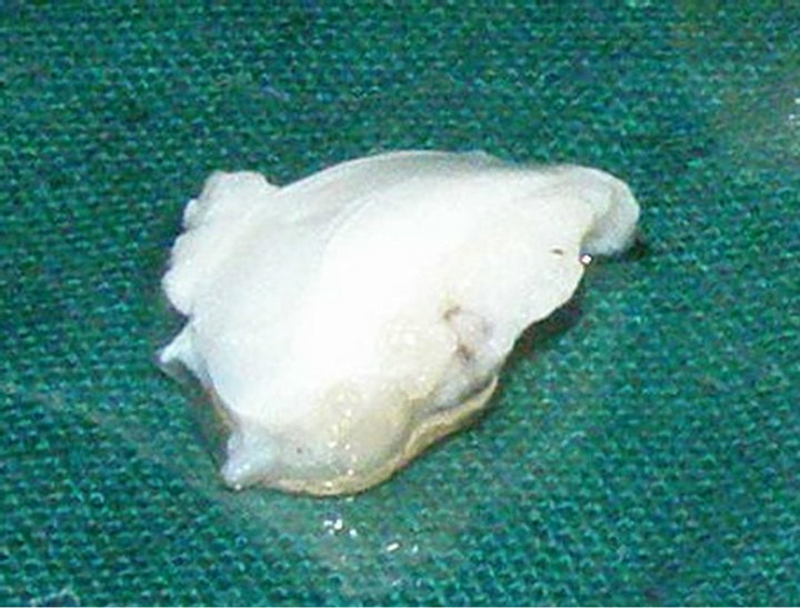
The articular disc, a mobile fossa for the condyle, adheres anteriorly, medially and laterally to the articular capsule. Posteriorly, it extends to the retro-articular connective tissue pad. The thickness of unloaded articular disc is 1. 5 mm, firm and slightly elastic in nature (Figure 3(a)). A particular feature of the joint is its retroarticular pad (bilaminar zone, genu vasculosa) comprising of vascular tufts, connective tissue and fatty tissue. It is hydro-pneumatic pad for dislocations (if any) within the joint and compensates for volume changes. It splits into two portions 1) upper portion attached to the glenoid fossa and 2) lower portion attached to the condyle as shown in Figure 3(b). Moreover, tendon ends of the muscle lateral pterygoid medially radiate to the anterior disc as seen in Figure 3(c). The disc is functionally divided into three parts: anterior part, intermediate part and posterior part.
4. The Articular Capsule
The articular capsule (Figure 4) is relatively wide, thus providing the condyle with large moving space. Laterally, it is reinforced by the lateral ligament, which restrains retrusive and downward movements of the condyle beyond the articular tubercle. On the medial part, the
 (a)
(a)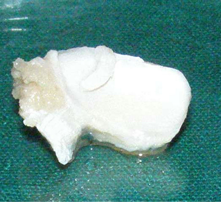 (b)
(b)
Figure 2. (a) Mandibular condyle articulating with the glenoid fossa; (b) Specimen of mandibular condyle fixed in 10% formalin.
 (a)
(a)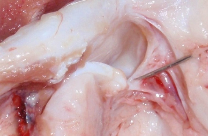 (b)
(b)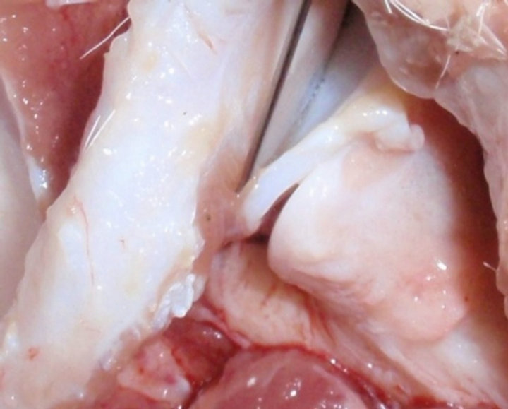 (c)
(c)
Figure 3. (a) Tissue specimen of articular disc fixed in 10% formalin; (b) Posterior retrodiscal lamina attached superiorly to the glenoid fossa; (c) Lateral pterygoid muscle attached to disc.

Figure 4. Articular capsule seen medially.
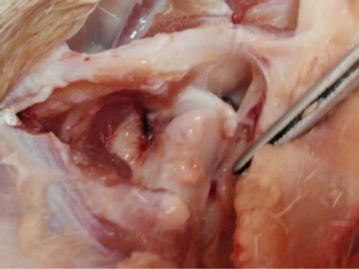
Figure 5. Glenoid fossa.
capsule is only slightly reinforced, which may account for articular bilaterality.
5. Glenoid Fossa
The glenoid fossa is antero-posteriorly larger than mediolaterally with a convexity downwards to articulate like a ball and socket joint, exactly opposite to that of glenoid fossa in human beings which has concavity upwards. It is formed by inferior part of the temporal bone, forming base of the skull (Figure 5). The glenoid fossa allows free movement of the condylar head medio-laterally for grinding of food.
6. Articular Tubercle
Articular tubercle, a special feature in the human beings, is rudimentary in the sheep, since the path of movement of the condyle is s-shaped, sloping upwards contradictory to as seen in humans beings.
7. Conclusion
Sheep is an excellent large experimental model for the study of condylar growth, with and without the use of functional appliances, because of the similarity in anatomy related to size, shape and position of the condyle. This review will help future investigators in the field of dentistry to consider the sheep (Ovis aries) as an experimental animal for application oriented research.
REFERENCES
- S. J Maynard and R. J. G. Savage, “The Mechanics of Mammalian Jaws,” School Science Review, Vol. 40, No. 4, 1959, pp. 289-301.
- F. Jochen and G. Tomasz, “On the Development, Morphology and Function of the Temporomandibular Joint in the Light of the Orofacial System,” Annals of Anatomy— Anatomischer Anzeiger, Vol. 189, No. 4, 2007, pp. 314- 319. doi:10.1016/j.aanat.2007.02.024
- R. Sprinz, “A Note on the Mandibular Intra-Articular Disc in the Joints of Marsupialia and Monotremata,” Proceedings of the Zoological Society of London, Vol. 144, No. 3, 1965, pp. 327-338. doi:10.1111/j.1469-7998.1965.tb05185.x
- S. W. Herring, “TMJ Anatomy and Animal Models,” Journal of Musculoskeletal and Neuronal Interactions, Vol. 3, No. 4, 2003, pp. 391-394.
- S. W. Herring, J. D. Decker, Z. J. Liu and T. Ma, “The Temporomandibular Joint in Miniature Pigs: Anatomy, Cell Replication, and Relation to Loading,” The Anatomical Record, Vol. 266, No. 3, 2002, pp. 152-166. doi:10.1002/ar.10049
- A. Bermejo, O. González and J. M. González, “The Pig as an Animal Model for Experimentation on the Temporomandibular Articular Complex,” Oral Surgery, Oral Medicine, Oral Pathology, Vol. 75, No. 1, 1995, pp. 18-23. doi:10.1016/0030-4220(93)90399-O
- J. J. Thomson, L. E. Grovum, A. G. Deswysen and W. W. Bignell, “In Vivo Surface Strain and Stereology of the Frontal and Maxillary Bones of Sheep: Implications for the Remodeling and Mechanical Optimization of Cranial Bones,” The Anatomical Record, Vol. 264, No. 4, 2001, pp. 325-338.
- W. Susan, Z. Herring and L. Jun, “Loading of the Temporomandibular Joint: Anatomical and in Vivo Evidence from the Bones,” Cells Tissues Organs, Vol. 169, No. 3, 2001, pp. 193-200. doi:10.1159/000047882
- F. de Vree and C, Gans, “Mastication in Pygmy Goats Capra hircus,” Annales de la Societe Royale Zoologique de Belgique, Vol. 195, No. 3, 1976, pp. 255-306.
NOTES
*Corresponding author.