Advances in Microbiology
Vol.3 No.1(2013), Article ID:29224,3 pages DOI:10.4236/aim.2013.31014
Growth Yields of Four Coxiella burnetii Isolates in Four Different Cell Culture Lines
1The Australian Rickettsial Reference Laboratory/Barwon Biomedical Research, The Geelong Hospital, Geelong, Australia
2Murdoch University, Murdoch, Australia
3Department of Microbiology, Pathology North-Hunter, John Hunter Hospital, New Lambton Heights, Australia
Email: michelle.g.lockhart@gmail.com
Received January 27, 2013; revised February 28, 2013; accepted March 15, 2012
Keywords: Bacterial Yield; Cell Culture; Coxiella burnetii; Intracellular Bacteria
ABSTRACT
Although Coxiella burnetii is considered to be an obligate intracellular bacterium and grows in embryonated eggs, laboratory animals and cell culture, recently it has been grown in cell-free media and on agar plates. This current study was conducted to compare four cell lines for their yield of C. burnetii. Four different isolates of C. burnetii (Henzerling, Arandale, Cumberland and Timony) were grown in DH82, L929, Vero and XTC-2 cell lines. The DH82 and XTC-2 cells lines produced the highest C. burnetii yield which was slightly less than the yields achieved in recently published studies using cell free media. The Arandale isolate of C. burnetii produced a significantly higher yield in DH82 cells compared to XTC-2 cells (P < 0.03).
1. Introduction
Q Fever is a worldwide zoonosis caused by Coxiella burnetii and serological testing by immunofluorescence assay (IFA) is generally used for diagnosis. C. burnetii was traditionally cultured in embryonated eggs or laboratory animals such as guinea pigs and mice. The use of cell culture has permitted the growth of C. burnetii, in flasks or multi-welled trays containing a monolayer of eukaryotic host cells [1]. Traditionally, C. burnetii has been considered an obligate intracellular bacterium. However, C. burnetii was recently grown without host cells [2].
In cell culture the infection does not generally destroy the host cells and infected cells have the same cell cycle progression as uninfected cells. This is a result of asymmetric division of infected cells producing one infected and one uninfected daughter cell. This ability of C. burnetii has allowed it to persistently infect cell cultures for over two years without the addition of uninfected cells [3]. The infected cell monolayer exhibits cytopathic effect (CPE) at the same rate as uninfected cultures. Thus infection of the culture must be observed through the use of other methods such as IFA or polymerase chain reaction (PCR). The optimal growth of C. burnetii is important if large numbers of bacteria are required for protein or DNA studies or vaccine production. In this study four different cell lines were compared to determine which produced the greatest yield of C. burnetii. The cell lines chosen for this study included cell lines used to grow C. burnetii previously; Vero (African green monkey epithetlial cells) [4] and L929 (mouse fibroblast cells) [5,6] and two other cell lines currently used in our laboratory; DH82 (canine macrophage cells) and XTC-2 (South African clawed frog epithelial cells). This study compares the yields obtained in cell lines to the yields obtained in cell-free media [7].
2. Materials and Methods
Vero, DH82 and L929 cells were grown in 10ml RPMI (Gibco, Australia) supplemented with 10% new born calf serum (NBCS) (Gibco, Australia) and 1% L-glutamine (Gibco, Australia). Cell lines were incubated at 35˚C with 5% CO2. The XTC-2 cell line was grown with 10 ml Leibovitz L-15 (Gibco, Australia) media supplemented with 10% NBCS (Gibco, Australia), 0.4% tryptose phosphate broth (Oxoid, England) and 1% L-glutamine (Gibco, Australia) and incubated at 28˚C. These were inoculated with suspensions of antigenic phase I C. burnetii of the Henzerling isolate (homogenised infected egg yolk sack, courtesy of Commonwealth Serum Laboratories CSL, Australia) and three Australian isolates Arandale, Cumberland and Timony (homogenised infected spleens from severe combined immunodeficiency [SCID] mice). The suspensions (0.5 mL) were first diluted in 9.5 mL of Hanks’ balanced salt solution (HBSS, Gibco, Australia) and filtered through a 0.45 μm filter to reduce the amount of host material. An aliquot of the filtrate (0.8 ml) was added to each flask. Two flasks of each confluent cell line were inoculated with each C. burnetii isolate. Cultures had fortnightly changes of media. At six weeks post infection it was assumed that maximal growth had occurred and the monolayer was harvested by scraping, pelleted and resuspended in 1 ml PBS. All cultures growing C. burnetii were performed in bio-containment level 3 laboratory at the Department of Microbiology, John Hunter Hospital, Newcastle.
DNA was extracted from a 200 μl aliquot of this suspension using the Qiagen QIAamp DNA Mini Kit (Qiagen, Germany) following the manufacturers instructions. Samples were then analysed by a real time PCR assay [8] targeting the Com1 gene, which codes for a highly conserved 27 kDa outer membrane protein. The cycling threshold (Ct) result was used to calculate the DNA concentration (μg/μl). This was achieved using a standard curved made with plasmids cloned with the resulting PCR amplicon as described previously [8].
3. Results
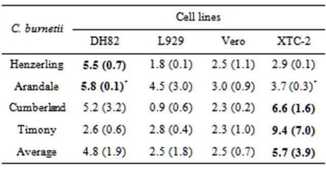
The results showed higher growth yields in DH82 cell line for two isolates (Henzerling and Arandale) and in XTC-2 cell line for the other two (Cumberland and Timony) (Table 1). The Arandale isolate grew to higher numbers in the DH82 cell line compared to the XTC-2 cell line (P = 0.03). Overall the XTC-2 cell line appeared to yield the highest concentration of C. burnetii DNA after six weeks of culture. To determine if the host cell density was proportional to the yield of intracellular bacteria the optical density (OD) of the cell monolayers harvested was determined (data not shown). While variation was found in OD between flasks this did not correlate with the yield of C. burnetii DNA. To compare these results with previous studies the data was converted into bacterial cell numbers per ml of media. The total yield of C. burnetii obtained was approximately 107/mL of media in a flask containing a monolayer of host cells and 10 mL of media.
4. Discussion
Obtaining C. burnetii isolates enables comparative studies to be made on different isolates. The yield of these bacteria produced in different cell lines is currently unknown. The findings of this study indicate that cell line DH82 and/or XTC-2 are best for growing C. burnetii to a high yield. The reasons for the preference of one cell line over another were not determined. This may be due to the ease with which C. burnetii can enter and multiply within that host cell, a feature of the C. burnetii strain itself, or the conditions under which it was grown; such as the lower temperature for the XTC-2 cell line or a component of the media.
The isolates used include the Henzerling isolate from an acute case of Q fever in Italy and which has been classified as group II [9]. The three other isolates used in this study were from acute cases of Q fever in Australia and were classified as group III (typing method from [10] (data not shown).
To determine if the lack of statistical significance between each cell line was due to high variability of host cell numbers, the optical density (OD) of the cells was determined (data not shown). It was presumed that a higher OD reading and a higher density of host cells would correlate with a higher yield as there would be more host cells with vacuoles full of bacteria. While variation was found in OD between flasks this did not correlate with the variability in the amount of C. burnetii DNA detected when compared across either isolate or cell line groupings.
Using these cell lines the total yield (in a flask containing host cells and 10 mL of media) was around 107/mL. This was less than when C. burnetii was grown without the use of host cells yeilding around 108/mL of media [7]. C. burnetii grown in cell lines consists of C. burnetii free in the media (having been released from their intracellular location) and in vacuoles within host cells in the monolayer. If the monolayer was growing in 1 mL of media (rather than 10 mL) the yield of bacteria per mL would have been the same as the cell free media. However, such a low media volume may not give optimal conditions for the host cells. Greater yields have been obtained through animal inoculation with severe combined immunodeficiency (SCID) mice producing 9 ´ 109/g of spleen (Michelle Lockhart, personal communication). However the use of animals is more costly to set up and maintain. The results of this study indicate that the cell free media may be preferable for producing C. burnetii with the added advantage of no contaminating host cells.
Table 1. Yield (μg/μl of DNA detected by Com1 PCR) of four isolates of C. burnetii grown in four different cell culture lines.

One standard deviation is given in brackets; highest yield of DNA from each isolate is given in bold. Statistical differences (by student t-test) *P = 0.03.
Higher yeilds would be useful for some studies and in the case of vaccine production.
In the human host C. burnetii infects monocytes and macrophages. Coxiella burnetii enters the host cell by inducing endocytosis in phagocytes. Unlike other intracellular bacteria that either escape the phagosome into the cytoplasm or prevent the binding of the lysosomes to the phagosome, C. burnetii survives in the highly acidic environment within the phagolysosome [6]. The intracellular environment gives the bacteria access to the host cell’s nutrients and molecular building blocks. Coxiella actively transports glucose, glutamate [11], proline [12] and other substrates from the intracellular environment. These transportation systems and the metabolism of nucleic acids and amino acids have been shown to be pH dependent [11-13]. The host-cell free media [2,7] appears to mimic this intra phagolysosome environment and reduces the need for growing and maintaining cell lines. We have shown that the use of cell lines produces a lower yield of C. burnetii than in cell free media [7].
REFERENCES
- W. Rumin, D. Kruszewska, W. Sadowski and S. Tylewska-Wierzbanowska, “Growth of Coxiella burnetii in Selected Cell Cultures,” Medycyna Doświadczalna i Mikrobiologia, Vol. 42, No. 1-2, 1990, pp. 44-49.
- A. Omsland, et al., “Host Cell-Free Growth of the Q Fever Bacterium Coxiella burnetii,” Proceedings of the National Academy of Sciences of the United States of America, Vol. 106, No. 11, 2009, pp. 4430-4434. doi:10.1073/pnas.0812074106
- M. J. Roman, P. D. Coriz and O. G. Baca, “A Proposed Model to Explain Persistent Infection of Host Cells with Coxiella burnetii,” Journal of General Microbiology, Vol. 132, No. 5, 1986, pp. 1415-1422. doi:10.1099/00221287-132-5-1415
- T. F. McCaul, T. Hackstadt and J. C. Williams, “Ultrastructural and Biological Aspects of Coxliella burnetii under Physical Disruptions,” In: W. Burgdorfer and R. L. Anacker, Eds., Rickettsiae and Rickettsial Diseases, Academic Press, New York, 1981, pp. 267-280.
- O. G. Baca, et al., “Cell Cycle Distribution Patterns and Generation Times of L929 Fibroblast Cells Persistantly Infected with Coxiella burnetii,” Infection and Immunity, Vol. 47, No. 2, 1985, pp. 366-369.
- P. R. Burton, et al., “Some Ultrastructural Effects of Persistant Infections by the Rickettsia Coxiella burnetii in Mouse L Cells and Green Monkey Kidney (Vero) Cells,” Infection and Immunity, Vol. 21, No. 2, 1978, pp. 556- 566.
- A. Omsland, et al., “Isolation From Animal Tissue and Genetic Transformation of Coxiella burnetii Are Facilitated by an Improved Axenic Growth Medium,” Applied and Environmental Microbiology, Vol. 77, No. 11, 2011, pp. 3720-3725. doi:10.1128/AEM.02826-10
- M. G. Lockhart, et al., “A Comparison of Methods for Extracting DNA from Coxiella burnetii as Measured by a Duplex qPCR Assay,” Letters in Applied Microbiology, Vol. 52, No. 5, 2011, pp. 514-520. doi:10.1111/j.1472-765X.2011.03034.x
- L. R. Hendrix, J. E. Samuel and L. P. Mallavia, “Differentiation of Coxiella burnetii Isolates by Analysis of Restriction Endonuclease—Digested DNA Separated by SDS-PAGE,” Journal of General Microbiology, Vol. 137, No. 2, 1991, pp. 269-276. doi:10.1099/00221287-137-2-269
- A. M. Denison, H. A. Thompson and R. F. Massung, “IS1111a Insertion Sequences of Coxiella burnetii: Charactarisation and Use For Repetitive Element PCR-Based Differentiation of Coxiella burnetii Isolates,” BMC Microbiology, Vol. 7, No. 91, 2007, pp. 91-98.
- T. Hackstadt and J. C. Williams, “Biochemical Stratagem for Obligate Parasitism of Eukaryotic Cells by Coxiella burnetii,” Proceedings of the National Academy of Sciences of the United States of America, Vol. 78, No. 5, 1981, pp. 3240-3244. doi:10.1073/pnas.78.5.3240
- L. Hendrix and L. P. Mallavia, “Active Transport of Proline by Coxiella burnetii,” Journal of General Microbiology, Vol. 130, No. 11, 1984, pp. 2857-2863. doi:10.1099/00221287-130-11-2857
- T. F. McCaul and J. C. Williams, “Developmental Cycle of Coxiella burnetii: Structure and Morphogenesis of Vegetative and Sporogenic Differentiations,” Journal of Bacteriology, Vol. 147, No. 3, 1981, pp. 1063-1076.

